Another study, published in the American Journal Of Sports Medicine, found that the following exercise routine helped participants perform better on the single leg hop test, improve self-reported knee function scores, improve quadriceps and hamstring strength, and even showed a positive trend for return to sports after surgery
According to the study:
“The program consisted of 4 exercise periods per week: 2 supervised gym sessions alternating with 2 supervised home sessions… Patients were allowed to perform 1 repetition of their maximal effort to determine starting weights in the gym. Each gym activity consisted of 3 sets of 12 repetitions and the weights were increased by 10% to 15% each week. During the last gym session, the weights were returned to the previous week’s value to prevent preoperative fatigue and to promote muscular endurance response and mass gain. Home exercises consisted of the same program as in the gym, but with the use of a TheraBand instead of weights. Patients used a DonJoy knee brace locked at 20 degrees of extension to reduce the risk of further ACL loading pre- or postoperatively during open kinetic chain (OKC) exercise. The brace also acts as a confidence factor during OKC exercises. Proprioception training was performed on a wobble cushion.”
The exercise routine, highlighted in the appendix of the study, consists of the next. Although the full text of the study is not freely available, you can access the appendix here by clicking on “Supplementary Material” in the left column.
Exercise group 1 – All exercises 3×12
Leg press
Leg curl
Leg extension
Exercise group 2 – All exercises 3×12
Half drop
45 degree squat
Calf raises
Exercise group 3 – All exercises 3×12
Hip abduction
Hip adduction
Hip flexion
Balancing/Proprioception – 3×30 seconds – Use a wobble cushion
Step 1: Stand on one leg with eyes open
Step 2: Stand on one leg with eyes closed
Step 3: Stand on one leg with eyes closed, head raised
Stretching – All stretching exercises 3×30 seconds
The largest study of CTE to date has found a new link between contact sports participation, chronic traumatic encephalopathy (CTE) and the development of a movement disorder known as parkinsonism.
The study of 481 deceased athletes by researchers from Boston University Chobanian and Avedisian School of Medicine and VA Boston Healthcare, published today in JAMA Neurologyit appears that most individuals with CTE developed parkinsonism, and CTE pathology appears to be the cause of the parkinsonism symptoms in most cases.
Parkinsonism is a condition characterized by symptoms similar to Parkinson’s disease, such as tremor, abnormal slowness of movement, or abnormal stiffness of the arms or legs. It has long been associated with traumatic brain injury (TBI) and CTE in boxers. However, the specific pathologies underlying these symptoms in CTE were unknown.
Parkinson’s disease is classically associated with the buildup of proteins called Lewy bodies in brain cells, but researchers found that 76% of individuals with CTE and Parkinsonism did not do Have Lewy body pathology.
“We were surprised to find that most individuals with CTE and parkinsonism did not have Lewy body pathology,” noted Thor Stein, MD, PhD, associate professor of pathology and laboratory medicine at BU and VA Boston Healthcare, and one of the study’s corresponding authors. “Instead,” Stein explained, “subjects with parkinsonism were more likely to have more severe CTE-related brain cell death in a region of the brainstem important for controlling movement.”
CTE is a degenerative brain disease whose only known cause is repeated head blows, such as those that occur in contact sports. A 2018 study by the same research team found that the duration of contact sports is associated with an increased risk of developing Lewy body disease. However, the current study is the first to describe a link between contact sports participation, brainstem pathology, and parkinsonism in CTE.
“Increased CTE severity has been shown to be associated with longer playing time,” noted Daniel Kirsch, an MD/PhD student at BU and one of the study’s first authors. “In this study, we found that eight additional years of contact sports play was associated with a 50 percent increased risk of more severe disease in a specific area of the brainstem that controls movement.”
The study subjects donated their brains to the Understanding Neurologic Injury and Traumatic Encephalopathy (UNITE) brain bank. People with parkinsonism were compared to those without to identify the types of pathologies that might explain why some people with CTE develop these symptoms and to examine relationships with the duration of contact sports.
This study underscores the importance of understanding the long-term effects of repeated head impacts and the need for preventive measures in contact sports to reduce the risk of neurodegenerative diseases such as CTE and parkinsonism.
Are you recovering from ACL surgery and wondering what exercises you can do to aid in your recovery? This video will cover the early stages of recovery, explain what factors are important during this stage, and outline key exercises you can do every day to aid in your recovery.
During the early stages of recovery after anterior cruciate ligament surgery, the main goals are to achieve full range of motion and regain neuromuscular control of the knee.[1] To achieve these goals, it is essential to manage pain, swelling, and inflammation of the knee to ensure that you can complete your rehabilitation exercises. Using ice, applying compression, elevating your foot, and doing ankle pumps are great ways to manage pain, swelling, and inflammation.
While achieving both flexion and extension range of motion is important for activities of daily living, achieving full knee extension is emphasized for people recovering from ACL surgery.[1] Knee extension is especially important in the early stages of recovery, as numerous studies have shown that loss of full knee extension can be a predictor of postoperative complications and poor outcomes.[2][3][4][5] Therefore, exercises to increase knee range of motion are especially important during the early phase of ACL recovery.
Achieving neuromuscular control of the knee is another important goal after ACL surgery. Neuromuscular control of the knee refers to the coordination of your brain, nerves, and muscles to create movement and stability.[6] Regaining neuromuscular control is essential to not only ensure proper movement and function, but also to prevent further injury to your knee.[6] Some ways to regain neuromuscular control of the knee include strengthening the supporting muscles around the knee and doing balance exercises.[1]
During the early recovery phase following ACL surgery, exercises that focus on knee flexion, knee extension, knee muscle strengthening, and proprioception are recommended for the first 4 weeks following surgery. Five safe and effective exercises that you can do daily to help you recover functionally include quadriceps and hamstring co-contractions, heel glides, knee extension over a roller, straight leg raises, and chair assisted squats. Click on this link for a more detailed description of these exercises. Keep in mind that everyone recovers at their own pace, so your ability to perform these exercises may vary depending on your condition. Consult with your healthcare provider to ensure that these exercises are safe for you.
Listen to Joey Wong, kinesiologist, explain the key exercises to focus on after ACL surgery, and learn about ACL recovery goals.
The Curovate Physical Therapy App has detailed videos and descriptions of these 5 essential exercises, as well as many other important exercises for your recovery. Click on one of the download links below to get started on your ACL recovery today! Curovate is an evidence-based app that provides guided physical therapy plans, daily guided video exercises, ways to measure and monitor your progress, and measures your knee range of motion! Download the Curovate App by clicking on the links below.
If you require further, tailored assistance during your ACL recovery, you can book a 1-on-1 video session with a physiotherapist on our Virtual Physiotherapy page.
When it comes to traumatic knee injuries, treatment options vary depending on the severity of the injury. Knee injuries can be caused by a variety of factors, including sports injuries, falls, and car accidents. Symptoms of a knee injury may include pain, swelling, stiffness, and difficulty moving the knee joint.
If you suspect that you have a knee injury, it is important to seek medical attention as soon as possible. A doctor can diagnose the injury and recommend the appropriate treatment. Diagnosis may involve a physical exam, imaging tests such as X-rays or MRI scans, and possibly arthroscopy, which involves inserting a small camera into the knee joint to examine the damage.
Treatment options for a traumatic knee injury may include rest, ice, compression, and elevation (RICE), physical therapy, medications, and surgery. The specific treatment plan will depend on the type and severity of the injury. In some cases, conservative treatment such as RICE and physical therapy may be sufficient to heal the injury. In more severe cases, surgery may be necessary to repair or replace damaged tissue.
Assessment and Immediate Care
When a traumatic knee injury occurs, prompt assessment and care are essential for optimal healing. In this section, we will discuss the initial evaluation, pain management, and swelling reduction.
Initial Evaluation
The initial evaluation of a traumatic knee injury includes a thorough physical exam and imaging studies. During the physical exam, we will assess the knee for tenderness, swelling, and range of motion. We may also perform special tests to evaluate the ligaments and menisci of the knee. X-rays may be obtained to evaluate for fractures or dislocations, while an MRI may be necessary to assess for soft tissue injuries.
Pain Management
Pain management is an important aspect of the immediate care of a traumatic knee injury. We recommend the use of non-steroidal anti-inflammatory drugs (NSAIDs) for pain relief. In addition, we may prescribe stronger pain medications if necessary. We also recommend the use of ice to reduce pain and inflammation. Applying ice to the knee for 20 minutes every 2-3 hours can help to alleviate pain.
Swelling Reduction
Swelling is a common symptom of a traumatic knee injury and can be reduced with the use of rest, ice, compression, and elevation (RICE). We recommend that patients rest the affected knee and avoid weight-bearing activities until the swelling has subsided. Applying ice to the knee can help to reduce swelling, while compression with an elastic bandage can help to prevent further swelling. Elevating the knee above the level of the heart can also help to reduce swelling.
In conclusion, prompt assessment and care are essential for the treatment of a traumatic knee injury. The initial evaluation should include a thorough physical exam and imaging studies. Pain management can be achieved with the use of NSAIDs and ice, while swelling can be reduced with RICE.
Rehabilitation and Treatment Options
When it comes to treating a traumatic knee injury, there are several rehabilitation and treatment options available. These options can be divided into two categories: non-surgical therapies and surgical interventions.
Non-Surgical Therapies
For less severe knee injuries, non-surgical therapies such as physical therapy and exercise can be effective in promoting healing and restoring function. Physical therapy can help to strengthen the muscles around the knee, improve range of motion, and reduce pain and swelling. Exercises such as straight leg raises, hamstring curls, and calf raises can also be beneficial in promoting strength and function.
In addition to physical therapy and exercise, medication such as nonsteroidal anti-inflammatory drugs (NSAIDs) can be used to reduce pain and inflammation. Bracing and compression can also be used to provide support and reduce swelling.
Surgical Interventions
For more severe knee injuries, surgical intervention may be necessary. The type of surgery required will depend on the nature and extent of the injury. Common surgical interventions for knee injuries include arthroscopic surgery, meniscus repair, ACL reconstruction, and ligament reconstruction.
Arthroscopic surgery is a minimally invasive procedure that uses small incisions and a camera to visualize and treat the injured area. Meniscus repair involves repairing or removing the damaged meniscus. ACL reconstruction involves replacing the torn ACL with a graft. Ligament reconstruction involves repairing or replacing the damaged ligament.
Recovery time for surgical interventions can vary depending on the type and extent of the surgery. Rehabilitation is an important part of the recovery process and typically involves physical therapy to restore strength and function to the knee.
In conclusion, there are several rehabilitation and treatment options available for traumatic knee injuries. Non-surgical therapies such as physical therapy and exercise can be effective for less severe injuries, while surgical interventions may be necessary for more severe injuries. It is important to work closely with a healthcare professional, such as an orthopedic surgeon, to determine the best course of treatment for your specific injury. Clinical trials may also be available for those interested in exploring new treatment options.
Knee injuries are common and can be caused by various factors such as sports, accidents, and repetitive motion. Traumatic knee injuries can be devastating and can lead to long-term complications. Therefore, it is essential to take preventive measures to avoid such injuries. In this article, we will discuss some effective ways to prevent traumatic knee injuries.
One of the most effective ways to prevent knee injuries is by strengthening the muscles around the knee. Strong muscles can help support the knee joint and reduce the risk of injury. Exercises such as lunges, squats, and leg presses can help strengthen the muscles around the knee. Additionally, stretching can help improve flexibility and reduce the risk of muscle strains.
Another way to prevent knee injuries is by wearing protective gear during sports or activities that involve jumping, running, or sudden changes in direction. Knee pads, braces, and proper footwear can help absorb shock and reduce the risk of injury. It is also essential to warm up before engaging in physical activities to prepare the muscles and joints for the stress they will endure.
In summary, taking preventive measures can help reduce the risk of traumatic knee injuries. By strengthening the muscles around the knee, wearing protective gear, and warming up before physical activities, we can protect our knees and avoid long-term complications.
Understanding Knee Injuries
When it comes to knee injuries, prevention is key. Knee injuries can be caused by various factors, including trauma, overuse, and degenerative conditions. Understanding the anatomy of the knee, common causes and types of knee injuries, as well as their symptoms and diagnosis can help you take steps to prevent knee injuries.
Anatomy of the Knee
The knee is a complex joint that connects the thigh bone (femur) to the shin bone (tibia). The knee joint also includes the kneecap (patella) and other structures such as ligaments, tendons, cartilage, and meniscus. The ligaments provide stability to the knee, while the tendons connect the muscles to the bones. The cartilage and meniscus act as shock absorbers and help to distribute weight evenly across the joint.
Common Causes and Types of Knee Injuries
Knee injuries can be caused by a variety of factors, including trauma, overuse, and degenerative conditions such as osteoarthritis and rheumatoid arthritis. Common types of knee injuries include fractures, dislocations, ligament tears, meniscus tears, and torn cartilage.
Fractures occur when there is a break in the bone, while dislocations occur when the bones in the joint are forced out of position. Ligament tears can occur when the ligaments that connect the bones in the knee are stretched or torn, while meniscus tears can occur when the cartilage in the knee is torn. Torn cartilage can also occur as a result of overuse or degenerative conditions.
Symptoms and Diagnosis
Symptoms of knee injuries can include pain, swelling, stiffness, redness, warmth, and fever. Chronic pain can also be a symptom of knee injuries. Diagnosis of knee injuries typically involves a physical examination, imaging tests such as X-rays and MRI scans, and sometimes arthroscopy, a minimally invasive surgical procedure.
In conclusion, knee injuries can be caused by various factors, and understanding the anatomy of the knee, common causes and types of knee injuries, as well as their symptoms and diagnosis can help you take steps to prevent knee injuries.
Prevention and Management Strategies
Exercise and Physical Activity
Engaging in regular physical activity and exercise can help prevent traumatic knee injuries. It is important to incorporate exercises that focus on strengthening the muscles around the knee joint, such as the quadriceps, hamstrings, and calf muscles. These exercises can include lunges, squats, leg presses, and step-ups.
Stretching is also important in preventing knee injuries. It is recommended to stretch before and after exercise to improve flexibility and reduce the risk of injury. Proper technique and form during exercise is crucial in preventing knee injuries. Coaches and trainers can provide guidance on proper technique and form during exercise.
Protective Gear and Techniques
Wearing proper protective gear, such as knee braces, can help prevent knee injuries during sports and physical activity. Knee braces can help provide support and stability to the knee joint, reducing the risk of injury.
Techniques such as jumping and landing properly can also help prevent knee injuries. It is important to land softly and use proper technique when jumping, especially during sports such as basketball or volleyball.
Treatment and Recovery
If a knee injury does occur, it is important to seek medical attention from a doctor or physical therapist. Treatment may include rest, compression, elevation, and ice to reduce swelling and pain. Anti-inflammatory drugs such as ibuprofen may also be prescribed to reduce pain and inflammation.
In some cases, surgery may be necessary to repair a torn ligament or tendon. Physical therapy may also be recommended to help with recovery and prevent future injuries.
Overall, taking steps to prevent knee injuries through exercise, proper technique, and protective gear can help reduce the risk of injury and promote long-term knee health.
Our knees are fundamental to our mobility and daily activities. Yet, knee pain is a widespread issue affecting millions worldwide. This comprehensive guide will delve into the causes, symptoms, and treatments for knee pain, providing you with the knowledge to better understand and manage this common condition.
Introduction
Knee pain is a pervasive problem that doesn’t discriminate by age or lifestyle. Whether you’re an athlete pushing your limits, an office worker with a sedentary job, or somewhere in between, knee pain can strike and significantly impact your quality of life. Understanding the complexities of knee pain is crucial for effective management and treatment.
According to recent studies, approximately 25% of adults suffer from frequent knee pain. This staggering statistic underscores the importance of knee health education and proactive care.
[Image: Anatomical diagram of a knee] Alt text: Detailed anatomical diagram showing the structure of a human knee joint
Anatomy of the Knee
To truly understand knee pain, we must first explore the intricate anatomy of the knee joint.
Basic Structure
The knee is one of the largest and most complex joints in the human body. It consists of:
Bones: The femur (thighbone), tibia (shinbone), and patella (kneecap)
Cartilage: Including the meniscus, which provides cushioning between bones
Ligaments: Connect bones and provide stability (e.g., ACL, PCL, MCL, LCL)
Tendons: Connect muscles to bones (e.g., patellar tendon)
Bursae: Small fluid-filled sacs that reduce friction between tissues
Function of Each Component
Bones: Provide the structural framework and enable movement
Cartilage: Acts as a shock absorber, reducing friction between bones
Ligaments: Stabilize the joint and guide motion
Tendons: Allow muscles to move the joint
Bursae: Reduce friction and provide smooth movement
The knee’s design allows for a range of motions, including flexion, extension, and a small degree of rotation. This complexity, while providing great functionality, also makes the knee susceptible to various types of injuries and conditions.
[Image: Labeled diagram of knee anatomy] Alt text: Labeled anatomical diagram of a knee showing bones, cartilage, ligaments, and tendons
Common Causes of Knee Pain
Knee pain can result from a wide array of factors, ranging from acute injuries to chronic conditions. Understanding these causes is crucial for proper diagnosis and treatment.
Injuries
Sprains and strains: Overstretching or tearing of ligaments or muscles
Meniscus tears: Damage to the cartilage that cushions the knee joint
Ligament tears: Such as ACL (Anterior Cruciate Ligament) or MCL (Medial Collateral Ligament) injuries
Fractures: Breaks in the kneecap, lower femur, or upper tibia
Arthritis
Osteoarthritis: The most common form, caused by wear and tear of cartilage
Rheumatoid arthritis: An autoimmune condition that causes joint inflammation
Post-traumatic arthritis: Develops after an injury to the knee
Overuse Conditions
Tendinitis: Inflammation of tendons, often due to repetitive activities
Bursitis: Inflammation of the bursae, frequently caused by prolonged kneeling
Patellofemoral pain syndrome: Pain around the kneecap, common in runners
Other Causes
Gout: Build-up of uric acid crystals in the joint
Infections: Bacterial or viral infections in the joint or surrounding tissues
Osgood-Schlatter disease: Common in adolescents, causing pain below the kneecap
Iliotibial band syndrome: Common in runners, causing outer knee pain
[Image: Infographic showing percentage breakdown of common knee pain causes] Alt text: Pie chart illustrating the percentage distribution of common causes of knee pain
Statistics on Knee Pain Causes
I have created a detailed table showing the prevalence of different causes of knee pain, including the age group most affected, common symptoms, and potential treatments. If you need any further modifications or additions, please let me know!
These statistics highlight the significant role that degenerative conditions like osteoarthritis play in knee pain, while also showing the substantial impact of acute injuries.
Symptoms Associated with Knee Pain
Knee pain can manifest in various ways, and understanding these symptoms can help in early identification and treatment.
Types of Pain
Sharp pain: Often associated with injuries or meniscus tears
Dull ache: Common in chronic conditions like arthritis
Burning pain: May indicate nerve issues or inflammation
Throbbing pain: Could be related to fluid buildup or inflammation
Other Symptoms
Swelling: Indicates inflammation or injury
Stiffness: Common in arthritis or after periods of inactivity
Instability: Feeling like the knee might give way, often due to ligament issues
Locking: The knee gets stuck in one position, often due to a meniscus tear
Popping or crunching noises: May indicate cartilage wear or loose bodies in the joint
Warning Signs
Severe pain or swelling
Inability to bear weight
Visible deformity
Fever accompanying knee pain
Redness and warmth around the joint
It’s important to note that symptoms can vary greatly depending on the underlying cause. For instance, osteoarthritis pain often worsens with activity, while rheumatoid arthritis may cause more pain in the morning.
[Image: Photo series showing visual symptoms like swelling] Alt text: Series of photos showing different visual symptoms of knee problems, including swelling and redness
Diagnosis of Knee Pain
Accurate diagnosis is crucial for effective treatment of knee pain. Healthcare providers use a combination of methods to determine the underlying cause.
Physical Examination
A thorough physical exam is often the first step in diagnosis. This may include:
Assessment of range of motion
Palpation for areas of tenderness
Stability tests to check ligament integrity
Gait analysis to observe walking patterns
Imaging Tests
Various imaging techniques can provide detailed views of the knee structures:
X-rays: Show bone structure and alignment, useful for diagnosing fractures or arthritis
MRI (Magnetic Resonance Imaging): Provides detailed images of soft tissues, excellent for diagnosing ligament or meniscus injuries
CT (Computed Tomography) scan: Offers cross-sectional images of the knee, useful for complex fractures
Ultrasound: Can show real-time images of tendons and ligaments in motion
Other Diagnostic Procedures
Blood tests: To check for infections or inflammatory conditions
Arthroscopy: A minimally invasive surgical procedure to view inside the joint
Synovial fluid analysis: Examination of fluid from the joint to diagnose infection or gout
The choice of diagnostic tools depends on the suspected condition and the information needed to make an accurate diagnosis.
[Image: Doctor examining a patient’s knee] Alt text: Healthcare professional performing a physical examination of a patient’s knee.
Treatment Options
Treatment for knee pain varies widely depending on the underlying cause, severity of symptoms, and individual patient factors. Here’s an overview of common treatment approaches:
Non-surgical Treatments
Rest and activity modification
Avoiding activities that exacerbate pain
Using assistive devices like canes or crutches when necessary
Physical therapy and exercises
Strengthening exercises for muscles supporting the knee
Flexibility exercises to improve range of motion
Balance and proprioception training
Medications
Non-steroidal anti-inflammatory drugs (NSAIDs) for pain and inflammation
Acetaminophen for pain relief
Topical medications like creams or patches
Corticosteroid injections for severe inflammation
Braces and supports
Knee sleeves for compression and support
Functional braces to support unstable knees
Unloader braces for arthritis
Alternative therapies
Acupuncture
Massage therapy
Cold and heat therapy
Surgical Treatments
When non-surgical treatments fail to provide relief, surgical options may be considered:
Arthroscopy
Minimally invasive procedure to repair damaged cartilage or ligaments
Remove loose bodies in the joint
Knee replacement
Partial knee replacement: Replaces only the damaged part of the knee
Total knee replacement: Replaces the entire knee joint
Other surgical procedures
Osteotomy: Realignment of the knee to relieve pressure
Cartilage grafting: To repair damaged cartilage
Synovectomy: Removal of inflamed synovial tissue
The choice of treatment depends on various factors, including the patient’s age, activity level, overall health, and the specific condition causing the knee pain.
[Image: Collage of various treatment methods] Alt text: Collage showing different knee pain treatment methods including medication, physical therapy, and surgery .
Home Remedies and Self-Care
Many knee pain issues can be effectively managed at home, especially in the early stages or for mild cases.
RICE Method
The RICE method is a well-established approach for managing acute knee injuries:
Rest: Avoid activities that aggravate pain
Ice: Apply cold packs for 15-20 minutes every 2-3 hours to reduce swelling
Compression: Use an elastic bandage to minimize swelling
Elevation: Raise the leg above heart level to reduce blood flow and swelling
Gentle Exercises and Stretches
Low-impact activities like swimming or cycling to maintain fitness without stressing the knee
Stretching exercises to improve flexibility of muscles around the knee
Strengthening exercises for quadriceps, hamstrings, and calf muscles
Proper Footwear and Posture
Wear shoes that provide good support and cushioning
Use orthotic inserts if recommended by a healthcare provider
Maintain good posture to reduce stress on knees
<Weight Management
Maintaining a healthy weight is crucial for knee health. Every pound of weight lost reduces the load on the knee by about 4 pounds during walking.
Dietary Considerations
Consume anti-inflammatory foods like fish, nuts, and leafy greens
Stay hydrated to maintain joint lubrication
Consider supplements like glucosamine and chondroitin (consult with a doctor first)
Prevention Strategies
Preventing knee pain is often easier and more effective than treating it. Here are some strategies to maintain healthy knees:
Maintain a Healthy Weight
Excess weight puts additional stress on your knees. Losing weight if you’re overweight or maintaining a healthy weight can significantly reduce your risk of knee pain and osteoarthritis.
Exercise Regularly
Engage in low-impact exercises like swimming, cycling, or elliptical training
Incorporate strength training to build muscles that support the knee
Practice flexibility exercises to maintain good range of motion
Use Proper Technique
Always warm up before and cool down after exercise
Learn and use proper form during physical activities
Gradually increase the intensity and duration of your workouts
Protect Your Knees
Wear appropriate protective gear during sports or high-risk activities
Use knee pads when kneeling for prolonged periods
Listen to Your Body
Don’t ignore pain or discomfort in your knees
Take breaks and rest when needed
Modify activities that cause knee pain
Maintain Good Overall Health
Stay hydrated to keep joints lubricated
Eat a balanced diet rich in nutrients that support joint health
Get enough sleep to allow your body to recover and repair
[Image: Person demonstrating proper exercise form] Alt text: Individual demonstrating proper form for knee-strengthening exercises
When to See a Doctor
While many cases of knee pain can be managed at home, there are situations where professional medical attention is necessary. Seek medical care if you experience:
Severe pain or swelling
Inability to bear weight on the affected leg
Visible deformity of the knee or leg
Signs of infection (fever, redness, warmth around the knee)
Pain that persists for several days despite home care
Instability or giving way of the knee
Locking of the knee joint
Numbness or tingling around the knee
Remember, early intervention can often prevent more serious problems from developing.
[Image: Infographic of warning signs] Alt text: Infographic listing key warning signs that indicate when to seek medical attention for knee pain
Living with Chronic Knee Pain
For some individuals, knee pain becomes a chronic condition. Learning to live with ongoing knee issues requires a multifaceted approach:
Lifestyle Adjustments
Modify your home or workplace to reduce stress on your knees
Use assistive devices when necessary (canes, walkers, raised toilet seats)
Adapt your activities to accommodate your knee condition
Pain Management Techniques
Learn relaxation techniques like deep breathing or meditation
Consider cognitive behavioral therapy to manage pain perception
Explore alternative therapies like acupuncture or tai chi
Emotional and Psychological Considerations
Recognize the emotional impact of chronic pain
Seek support from friends, family, or support groups
Consider counseling if you’re struggling with depression or anxiety related to your knee pain
Stay Active
Work with a physical therapist to develop a safe exercise routine
Focus on low-impact activities that don’t exacerbate your knee pain
Maintain social activities and hobbies as much as possible
Ongoing Medical Care
Attend regular check-ups with your healthcare provider
Be proactive about discussing new treatments or management strategies
Stay informed about advancements in knee pain treatment
Living with chronic knee pain can be challenging, but with the right approach, many people can maintain a good quality of life and continue to engage in meaningful activities.
[Image: Person using assistive devices] Alt text: Individual using a cane or walker to assist with mobility due to knee pain
Conclusion
Knee pain is a complex issue that affects millions of people worldwide. By understanding the anatomy of the knee, recognizing common causes and symptoms of knee pain, and familiarizing yourself with various treatment and prevention strategies, you can take proactive steps to maintain healthy knees and manage pain effectively when it occurs.
Remember that each person’s experience with knee pain is unique, and what works for one individual may not work for another. It’s essential to work closely with healthcare professionals to develop a personalized approach to knee health and pain management.
By implementing preventive measures, seeking timely medical attention when needed, and following appropriate treatment plans, you can optimize your knee health and maintain an active, fulfilling lifestyle. Your knees play a crucial role in your mobility and independence – taking care of them is an investment in your overall quality of life.
Titanium knee replacement surgery has revolutionized the field of joint health, offering a durable solution for individuals suffering from knee-related issues. With the evolution of knee replacement surgery, the use of titanium implants has significantly impacted the longevity and overall well-being of patients. This article explores the benefits, risks, surgical procedures, and lifestyle recommendations associated with titanium knee replacements, providing valuable insights into this innovative medical approach.
Key Takeaways
Titanium knee replacements offer long-term durability and stability for joint health.
Potential complications and risks of titanium knee replacements should be carefully evaluated and discussed with healthcare professionals.
Comparative analysis reveals the advantages of titanium knee replacements over other materials, emphasizing their superior performance.
Preoperative preparation, surgical techniques, and postoperative rehabilitation play crucial roles in the success of titanium knee replacement surgery.
Following surgery, recipients of titanium knee replacements should adhere to physical activity guidelines and dietary considerations to maintain joint health and overall well-being.
The Evolution of Knee Replacement Surgery
Historical Background of Knee Replacement
Knee replacement surgery has a rich history dating back to the early 20th century. Our understanding of joint health and surgical techniques has evolved significantly over the years, leading to the development of advanced materials and procedures. The introduction of titanium as a primary material for knee replacements marked a significant milestone in the field of orthopedic surgery. This transition to titanium implants revolutionized the durability and longevity of knee replacements, offering enhanced stability and reduced wear and tear over time. The use of titanium has reshaped the landscape of joint health and paved the way for improved patient outcomes and quality of life.
Advancements in Knee Replacement Materials
As we delve into the realm of knee replacement materials, it’s crucial to acknowledge the significant strides made in this field. The journey from early materials like stainless steel and polyethylene to the modern use of titanium has been transformative. Titanium stands out for its exceptional strength-to-weight ratio and biocompatibility, making it an ideal choice for implants.
In our pursuit of improved patient outcomes, we’ve seen a shift towards materials that better mimic the natural behavior of bone. This is where titanium’s ability to osseointegrate, or bond with bone tissue, becomes a pivotal advantage. It’s not just about durability; it’s about creating a harmonious relationship between implant and body.
Consider the following points highlighting the evolution of materials used in knee replacements:
Early materials included metals like stainless steel and cobalt-chromium alloys.
Polyethylene was introduced for its cushioning effect but had wear issues.
The introduction of titanium revolutionized the field with its durability and compatibility.
Ongoing research focuses on surface treatments and coatings to enhance the performance of titanium implants.
Tip: Patients should discuss with their surgeons the type of material used in their knee implants to understand the implications for longevity and lifestyle.
Impact of Titanium Knee Replacements on Joint Health
The use of titanium in knee replacements has revolutionized the field of joint health. Its exceptional strength and biocompatibility make it an ideal material for ensuring long-term durability and stability of the implant. Moreover, the low incidence of adverse reactions and the ability to promote bone growth around the implant contribute to improved joint health and function.
Tip: Engaging in regular physical therapy and following a balanced exercise regimen can further enhance the benefits of titanium knee replacements, promoting flexibility and strength in the affected joint.
Benefits and Risks of Titanium Knee Replacement
Long-Term Durability of Titanium Implants
We recognize the significance of long-term outcomes in knee replacement surgeries, and it’s here that titanium implants truly shine. Their exceptional durability stems from titanium’s inherent properties, including its high strength-to-weight ratio and corrosion resistance. These characteristics ensure that titanium implants can withstand the rigors of daily activity while minimizing wear and tear.
Titanium’s biocompatibility is another key factor contributing to its longevity. This compatibility with the human body reduces the risk of rejection and facilitates the integration of the implant with bone tissue. To illustrate the durability of titanium knee implants, consider the following data:
Years Post-Surgery
Implant Success Rate
5
95%
10
93%
15
90%
20
85%
Tip: Regular follow-up appointments are crucial to monitor the condition of the implant and to address any potential issues early on.
When comparing titanium to other materials, it is evident that titanium implants offer a reliable solution for those seeking a long-term fix for knee pain. Their ability to maintain structural integrity over time is a testament to the material’s superiority in the realm of joint replacement.
Potential Complications and Risks
While we recognize the transformative impact of titanium knee replacements, it is our responsibility to acknowledge the potential complications and risks associated with the procedure. Infections, though rare, remain a concern and can occur at the site of the surgery. We also consider the possibility of implant failure, which, despite the durability of titanium, can arise from factors such as incorrect placement or the patient’s activity level.
Prosthesis wear and tear is another risk, albeit a long-term one, as even the sturdiest materials eventually degrade. Allergic reactions to the metal, though uncommon, are also a potential risk that must be monitored. It is crucial for patients to understand these risks to make informed decisions about their health care.
Tip: Regular follow-up appointments with your healthcare provider are essential to monitor the condition of your titanium knee replacement and to address any concerns promptly.
To provide a clearer picture, here’s a list of potential risks associated with titanium knee replacements:
Infection at the surgery site
Implant failure
Prosthesis wear and tear
Allergic reactions to titanium
Blood clots or deep vein thrombosis (DVT)
Nerve damage around the knee area
Each of these risks carries its own set of challenges and requires careful consideration when opting for a titanium knee replacement.
Comparative Analysis of Titanium vs. Other Materials
When comparing titanium knee replacements with other materials, long-term durability is a key factor to consider. Titanium implants have demonstrated exceptional longevity and resistance to wear, making them a preferred choice for many patients. In addition, titanium’s compatibility with the body’s natural bone structure promotes osseointegration, leading to improved stability and reduced risk of implant loosening over time. This is further supported by clinical studies that have shown a significantly lower rate of revision surgeries for titanium knee replacements compared to alternative materials.
Surgical Procedure and Rehabilitation Process
Preoperative Preparation and Evaluation
Before undergoing the titanium knee replacement surgery, we must undergo a thorough preoperative evaluation to ensure that we are in optimal health for the procedure. This evaluation will include a comprehensive medical history review, physical examination, and diagnostic tests such as X-rays and blood work. Additionally, we may need to make certain lifestyle adjustments and adhere to specific preoperative guidelines to enhance the success of the surgery.
Furthermore, it is important to maintain open communication with the surgical team and ask any questions or express concerns we may have. This collaborative approach will help us feel more confident and informed about the upcoming procedure.
As part of the preoperative preparation, we should also be aware of the potential risks and complications associated with the surgery. This awareness will allow us to make well-informed decisions and actively participate in the decision-making process alongside the healthcare professionals.
In addition, here is a brief overview of the preoperative preparation and evaluation process:
Process
Description
Medical History Review
Comprehensive review of our medical history to assess overall health and potential risks.
Physical Examination
Thorough examination to evaluate the condition of the knee joint and overall physical health.
Diagnostic Tests
X-rays, blood work, and other tests to provide detailed information for surgical planning.
Lifestyle Adjustments and Guidelines
Recommendations for lifestyle modifications and preoperative preparations.
Surgical Techniques for Titanium Knee Replacement
When undergoing surgical techniques for titanium knee replacement, meticulous precision is crucial for ensuring the proper alignment and stability of the implant. Our team emphasizes the use of advanced imaging technology to guide the placement of the titanium implant, resulting in improved postoperative outcomes. Additionally, the surgical process involves a comprehensive approach to soft tissue management, which is essential for promoting efficient healing and reducing the risk of complications. Furthermore, the integration of minimally invasive techniques allows for reduced tissue trauma and faster recovery times, contributing to an overall positive surgical experience for patients.
Postoperative Rehabilitation and Recovery
Following the surgical implantation of a titanium knee replacement, we place a significant emphasis on the postoperative rehabilitation and recovery process. This phase is crucial for ensuring the longevity of the implant and the overall success of the surgery.
Rehabilitation typically begins within 24 hours after surgery, aiming to reduce recovery time and improve functional outcomes. Patients are guided through a series of exercises designed to restore movement and strengthen the knee.
Recovery protocols are tailored to individual needs, but generally include a combination of physical therapy, pain management, and gradual return to activities. It’s important to monitor treatment effects and compare pre- and postoperative functional scores, such as the KOOS (Knee injury and Osteoarthritis Outcome Score), to gauge progress.
Tip: Consistent physiotherapy sessions and adherence to prescribed exercises are key to a successful recovery. Utilizing tools like the Curovate app can help track rehabilitation milestones and ensure a structured recovery plan.
As patients navigate through the recovery journey, we continuously assess and adjust the rehabilitation program to align with their healing progress and personal goals.
Lifestyle and Activity Recommendations After Surgery
Physical Activity Guidelines for Titanium Knee Recipients
After undergoing titanium knee replacement surgery, regular physical activity is crucial for maintaining joint flexibility and strength. Our rehabilitation program emphasizes a gradual increase in activity levels, starting with low-impact exercises such as walking and swimming. As we progress, we incorporate strengthening exercises to improve muscle tone and support the knee joint. It’s important to consult with our healthcare team to develop a personalized exercise plan that aligns with our recovery goals and physical capabilities. Additionally, maintaining a healthy body weight through a balanced diet and regular exercise can further enhance the long-term success of the titanium knee replacement. Remember, consistency and patience are key to achieving optimal results in our recovery journey.
Dietary Considerations for Joint Health
Maintaining a balanced and nutrient-rich diet is crucial for supporting the long-term health of our titanium knee replacements. We emphasize the consumption of foods rich in calcium, vitamin D, and omega-3 fatty acids to promote bone strength and overall joint function. Additionally, we recommend limiting the intake of processed foods and sugary beverages, as they can contribute to inflammation and joint discomfort.
Incorporating a variety of colorful fruits and vegetables into our diet provides essential antioxidants and anti-inflammatory compounds, which can aid in reducing oxidative stress and supporting joint health. Furthermore, staying hydrated by consuming an adequate amount of water throughout the day is essential for maintaining the elasticity of connective tissues and lubricating the joints.
For a comprehensive overview of dietary considerations, please refer to the following table:
Nutrient
Food Sources
Calcium
Dairy products, leafy greens, almonds
Vitamin D
Fatty fish, fortified dairy products
Omega-3 Fatty Acids
Flaxseeds, walnuts, salmon
It is important to consult with a healthcare professional or a registered dietitian to personalize dietary recommendations based on individual health needs and considerations.
Impact of Titanium Knee Replacement on Daily Activities
After undergoing titanium knee replacement surgery, we experienced a significant improvement in mobility and comfort. We were able to resume daily activities such as walking, climbing stairs, and standing for longer periods without experiencing discomfort or pain. Additionally, we found that engaging in low-impact exercises such as swimming and cycling helped to further strengthen the knee and improve overall joint health.
Furthermore, we noticed that the titanium knee replacement allowed us to participate in activities that were previously challenging, such as gardening, light sports, and recreational activities. This enhanced our quality of life and provided a sense of normalcy in our daily routines.
We also found it beneficial to maintain a balanced diet rich in nutrients that support joint health, such as omega-3 fatty acids, vitamin D, and antioxidants. This, combined with regular physical activity, contributed to the long-term success of the titanium knee replacement.
It’s important to consult with your healthcare provider before engaging in any new physical activities post-surgery, and to follow the recommended guidelines for a safe and effective recovery.
Conclusion
In conclusion, the titanium knee replacement offers a durable and effective solution for improving joint health. With its remarkable strength and longevity, this advanced medical technology provides patients with the opportunity to regain mobility and enhance their quality of life. As research and innovation continue to drive advancements in orthopedic care, the titanium knee replacement stands as a testament to the ongoing pursuit of excellence in medical science and patient care.
Frequently Asked Questions
What is the typical lifespan of a titanium knee replacement?
The typical lifespan of a titanium knee replacement is around 15-20 years, but it can vary based on individual factors and activity level.
Are there any restrictions on physical activities after getting a titanium knee replacement?
While low-impact activities like walking and swimming are generally encouraged, high-impact activities like running and jumping may need to be limited to protect the implant.
What are the common complications associated with titanium knee replacements?
Common complications include infection, blood clots, implant loosening, and allergic reactions to the implant materials. However, these risks are relatively low.
Can a titanium knee replacement set off metal detectors at airports or security checkpoints?
Yes, titanium knee replacements can trigger metal detectors. Patients are advised to carry a medical alert card or inform security personnel about the implant.
How soon can I return to work after undergoing a titanium knee replacement surgery?
The timeline for returning to work varies, but most patients can resume light work duties within 4-6 weeks after surgery, with full recovery taking several months.
What dietary changes should I make to support the longevity of my titanium knee replacement?
A balanced diet rich in vitamins, minerals, and protein can support the health of the implant and surrounding tissues. Maintaining a healthy weight is also important for joint health.
Knee joint replacement is a surgical procedure that aims to relieve pain and improve function in individuals with severe knee joint damage. This article will provide a comprehensive overview of the anatomy of the knee joint, indications for knee joint replacement, preoperative evaluation, the surgical procedure, and postoperative care. Understanding the process of knee joint replacement is essential for patients and caregivers to make informed decisions about treatment options and recovery.
Key Takeaways
Knee joint replacement is indicated for conditions such as degenerative joint disease, rheumatoid arthritis, and post-traumatic arthritis.
Preoperative evaluation includes a thorough medical history assessment, physical examination, and imaging studies to determine the need for knee joint replacement.
The surgical procedure involves anesthesia and incision, resurfacing the joint, and implantation of the prosthesis to restore knee function.
Postoperative care includes a structured rehabilitation program, effective pain management, and monitoring for potential complications.
Understanding the process of knee joint replacement is crucial for patients and caregivers to make informed decisions and ensure successful recovery.
Anatomy of the Knee Joint
Structure of the Knee Joint
We understand the knee joint as a complex hinge that connects the thigh bone (femur) to the shin bone (tibia). This articulation allows for both stability and mobility, which are essential for standing, walking, and running. The knee joint is reinforced by ligaments and cushioned by cartilage, particularly the menisci, which act as shock absorbers.
The primary components of the knee joint include the bones, cartilage, ligaments, and tendons. Each plays a vital role in maintaining the joint’s integrity and function:
Bones: The femur, tibia, and patella (kneecap) form the bony structures of the knee.
Cartilage: A smooth tissue that covers the ends of the bones, allowing for frictionless movement.
Ligaments: Bands of tough, elastic tissue that connect bones and stabilize the joint.
Tendons: Connect muscles to bones, facilitating movement.
Tip: Maintaining strong muscles around the knee joint through regular exercise can help support and protect the joint, potentially delaying the need for surgical intervention.
Function of the Knee Joint
The function of the knee joint is crucial for mobility and stability. It allows us to perform activities such as walking, running, and climbing stairs. Additionally, the knee joint provides support for the body’s weight and facilitates smooth movement. Maintaining the integrity of the knee joint is essential for overall musculoskeletal health.
The knee joint is a complex structure comprising bones, ligaments, and cartilage.
It is responsible for flexion, extension, and rotation of the lower limb.
It is important to prioritize the health and functionality of the knee joint through regular exercise and proper care to prevent degenerative conditions and injuries.
Indications for Knee Joint Replacement
Degenerative Joint Disease
We often encounter patients whose knee joint has deteriorated due to degenerative joint disease, commonly known as osteoarthritis. This condition is characterized by the breakdown of cartilage, the cushioning substance between bones, which leads to pain, stiffness, and reduced mobility.
Osteoarthritis is the most prevalent reason for knee replacement surgery. As the disease progresses, conservative treatments such as medications and physical therapy may no longer provide relief, making surgical intervention a necessary consideration.
It is crucial to recognize the symptoms early and seek medical advice to manage the condition effectively and delay the need for surgery.
The following list outlines the typical symptoms associated with degenerative joint disease:
Knee stiffness, especially in the morning or after a period of inactivity
Rheumatoid Arthritis
We recognize rheumatoid arthritis as a chronic autoimmune condition that significantly impacts the knee joint. Unlike degenerative joint diseases that result from wear and tear, rheumatoid arthritis involves the body’s immune system mistakenly attacking the joint lining. This leads to inflammation, pain, and eventually joint damage that may necessitate a knee joint replacement.
The symptoms of rheumatoid arthritis in the knee can vary in severity, but typically include:
Persistent pain
Swelling and tenderness
Stiffness, especially in the morning or after periods of inactivity
Loss of joint function
Tip: Early intervention with disease-modifying antirheumatic drugs (DMARDs) can slow the progression of rheumatoid arthritis and may delay the need for joint replacement surgery.
In our approach to treating rheumatoid arthritis, we aim to alleviate symptoms and improve joint function. When conservative treatments fail to provide relief, and significant joint damage is evident, knee joint replacement becomes a viable option to restore mobility and enhance quality of life.
Post-Traumatic Arthritis
Following the discussion on post-traumatic arthritis, we recognize this condition as a significant factor necessitating knee joint replacement. This form of arthritis develops after an injury to the knee, such as a fracture, ligament damage, or meniscal tear. The trauma can lead to a breakdown of cartilage and the onset of arthritis over time.
We understand that not all injuries will result in post-traumatic arthritis. However, certain factors increase the risk, including the severity of the injury and the presence of pre-existing knee problems. It’s crucial to identify and address these factors early to potentially mitigate the progression of arthritis.
Early intervention and proper management of knee injuries are essential to prevent or delay the onset of post-traumatic arthritis. *
Tip: Maintaining a healthy weight and engaging in regular, low-impact exercise can help reduce stress on the knee joint and may slow the progression of post-traumatic arthritis.
Preoperative Evaluation
Medical History Assessment
Following the assessment of the patient’s medical history, we delve into the specifics of their previous health conditions and surgeries. This step is crucial as it helps us understand the patient’s baseline health and identify any potential risks that may affect the surgical outcome. For instance, a history of osteoporosis may influence the choice of implant or surgical technique.
Irma Jennings’ case is a poignant example of the importance of comprehensive bone health assessment. Her DXA and TBS report, which are critical in evaluating bone density, underscore the need for a tailored approach to each patient’s unique situation.
Tip: Always ensure that the patient’s medical history is meticulously documented and reviewed. This information is invaluable in planning a safe and effective knee joint replacement surgery.
We also consider factors such as allergies, medication use, and previous reactions to anesthesia. These details are systematically organized in the patient’s chart to ensure that all team members are aware of the patient’s medical background:
Physical Examination
After the medical history assessment, we proceed with a thorough physical examination to evaluate the range of motion, stability, and alignment of the knee joint. This examination involves assessing the ligaments, tendons, and muscles surrounding the knee. Additionally, we perform specific tests to check for signs of joint inflammation and any limitations in movement. Our findings from the physical examination guide the next steps in the preoperative evaluation process.
In some cases, a detailed table of the physical examination findings may be utilized to document and track the patient’s progress. This table includes quantitative measurements of joint flexibility, muscle strength, and any specific clinical signs observed during the examination. The use of a structured table allows for easy comparison of preoperative and postoperative results, aiding in the assessment of surgical outcomes and rehabilitation progress.
It is important to note that the physical examination is a critical component of the preoperative evaluation, providing valuable insights into the patient’s knee joint health and function. Our team emphasizes the significance of a comprehensive physical examination to ensure thorough assessment and accurate planning for the surgical procedure.
Imaging Studies
After completing the Imaging Studies, we carefully analyze the results to assess the condition of the knee joint. This evaluation guides us in determining the most suitable approach for the surgical procedure. Our team collaborates to review the imaging data, including X-rays, MRI scans, and CT scans, to ensure comprehensive understanding of the joint’s structure and any abnormalities. The findings from these studies inform our decision-making process and contribute to the personalized care we provide to each patient.
Surgical Procedure
Anesthesia and Incision
After the anesthesia is administered, the surgical team carefully makes the incision to access the knee joint. This step is crucial for ensuring proper visibility and access to the joint. Once the incision is made, we proceed with the meticulous process of resurfacing the joint and implanting the prosthesis. This involves precise measurements and alignment to ensure optimal functionality and longevity of the implant. The success of this stage is critical for the overall outcome of the knee joint replacement surgery.
It is important to note that the anesthesia and incision are the initial steps that set the foundation for the rest of the surgical procedure.
The resurfacing and implantation process requires a high level of precision and attention to detail to achieve the desired results.
Careful monitoring and coordination among the surgical team are essential throughout this phase to minimize the risk of complications and ensure a successful outcome.
Resurfacing the Joint
Once we have accessed the knee joint, our next step is to resurface the damaged areas. We meticulously remove the damaged cartilage and bone from the surface of the knee joint, preparing it for the new prosthesis. This process is critical as it ensures that the prosthesis fits perfectly and functions seamlessly.
Resurfacing involves shaping the ends of the femur and tibia to match the corresponding parts of the artificial joint. This may include cutting the bone with precision instruments to achieve the correct alignment and stability. The following table summarizes the common tools we use during this phase:
Tool
Purpose
Oscillating Saw
Cuts bone
Chisels
Refines bone edges
Bone Files
Smoothens bone surface
Tip: The accuracy of bone cuts is paramount to the success of the knee replacement. It is essential to ensure that all cuts are made in alignment with the patient’s natural knee mechanics.
After the bone surfaces are prepared, we test the fit of the trial components to confirm that the knee can move through its full range of motion without any obstruction. This step is crucial to prevent any postoperative complications related to mobility or fit.
Implantation of Prosthesis
Once the knee joint surfaces have been meticulously prepared, we proceed with the implantation of the prosthesis. This step is crucial as it involves the placement of the artificial components that will restore the knee’s function. We ensure that the implants are aligned correctly to match the patient’s anatomy, optimizing the joint’s biomechanics.
Implant fixation can be achieved either through the use of bone cement or a cementless technique that relies on bone growth into the surface of the implant for stability. The choice between these methods depends on various factors, including the patient’s bone quality and the surgeon’s preference.
After the prosthesis is in place, we conduct a thorough assessment of the knee’s range of motion and stability. This includes testing for any potential impingement and ensuring that the ligaments surrounding the knee are balanced. If adjustments are necessary, we make them at this stage to ensure the best possible outcome.
Tip: It’s essential to follow the rehabilitation plan closely after surgery to facilitate optimal recovery and function of the new joint.
The table below summarizes the key aspects of prosthesis implantation:
Aspect
Consideration
Alignment
Correct alignment with patient’s anatomy
Fixation
Choice of bone cement or cementless technique
Assessment
Range of motion and ligament balance
Adjustments
Made if necessary for optimal function
Postoperative Care
Rehabilitation Program
After completing the rehabilitation program, we emphasize the importance of gradual increase in physical activity. This helps in preventing complications and ensuring a smooth recovery process. Additionally, we recommend regular follow-up appointments to monitor the progress and address any concerns. It’s crucial to maintain a balance between rest and activity to achieve optimal outcomes. We also advise patients to adhere to the prescribed pain management regimen to alleviate discomfort and facilitate the healing process.
Pain Management
After the initial phase of acute pain management, physical therapy becomes a crucial component of our rehabilitation program. Our team focuses on personalized care to ensure optimal recovery and improved mobility. Additionally, we emphasize the importance of consistent monitoring to identify any potential complications early on. This proactive approach allows us to address any issues promptly, minimizing the risk of prolonged recovery periods and enhancing overall patient outcomes.
Complications and Monitoring
After knee joint replacement surgery, it is crucial for patients to be aware of potential complications and the importance of diligent monitoring. Complications may include infection, blood clots, and implant failure, which require immediate medical attention. Monitoring for these complications involves regular follow-up appointments, diagnostic tests, and ongoing communication with the healthcare team.
In addition, it is essential to be mindful of the signs and symptoms that may indicate a complication. These may include persistent pain, swelling, warmth, or redness around the surgical site. Any unusual symptoms should be promptly reported to the healthcare provider. Early detection and intervention are key to ensuring successful recovery and long-term joint function.
For a structured overview of postoperative complications and monitoring, refer to the following table:
It is important to note that the information provided here is for general guidance and should not replace personalized medical advice. Patients should always consult their healthcare provider for specific recommendations and individualized care.
Tip: Maintaining open communication with the healthcare team and promptly reporting any concerns can significantly contribute to a successful recovery process.
Conclusion
In conclusion, the process of knee joint replacement is a complex and intricate procedure that requires careful consideration and expert medical intervention. With advancements in medical technology and surgical techniques, patients can now benefit from improved mobility and quality of life following knee joint replacement surgery. It is imperative for individuals considering this procedure to consult with a qualified orthopedic surgeon and thoroughly understand the potential risks and benefits associated with the surgery.
Frequently Asked Questions
What is the recovery time after knee joint replacement surgery?
The recovery time varies for each individual, but most people can expect to resume normal activities within 6 to 8 weeks after surgery.
Is knee joint replacement surgery painful?
Patients may experience some discomfort and pain following the surgery, but pain management techniques are used to minimize this discomfort.
What are the risks associated with knee joint replacement surgery?
Some potential risks include infection, blood clots, implant failure, and damage to surrounding tissues. However, these risks are minimized through careful preoperative evaluation and surgical techniques.
Can I return to sports and physical activities after knee joint replacement surgery?
While high-impact activities may be limited, many patients are able to engage in low-impact sports and activities after fully recovering from the surgery.
How long does a knee joint prosthesis last?
The longevity of a knee joint prosthesis varies, but many patients can expect it to last for 15 to 20 years or more with proper care and regular follow-up appointments.
What is the success rate of knee joint replacement surgery?
The success rate of knee joint replacement surgery is generally high, with the majority of patients experiencing significant improvement in pain relief and joint function.
After undergoing knee replacement surgery, it’s important to consider the recovery process, adapting to lifestyle changes, and long-term care and maintenance. This article will provide valuable insights into these key aspects of post-surgery life and offer helpful tips for a successful recovery.
Key Takeaways
Follow the rehabilitation timeline as advised by your healthcare provider.
Manage pain and discomfort with prescribed medications and ice therapy.
Consistent participation in physical therapy and exercise is crucial for regaining strength and mobility.
Modify daily activities to reduce strain on the knee joint and promote healing.
Maintain a balanced and nutritious diet to support overall health and aid in the recovery process.
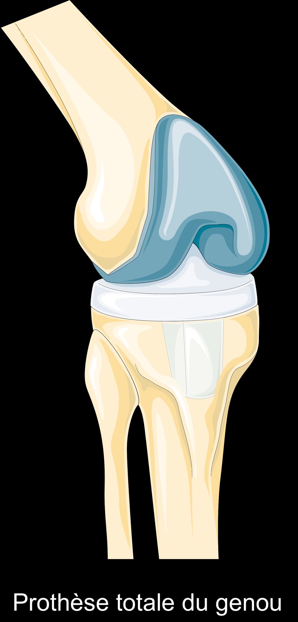
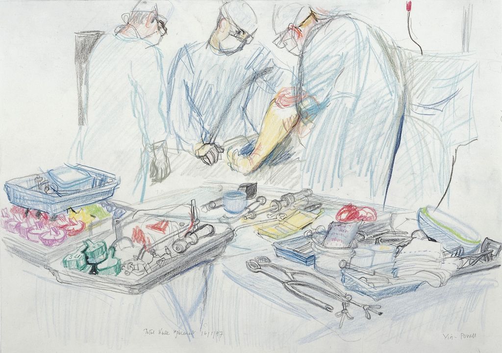
Recovery Process After Knee Replacement Surgery
Understanding the Rehabilitation Timeline
The rehabilitation timeline after knee replacement surgery is a crucial phase in our journey to recovery. It is a period of gradual progress and adjustment, where we must remain patient and committed to the prescribed regimen. Consistency in following the rehabilitation plan is key to achieving optimal results. It’s important to note that each individual’s timeline may vary based on factors such as age, overall health, and the type of surgery performed.
During this phase, we focus on building strength, flexibility, and mobility. Here’s a brief overview of the rehabilitation timeline:
Milestone
Timeframe
Initial recovery
1-2 weeks
Regaining mobility
2-6 weeks
Strengthening
6-12 weeks
Return to activities
3-6 months
It’s important to consult with our healthcare team to ensure that we are progressing according to the expected timeline and to address any concerns or challenges that may arise. Open communication with our healthcare providers is essential for a successful recovery.
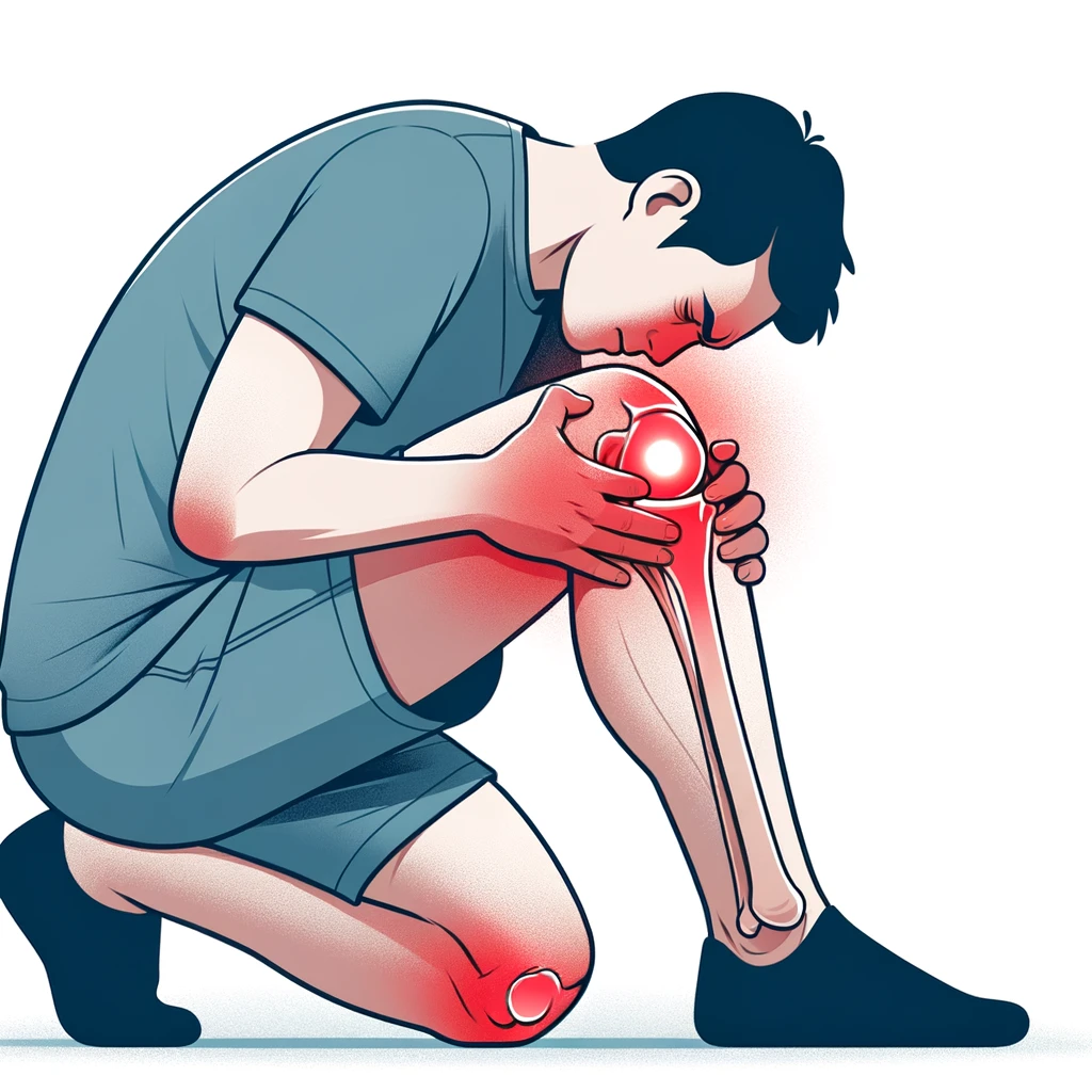
Managing Pain and Discomfort
After undergoing knee replacement surgery, we must be vigilant in managing pain and discomfort to facilitate a smoother recovery. It’s essential to follow the pain management plan prescribed by our healthcare provider, which may include medications, ice therapy, and rest. We should be aware that pain levels can vary and are influenced by activity levels and rehabilitation exercises.
Medication adherence is crucial for effective pain control. We must take prescribed pain relievers at the recommended times to maintain consistent pain relief. Here’s a simple guide to help us track our medication schedule:
Take medication before physical therapy sessions to reduce discomfort during exercises.
Set alarms as reminders to take medications on time.
Keep a log of pain levels and medication effects to discuss with our healthcare provider.
Tip: Always communicate any concerns about pain or medication side effects with our healthcare team. They can adjust our plan to better suit our needs and improve our comfort.
As we progress through recovery, we’ll gradually notice a decrease in pain and an increase in mobility. It’s important to stay patient and positive, as this is a key component of our overall well-being during the rehabilitation process.
Physical Therapy and Exercise Regimen
After completing the prescribed physical therapy sessions, we must continue to prioritize regular exercise to maintain joint flexibility and strength. It’s important to strike a balance between rest and activity, gradually increasing the intensity of workouts. We can refer to the following table for a basic exercise regimen:
Exercise Type
Frequency
Duration
Stretching
Daily
10-15 min
Low-impact Aerobics
3-4 times per week
20-30 min
Strength Training
Every other day
15-20 min
In addition to the table, we should also focus on maintaining a healthy weight and incorporating joint-friendly activities into our daily routine. It’s essential to listen to our body and communicate any concerns or setbacks to our healthcare provider for timely intervention and support.
Adapting to Lifestyle Changes
Modifying Daily Activities
After knee replacement surgery, we must recognize the importance of modifying our daily activities to accommodate our new joint’s capabilities. It’s essential to prioritize safety and prevent strain on the knee, which means rethinking how we perform routine tasks. For instance, we should avoid activities that involve high impact or twisting motions that can stress the knee joint.
Sit down to dress, using a chair or bench to support yourself.
Install grab bars in the bathroom to aid in stability.
Use assistive devices like a reacher to pick up items without bending.
Tip: Always listen to your body and avoid pushing through pain during daily activities. It’s a signal to stop and rest.
We should also consider the layout of our living spaces. Rearranging furniture to create clear pathways can minimize the risk of falls, and ensuring that commonly used items are within easy reach reduces the need to bend or stretch. These small adjustments can make a significant difference in our recovery and long-term joint health.
Nutrition and Diet Considerations
After knee replacement surgery, maintaining a balanced diet is crucial for promoting healing and overall well-being. Our dietary choices play a significant role in supporting the body’s recovery process and ensuring optimal health. It’s important to focus on consuming a variety of nutrient-dense foods, including lean proteins, healthy fats, and vitamin-rich fruits and vegetables. Additionally, staying hydrated is essential for aiding in the body’s healing and promoting joint health.
When considering dietary adjustments, it’s beneficial to consult with a healthcare professional or a registered dietitian to develop a personalized nutrition plan that aligns with our individual needs and supports our recovery journey. This plan may include specific dietary recommendations, such as increasing intake of calcium and vitamin D to support bone health and aid in the healing process. Furthermore, incorporating anti-inflammatory foods, such as turmeric and omega-3 fatty acids, can help reduce inflammation and support the body’s recovery.
It’s important to note that our dietary choices can impact our overall health and well-being, and making informed decisions about nutrition can contribute to a successful recovery and long-term joint health. By prioritizing a balanced diet and making mindful food choices, we can support our body’s healing process and enhance our quality of life after knee replacement surgery.
Mental and Emotional Well-being
After addressing the physical aspects of recovery, we must not overlook the importance of mental and emotional well-being. The journey to recuperation is not solely a physical challenge; it is intertwined with psychological resilience and stability. It is common to experience a range of emotions, from relief and optimism to anxiety and frustration.
To aid in this aspect of recovery, we recommend a series of steps to foster a positive mental state:
Acknowledge your feelings and understand that they are a normal part of the healing process.
Maintain open communication with family, friends, and your healthcare team.
Set realistic goals and celebrate small victories along the way.
Engage in activities that bring joy and relaxation, such as reading, listening to music, or meditating.
Remember, it’s crucial to give yourself grace and time to adjust to the changes. Patience and self-care are key components in building a strong foundation for your mental health post-surgery.
Long-Term Care and Maintenance
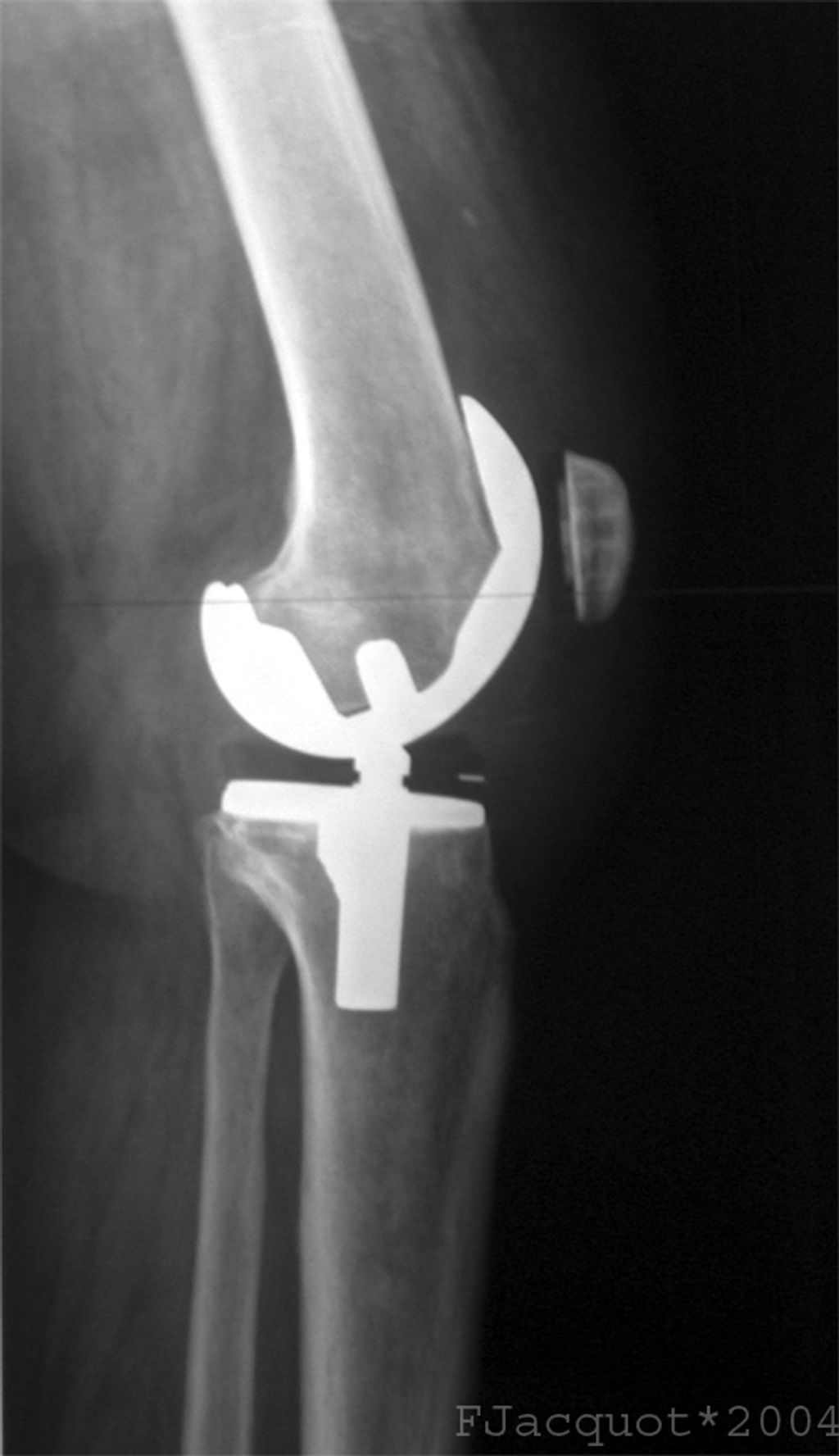
Monitoring Joint Health
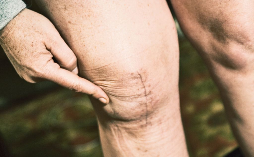
After knee replacement surgery, monitoring joint health is crucial for ensuring long-term success. Regular check-ups and consultations with a healthcare professional can help us track the progress of our joint health and identify any potential issues early on. Additionally, maintaining a healthy weight and following a balanced exercise routine can contribute to the overall well-being of our new joint.
When monitoring joint health, it’s important to pay attention to any changes in mobility, discomfort, or swelling. These could be indicators of underlying issues that require attention. Furthermore, staying informed about the recommended activities and restrictions for our specific condition is essential for preventing unnecessary strain on the joint.
Engaging in low-impact activities such as swimming, cycling, and walking can help us maintain joint flexibility and strength. These activities are gentle on the joint and can be incorporated into our regular routine to promote long-term joint health and mobility.
It’s important to remember that regular monitoring and care of our joint health can significantly impact our quality of life and the longevity of the knee replacement. By staying proactive and attentive to any changes, we can address potential issues early and maintain the health of our new joint.
Potential Complications and Warning Signs
After knee replacement surgery, we must remain vigilant to ensure the longevity of the joint and our overall health. Recognizing potential complications and understanding the warning signs is crucial for preventing serious issues.
Infection and thromboembolism are among the most serious complications that can occur after surgery. It’s imperative to monitor for signs of infection such as redness, swelling, or unusual discharge around the surgical site, as well as systemic symptoms like fever or chills. For thromboembolism, watch for leg pain or swelling, which could indicate a blood clot.
Tip: Regular check-ups with your healthcare provider are essential for early detection and management of any complications.
We should also be aware of more common, but less severe, issues such as stiffness or persistent pain. These symptoms might suggest the need for adjustments in our physical therapy regimen or lifestyle. Here’s a list of warning signs to be mindful of:
Persistent pain or swelling
Increased redness around the incision
Unusual warmth at the joint
Difficulty bearing weight
Fever or chills
By staying informed and proactive, we can address complications promptly and maintain a healthy, active lifestyle post-surgery.
Engaging in Low-Impact Activities
After knee replacement surgery, it is crucial for us to engage in low-impact activities to maintain joint health. These activities include walking, swimming, and cycling. Regular exercise helps to strengthen the muscles around the knee and improve overall mobility. It is important to consult with a healthcare professional to determine the most suitable low-impact activities based on individual circumstances.
Additionally, monitoring joint health is essential for long-term care. This involves regular check-ups with the orthopedic surgeon to assess the condition of the replaced knee. We should be vigilant for any signs of discomfort, swelling, or unusual sensations around the knee area, and report them to the healthcare provider promptly.
Furthermore, it is important to be aware of potential complications and warning signs. We should be informed about the signs of infection, blood clots, and other complications that may arise after knee replacement surgery. Early recognition of warning signs can lead to timely intervention and better outcomes.
In summary, engaging in low-impact activities, monitoring joint health, and being aware of potential complications are essential aspects of long-term care and maintenance after knee replacement surgery.
Conclusion
In conclusion, life after knee replacement surgery requires careful consideration and thoughtful planning. It is important to prioritize rehabilitation and recovery, while also being mindful of potential challenges and limitations. With the right support and commitment to self-care, individuals can navigate the post-surgery phase with resilience and determination.
Frequently Asked Questions
How long does it take to recover from knee replacement surgery?
The recovery timeline varies for each individual, but most people can expect to see significant improvement within 3 to 6 months post-surgery.
What are some common ways to manage pain and discomfort after knee replacement surgery?
Pain management techniques may include medication, icing, elevation, and using assistive devices such as a walker or cane.
How important is physical therapy in the recovery process?
Physical therapy plays a crucial role in restoring strength, flexibility, and mobility in the knee joint. It is essential for a successful recovery.
What daily activities may need to be modified after knee replacement surgery?
Activities such as kneeling, high-impact sports, and heavy lifting should be approached with caution or modified to reduce stress on the knee joint.
Are there specific dietary recommendations for individuals post-knee replacement surgery?
A balanced diet rich in nutrients such as calcium, vitamin D, and protein can support bone health and aid in the healing process.
What are some warning signs of potential complications after knee replacement surgery?
Warning signs may include persistent pain, swelling, warmth, redness, and difficulty bearing weight on the affected leg. It’s important to seek medical attention if any of these symptoms occur.
Meniscus tears are a common knee injury that can cause significant pain and discomfort. Understanding the symptoms and treatment options for meniscus tears is essential for effective management and recovery. This article provides an in-depth look at the anatomy of the meniscus, causes of meniscus tears, symptoms, diagnosis, and both non-surgical and surgical treatment options.
Key Takeaways
Meniscus tears can result from traumatic injuries or degenerative conditions.
Symptoms of meniscus tears include pain, swelling, and limited range of motion.
Physical examination and MRI evaluation are common methods for diagnosing meniscus tears.
Non-surgical treatment options for meniscus tears include rest, ice, and physical therapy.
Surgical treatment options for meniscus tears may involve arthroscopic surgery and meniscus repair or meniscectomy.
Anatomy of the Meniscus
Structure of the Meniscus
The meniscus is a C-shaped piece of cartilage that acts as a cushion between the femur and tibia. It plays a crucial role in load distribution and stability within the knee joint. The structure of the meniscus allows for smooth movement and shock absorption during weight-bearing activities.
The inner two-thirds of the meniscus has a poor blood supply, making it less likely to heal on its own.
The outer one-third of the meniscus has a better blood supply, which may facilitate healing in certain cases.
Function of the Meniscus
The meniscus plays a crucial role in distributing weight and absorbing shock in the knee joint. It also contributes to stabilizing the knee during movement. These functions are essential for maintaining proper joint mechanics and preventing excessive wear and tear on the articular cartilage.
In a study conducted on meniscus function, it was found that the meniscus contributes to approximately 50-70% of load transmission across the knee joint. This emphasizes the significant role of the meniscus in supporting the structural integrity of the knee.
Proper understanding of the function of the meniscus is crucial for developing effective treatment strategies for meniscus tears. It guides both non-surgical and surgical interventions, ensuring that the appropriate approach is chosen based on the specific needs of the patient.
Causes of Meniscus Tears
Traumatic Injuries
In our experience, meniscus tears often result from traumatic injuries, especially in athletic contexts. These injuries typically occur during activities that involve forceful twisting or rotation of the knee joint, such as when playing sports like football, basketball, or soccer.
Common mechanisms of injury include:
Sudden pivoting or stopping
Direct contact or collision
Deep squatting or kneeling
Tip: Immediate application of ice and elevation of the leg can help reduce swelling and pain following a traumatic knee injury.
It’s crucial to recognize the signs of a meniscus tear early to prevent further damage. While some athletes may ‘play through the pain,’ we advise against ignoring symptoms that may indicate a more serious condition.
Degenerative Conditions
Degenerative conditions such as osteoarthritis can lead to gradual wear and tear of the meniscus, resulting in increased susceptibility to tears. Age-related changes in the meniscus structure and composition contribute to this degeneration. In some cases, the degenerative process may be accelerated by repetitive stress or overuse.
Table: Degenerative Conditions and Meniscus Tears
Condition
Risk of Meniscus Tear
Osteoarthritis
High
Age-related changes
Moderate
Repetitive stress
Low
Tip: Engaging in low-impact exercises and maintaining a healthy weight can help reduce the risk of meniscus tears in individuals with degenerative conditions.
Symptoms of Meniscus Tears
Pain and Swelling
We often find that pain and swelling are the initial indicators that a patient may have a meniscus tear. The pain is typically localized on the side of the knee where the tear has occurred and can range from mild to severe, depending on the extent of the injury. Swelling usually develops within the first few hours post-injury and can persist for days.
To manage these symptoms, we recommend a structured approach:
Rest: Avoid activities that exacerbate knee pain.
Ice: Apply cold packs to reduce swelling and numb the painful area.
Compression: Use an elastic bandage or knee sleeve for support.
Elevation: Keep the injured leg elevated to decrease swelling.
Tip: Early intervention with these steps can significantly reduce discomfort and expedite the healing process.
It’s important to monitor the pain and swelling closely. If symptoms persist or worsen, it may be indicative of a more complex issue or a different injury altogether.
Limited Range of Motion
We often observe that individuals with meniscus tears experience a limited range of motion in the affected knee. This restriction can manifest as difficulty in fully straightening or bending the knee, which can severely impact daily activities and quality of life.
The following list outlines common movements that may be hindered by a meniscus tear:
Full knee extension
Deep knee flexion
Twisting or pivoting motions
It’s crucial to address this symptom promptly to prevent further complications. Engaging in targeted exercises under proper guidance can aid in restoring mobility and ensuring a smoother recovery process. We emphasize the importance of exercises and proper guidance for recovery and prevention of future injuries.
Tip: Early intervention with physical therapy can significantly improve the range of motion and facilitate a quicker return to normal activities.
Diagnosis and Imaging
Physical Examination
In our clinical practice, we begin the diagnosis of a meniscus tear with a thorough physical examination. We palpate the knee joint to identify areas of tenderness that may suggest a tear in the meniscus. Specific maneuvers, such as the McMurray test, are performed to elicit pain or clicking, which can be indicative of a meniscal injury.
We assess the knee’s range of motion and stability, looking for signs of joint effusion or locking, which are common in meniscal tears. It’s important to note that while these tests are valuable, they are not definitive, and further imaging may be necessary to confirm the diagnosis.
Remember, a careful physical examination by a healthcare professional is crucial for an accurate diagnosis, as symptoms of meniscus tears can mimic other knee problems.
MRI Evaluation
Following a thorough physical examination, we often turn to MRI evaluation to confirm the presence of a meniscus tear. This imaging technique provides us with detailed images of the soft tissues within the knee, allowing for a precise diagnosis.
An MRI not only confirms the tear but also helps us assess the extent of the injury. We can determine whether the tear is radial, horizontal, or complex, and the specific location within the meniscus. This information is crucial for planning the appropriate treatment strategy.
MRI evaluation is particularly useful in cases where physical examination results are inconclusive or when multiple knee injuries are suspected.
We categorize meniscus tears based on their appearance on MRI scans as follows:
Grade 1: Small, inconsequential tears
Grade 2: Larger but stable tears, possible pain and swelling
Grade 3: Full-thickness tears, often requiring surgical intervention
Non-Surgical Treatment Options
Rest and Ice
After implementing the Rest and Ice protocol, physical therapy becomes a crucial component of the recovery process. It focuses on strengthening the muscles around the knee joint and improving flexibility. Additionally, physical therapy provides individualized care for all ages with various conditions. Schedule a free assessment with highly trained staff at multiple locations. This personalized approach ensures that each patient receives the specific treatment they need to regain strength and mobility. In some cases, a structured exercise program may be recommended to aid in the rehabilitation process. It’s important to follow the guidance of the physical therapist to achieve the best possible outcome.
Physical Therapy
After discussing the benefits of physical therapy, it is important to note that physical therapy is not limited to athletes and is beneficial for all ages. It aims to relieve pain, improve flexibility, and reduce the risk of future injuries. Myth 1: Physical therapy is only for athletes. Myth 2: Physical therapy is painful. Myth 3: It is only for patients after surgery. Implementing a structured approach to physical therapy can yield positive outcomes. Here is a brief overview of the key components of an effective physical therapy program:
Pain Relief: Targeted exercises and modalities to alleviate pain and discomfort.
Flexibility Training: Techniques to improve range of motion and joint flexibility.
Strength Building: Progressive exercises to strengthen the muscles supporting the knee.
Functional Training: Activities to improve balance, coordination, and functional movement.
Education and Prevention: Guidance on injury prevention and self-management strategies.
It is important to adhere to the prescribed physical therapy regimen and communicate any concerns or progress to the healthcare provider for optimal results.
Surgical Treatment for Meniscus Tears
Arthroscopic Surgery
In our exploration of treatment options for meniscus tears, we find that arthroscopic surgery stands out as a minimally invasive procedure that allows us to address the injury directly. Utilizing small incisions and specialized instruments, we can visualize and repair the damaged meniscus with precision.
The steps involved in arthroscopic surgery typically include:
Administration of anesthesia.
Making small incisions around the knee joint.
Inserting the arthroscope to assess the damage.
Repairing or removing the torn meniscal tissue.
Closing the incisions and beginning the rehabilitation process.
Postoperative care is crucial for a successful recovery. We emphasize the importance of following a structured rehabilitation program, which often includes controlled exercises to restore strength and mobility.
Remember, the goal of arthroscopic surgery is not only to relieve pain but also to restore the knee’s functionality. A tailored approach to rehabilitation can significantly enhance the outcome.
Meniscus Repair vs. Meniscectomy
When considering surgical treatment for meniscus tears, it is important to weigh the options of meniscus repair and meniscectomy. Each procedure has its own benefits and considerations, and the decision should be made in consultation with a qualified orthopedic surgeon. Meniscus repair aims to preserve the natural structure of the meniscus, promoting long-term joint health and stability. On the other hand, meniscectomy involves the removal of damaged tissue, providing immediate relief from pain and discomfort. It is essential to understand the potential outcomes and recovery process associated with each procedure before making a decision. Here are some key points to consider:
Meniscus Repair
Suitable for younger patients with good tissue quality
Longer recovery time but potential for sustained joint function
Meniscectomy
Commonly performed for older patients with degenerative tears
Shorter recovery time but increased risk of future joint issues
Tip: It is crucial to follow post-operative care instructions diligently to optimize the outcomes of either procedure.
Conclusion
In conclusion, understanding the symptoms and treatment options for meniscus tear pain is crucial for effective management and recovery. By recognizing the signs of a meniscus tear and exploring the various treatment modalities available, individuals can make informed decisions about their healthcare and take proactive steps towards healing and rehabilitation.
Frequently Asked Questions
What are the common causes of meniscus tears?
Meniscus tears can be caused by traumatic injuries, such as sudden twisting or forceful impact, as well as degenerative conditions, such as osteoarthritis or aging-related wear and tear.
What are the typical symptoms of a meniscus tear?
The typical symptoms of a meniscus tear include pain, swelling, and limited range of motion in the affected knee. Some individuals may also experience a clicking or locking sensation.
How is a meniscus tear diagnosed?
A meniscus tear is diagnosed through a physical examination by a healthcare professional, as well as through imaging tests such as MRI evaluation to assess the extent and location of the tear.
What non-surgical treatment options are available for meniscus tears?
Non-surgical treatment options for meniscus tears include rest, ice therapy, physical therapy, and the use of supportive braces to aid in the healing process and improve knee function.
What is arthroscopic surgery for meniscus tears?
Arthroscopic surgery for meniscus tears involves the use of a small camera and specialized instruments to repair or remove the damaged portion of the meniscus, typically performed through small incisions in the knee.
What is the difference between meniscus repair and meniscectomy?
Meniscus repair aims to preserve the meniscus by suturing the torn edges together, while meniscectomy involves the partial or complete removal of the damaged meniscus tissue, depending on the severity of the tear and the patient’s condition.