Ever notice how stiff your legs feel after hours behind the wheel? You’re not alone. Millions of Americans experience discomfort from repetitive driving motions and fixed sitting positions. What if small changes to your routine could help you arrive feeling refreshed instead of achy?
Extended time in vehicles strains joints and soft tissues through limited movement. Tightness in the legs often stems from reduced blood flow and muscle fatigue. We’ve designed this guide to address these challenges with science-backed solutions.
Our approach focuses on simple movements that fit seamlessly into your schedule. Whether you’re navigating cross-country routes or daily traffic, these strategies promote flexibility without complicated equipment. Consistency matters more than intensity – even brief sessions can make a difference.
Key Takeaways
- Common driving habits contribute to stiffness in lower extremities
- Targeted movements improve circulation and joint function
- Quick exercises can be performed during rest stops or fuel breaks
- Proper technique prevents strain during physical activity
- Persistent discomfort warrants professional medical evaluation
Understanding Knee Pain from Long Drives
Many drivers experience a nagging ache after prolonged hours on the road. This discomfort often stems from repetitive pedal movements and fixed seating positions that strain soft tissues. Over time, these patterns can lead to chronic issues requiring attention.
What Is Driver’s Knee?
Commonly called gas pedal syndrome, this condition develops when constant pedal use irritates the tendon below the kneecap. Medical professionals classify it as patellar tendonitis – inflammation caused by repeated stress. Like a tennis player’s elbow, it results from small motions performed hundreds of times daily.
Common Causes and Symptoms
Improper foot placement creates uneven pressure across the joint. Angling your heel too high or twisting your ankle while braking forces the tendon to work at unnatural angles. Early signs include:
| Condition | Affected Area | Common Causes |
|---|---|---|
| Driver’s Knee | Patellar Tendon | Pedal repetition, poor posture |
| Tennis Elbow | Forearm Muscles | Racquet swings, gripping |
| Writer’s Cramp | Hand Tendons | Extended writing sessions |
Ignoring initial tenderness allows inflammation to worsen. Some drivers report sharp stabs when exiting vehicles or climbing stairs. Without intervention, cartilage deterioration called chondromalacia patella may develop – a key reason early action matters.
Benefits of Regular Knee Stretches
Consistent movement routines transform how your body handles road trips. Recent studies reveal targeted exercises boost joint resilience – particularly for those managing conditions like osteoarthritis. A 2022 review found dedicated flexibility work alone shows measurable improvements in mobility.
Strengthening key muscle groups creates natural support systems. The AAOS emphasizes this approach:
“Balanced development in quads, hamstrings, and calves reduces joint strain by up to 30% during repetitive motions.”
This protective effect matters most for drivers maintaining fixed positions for hours.
Improved Mobility and Reduced Discomfort
Dynamic movements counteract stiffness from prolonged sitting. Gentle stretches enhance blood flow, flushing out metabolic waste that causes fatigue. Over time, tissues regain their elastic quality – like breaking in new shoes.
Properly aligned joints distribute weight evenly across surfaces. This prevents hotspots where pressure builds up. Think of it as traffic management for your lower body – no more gridlocked tendons.
Regular routines build lasting protection. Flexible tissues absorb shocks better during sudden stops or rough terrain. Consistency beats intensity – even five-minute sessions between drives maintain progress. Your future self will thank you during mountain hikes or airport sprints.
Effective Post-drive knee pain stretches to Ease Discomfort

Maintaining comfort during extended road trips requires proactive measures. Smart movement strategies combat stiffness before it becomes problematic. We focus on two phases: adjustments during driving and targeted recovery afterward.
In-Car Simple Stretches
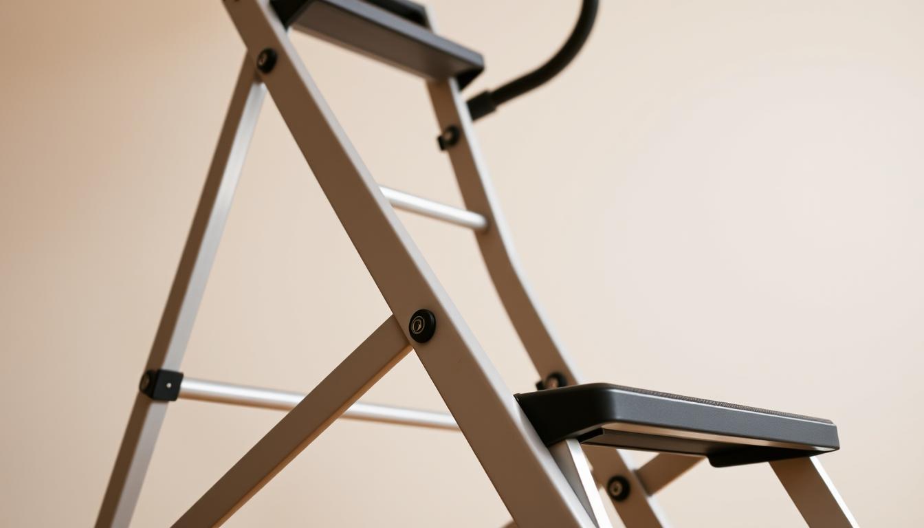
Adjust your seat position at safe stops. Push the seat back until your leg nearly straightens – this creates natural ankle flexion. Rotate your feet clockwise 10 times, then reverse. These micro-movements boost circulation without leaving your vehicle.
Try gentle extensions while parked. Lift one leg parallel to the floor, hold 5 seconds, then switch. Follow with seated calf stretches: press heels downward while flexing toes upward. Consistent practice prevents fluid buildup in lower extremities.
After-Drive Floor Exercises
Post-travel routines reset muscle balance. Lie flat and raise legs vertically against a wall for 2 minutes. This inverted position drains excess fluid from tired limbs. Follow with controlled movements:
- Straight leg raises (10 reps per side)
- Calf raises with 3-second holds
- Half-squats maintaining proper spine alignment
For lateral support, practice hip abductions. Stand sideways near a wall, lift outer leg sideways 12 times, then switch. These movements counteract the compressed positions from driving. Remember: Discomfort signals to stop – gradual progress yields lasting results.
Stretch Routines for the Road
Road warriors know the importance of strategic movement breaks. Regular stops help maintain circulation and joint health during extended trips. Our routines require no special equipment – just a few minutes of focused effort.
Stretches at Rest Stops
Try forward bends with palms flat on your vehicle’s roof. Hold for ten seconds to release hamstring tension. For quads, stand on one foot and gently pull your other ankle toward your glutes.
Figure-8 leg swings improve hip mobility. Lean against your car and move each limb in smooth, controlled patterns. This motion counteracts stiffness from accelerator repetition.
Bodyweight and Calf Exercises
Bodyweight squats strengthen multiple muscle groups simultaneously. Lower slowly until thighs parallel the ground, then push through your heels. Pair these with elevated calf raises – lift onto toes for three seconds before releasing.
Walk briskly around parking areas during breaks. Five minutes of movement pumps fresh blood through compressed tissues. Combine steps with deep breathing for enhanced oxygen flow.
Ergonomic Adjustments for Better Alignment
Adjust seat height so knees stay slightly bent when pressing pedals. Use lumbar support to maintain natural spinal curves. Position steering wheel within easy reach to prevent shoulder hunching.
Rotate footwear if possible – alternate between supportive shoes to vary pressure points. These tweaks help distribute weight evenly, reducing strain on specific joints during long hauls.
Enhancing Flexibility and Mobility with Warm-Ups
What’s the secret to maintaining comfortable movement during long drives? Proper preparation. The American Academy of Orthopaedic Surgeons recommends 5-10 minutes of light activity before exercise – think brisk walking or air cycling. This gentle approach primes your body for action like tuning an engine before a road trip.
Essential Preparation Techniques
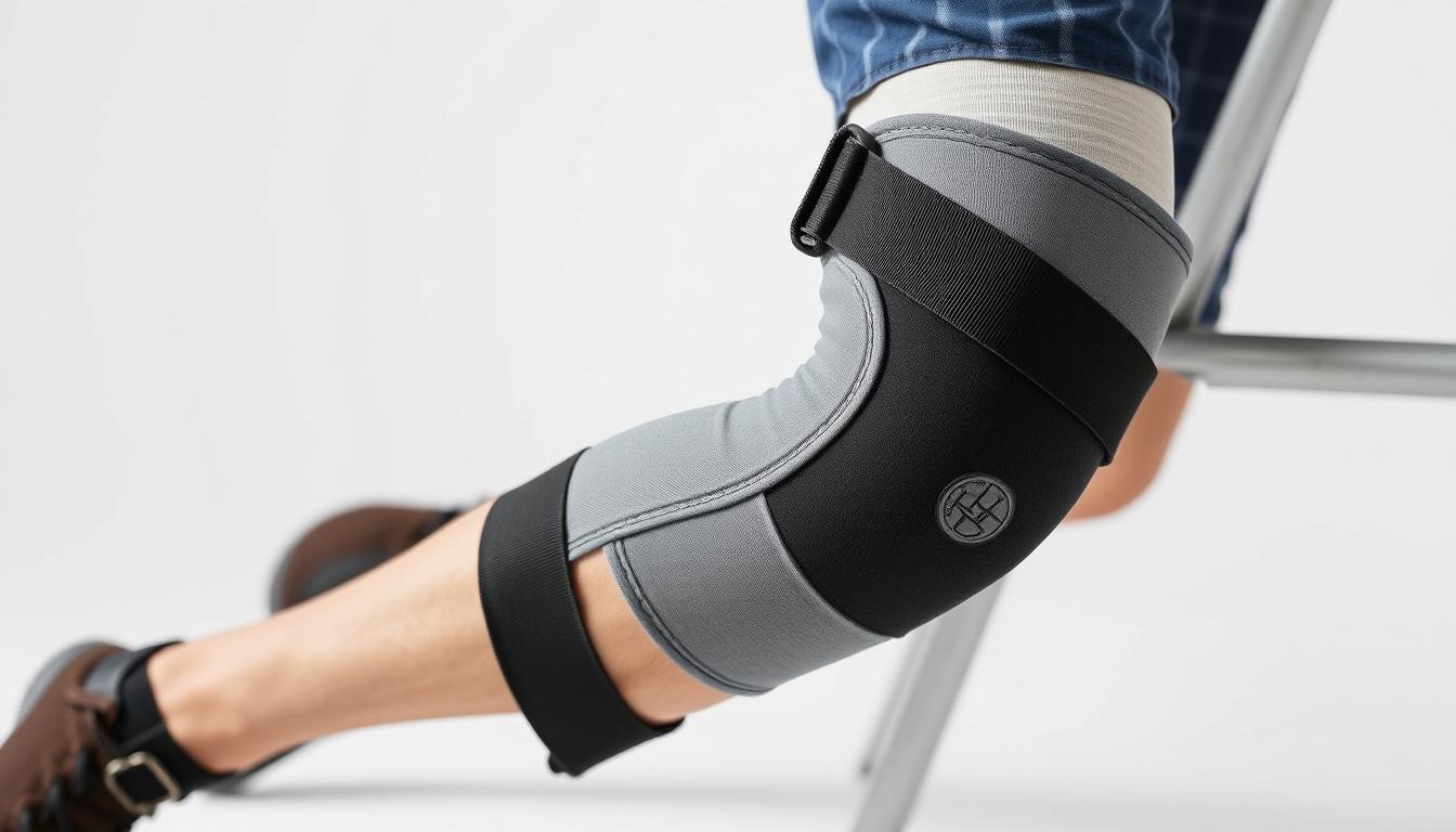
Start with lunging hip flexor stretches. Kneel on one leg, lean forward until you feel tension in the front hip. Hold 20 seconds per side. This counteracts tightness from sitting – a common contributor to restricted motion.
For hamstring care, try the supine wall stretch. Lie on your back, extend one leg upward against a wall. Keep your hips flat as you gently reach toward your ankle. Slow movements prevent strain while improving flexibility in these critical muscles.
Address calf tightness with step stretches. Stand on a curb or stair, let your heels dip below edge level. Rise onto toes, then lower slowly. Repeat 10 times to maintain ankle mobility and reduce heel pressure.
“Dynamic warm-ups increase blood flow by 40% compared to static stretching alone,” notes recent AAOS research.
The figure-four stretch targets hip stability. Cross one ankle over the opposite thigh, press knee outward. Hold 30 seconds per side. Finish with foam roller work along outer thighs – this supports proper knee alignment by releasing tight connective tissues.
Remember: Warm-ups should feel challenging but manageable. Breathe deeply through each motion, allowing muscles to gradually adapt. Consistent preparation builds lasting mobility that protects your joints during repetitive activities.
Preventing Knee Strain with Improved Driving Posture
Your driving position directly impacts joint health during long hauls. Start by adjusting your seat so hips sit slightly higher than your knees. This alignment reduces pressure on the front of your legs while maintaining natural spinal curves. Maintain 2-3 inches between the seat edge and your calves to prevent restricted blood flow.
Foot placement matters more than most drivers realize. Keep heels grounded while pressing pedals with the ball of your foot. This distributes weight evenly across ankles and lower limbs. Avoid overextending – your legs should never fully straighten when accelerating or braking.
Supportive accessories make a surprising difference. Lumbar cushions help maintain proper spinal alignment, which cascades down to hip and leg positioning. Pair these with non-slip seat covers to prevent sliding that strains joints. Remember: Compensatory movements from poor posture create long-term issues.
Choose footwear with arch support and shock-absorbing soles. Flexible shoes allow natural foot movement, while stiff soles force ankles into awkward angles. Rotate between pairs during multi-day trips to vary pressure points.
Lastly, avoid crossing legs or leaning to one side. These habits shift body weight unevenly, overloading specific joints. Regular posture checks every 30-60 minutes help reinforce proper alignment until it becomes automatic.
Professional Insights and When to Seek Medical Advice
Persistent discomfort deserves more than temporary fixes. While minor soreness often resolves with rest, certain patterns signal deeper issues. We recommend monitoring symptoms closely and acting when self-care falls short.
Recognizing When Pain Persists
Sharp twinges during daily activities often indicate overuse injuries. Swelling or redness around joints could suggest arthritis flare-ups. If discomfort disrupts sleep or lasts over 72 hours, consult a doctor. Early intervention prevents chronic conditions – especially for those with existing knee concerns.
Watch for these warning signs:
- Difficulty bearing weight on affected limbs
- Clicking sounds with movement
- Reduced range of motion compared to other leg
Benefits of Consultation with a Physical Therapist
Licensed therapists assess movement patterns causing strain. They create tailored plans addressing muscle imbalances – a common driver’s issue. For arthritis management, specific exercises may help preserve joint function better than generic stretches.
Research shows customized programs reduce re-injury risks by 40%. Therapists also teach proper body mechanics for driving tasks. Knowledge is power when protecting your musculoskeletal health long-term.
FAQ
What is driver’s knee?
Driver’s knee refers to discomfort caused by prolonged pressure on the leg during driving. It often affects muscles, tendons, or joints due to limited motion, leading to stiffness or swelling around the kneecap.
How can stretching help after long drives?
Gentle movements improve blood flow, reduce tension in tight muscles like hamstrings or calves, and restore flexibility. Consistent routines may also lower the risk of chronic joint stress or injuries.
What are quick in-car stretches?
While seated, try ankle circles, heel lifts, or extending one leg at a time to engage the thigh. Hold each motion for 15–20 seconds to ease stiffness without leaving your seat.
Are rest stop exercises effective?
Yes! Bodyweight squats, calf raises, or lunges during breaks activate muscles and improve alignment. Pair these with shoulder rolls to counteract slouching behind the wheel.
How does posture prevent knee strain?
Adjusting seat height and distance ensures hips stay level, reducing pressure on joints. Sitting too close or slumping can tighten hip flexors, worsening discomfort over time.
When should I consult a professional?
If soreness lasts beyond a few days or includes swelling, a physical therapist can assess imbalances. They may recommend tailored exercises or ergonomic tweaks for safer driving habits.