Category: kenee Management and treatments

  • What does my KOOS score mean and is my KOOS score normal?

    What does my KOOS score mean and is my KOOS score normal?

    If you weigh yourself on a scale and receive a number, how do you understand the value? How do you know if you are overweight, underweight or normal? Who do you compare yourself to?

    To determine what’s normal and understand your own score, you’ll likely compare your weight to other people who have similar characteristics to you, such as gender, age, and height. There would be no point in comparing yourself to someone with different characteristics, since what is normal for him or her is likely different for you. This same concept applies to normative values.

    Normative data identify what is common or typical and describe observed characteristics of a specific population at a specific time.[1] Using normative values ​​allows you to give meaning to your test scores by comparing your score to scores of people with similar characteristics to you.[2]

    As explained in part 1 of the KOOS blog series, the Knee Injury and Osteoarthritis Score (KOOS) is a questionnaire specifically designed for people with various knee conditions.[3] By completing the KOOS you will gain insight into the course of your knee injury and you and your healthcare provider can monitor the effects of the treatment over time.[3] While comparing your own preoperative and postoperative KOOS scores provides insight into your recovery process, you can also compare your scores to normative KOOS values ​​to determine your degree of disability or your level of progress compared to people who have had a similar injury, surgery, or have undergone treatment. .

    View the normal KOOS scoring blog for populations that have suffered an ACL injury,[4][5][6] total knee replacement (TKR) surgery, [7][8][9] as well as those who have knee osteoarthritis,[10] and populations without known knee disorders [11] in part 2 of the KOOS blog series.

    If you have had a knee injury or surgery, try our Curovate app for your daily recovery. Curovate offers video-guided daily exercises, progress tracking, the ability to measure the range of motion of your knee and hip, and the ability to complete the KOOS outcome measurement, all within the app.

    If you need more tailored help during your surgery or recovery from your injury, check out our Virtual Physiotherapy page to book your 1-on-1 video session with a physiotherapist.

    Learn more about the KOOS and what normative values ​​are. Also learn how to interpret your KOOS score in this YouTube video presented by Joey Wong, kinesiologist.

    acl knee physical therapy 1080x1080 2
    Download it on Google Play


    Other recommended blogs


    References

    Source link

  • Research Fellow – Rubin Institute for Advanced Orthopedics (RIAO) at Sinai Hospital in Baltimore


    This new position is intended for medical students who wish to strengthen their competitive position to obtain a residency position in the field of orthopedic surgery. Research assistants will work with national and international leaders in the field of total joint replacement to assist in preparing manuscripts for publication in peer-reviewed journals, to collect and analyze data, to attend and present your work at regional and national conferences, and to assist with FDA investigations of new drugs and devices. This is a large-scale research center where numerous projects have not only been published in PubMed-indexed journals, but have also been selected to present at national orthopedic meetings, namely the AAOS and AAHKS.

    Research assistants have the opportunity to attend daily morning conferences at the orthopedic surgery residency, led by program director Dr. Jack Ingari, MD. This is an excellent opportunity to strengthen your resume and increase your chances of matching with an orthopedic surgery residency.

    Student researchers will work under the guidance of:

    -Ronald E. Delanois, MD

    -Michael A. Mont, MD

    -James Nace, DO, MPT

    GOALS:

    – Research into basic scientific and clinical aspects of total joint arthroplasty and preservation.

    – Learn to create and extract clinical data from institutional and national databases

    – Develop a better appreciation for biostatistics in the orthopedics setting

    – Prepare manuscripts and textbook chapters.

    – Present at research meetings, seminars, journal clubs, symposia and professional association meetings.

    EDUCATIONAL LEVEL REQUIREMENT OR REASONS: All candidates who have completed their third year and passed their USMLE Step 1 and/or COMLEX Level 1 board exams. It is preferable that USMLE Step 2 be completed before beginning the research program. Experience in writing and biostatistics is a plus.

    APPLICATION: Attach the following materials This email address is being protected from spambots. You need JavaScript enabled to view it.:

    – Application letter OR letter of interest

    – PDF of Curriculum Vitae

    – All available USMLE AND/OR COMLEX score reports

    – ≥ 1 letters of recommendation

    RIAO website: www.lifebridgehealth.org/RIAO/RIAO.aspx

    Twitter handle: @RIAOResearch Instagram handle: @RIAOorthopedics

    US/Canadian MD/DO preferred. US IMGs will be considered on a case-by-case basis. J-1 visa is possible.

    Source link

  • The histone acetyltransferases CBP and p300 regulate stress response pathways in synovial fibroblasts at transcriptional and functional levels


    Hypoxia, the formation of reactive oxygen species (ROS) and subsequent oxidative stress in synovial tissues are key events in the pathogenesis of RA1. We provide evidence here that CBP and p300 are critical regulators of the adaptive response of SF that integrate the transcriptional and functional regulation of stress response pathways throughout the cell.

    CBP and p300 are HATs and H3K27ac mark writers, which activate post-translational histone modifications present in enhancers and promoters20. Among the CBP/p300 target proteins are several non-histone proteins in addition to histones, including many transcription factors and signaling effectors16.21. Despite their high protein sequence homology, CBP and p300 have distinct individual functions identified by our own and other studies12,13,14.

    In SF, persistent H3K27ac in inflammatory gene promoters was associated with long-lasting and persistent expression of the corresponding genes22. We recently demonstrated that p300 is the major HAT in SF that exerts a pro- and anti-inflammatory role. This is in contrast to CBP, which exerted anti-inflammatory effects upon silencing, and specifically regulated TNF-induced interferon signature gene expression14. Although we have ruled out that silencing p300 additionally reduced CBP and vice versa, we cannot completely rule out other potential off-target effects after our silencing approach. Krosel etc already. have shown that inhibitors targeting the HAT or bromodomain of CBP/p300 closely resemble the effects of p300 silencing, including increased expression of TNF-induced proinflammatory gene expression14.

    Our RNAseq data provide evidence that the number of p300-regulated target genes is greater than that of CBP-regulated target genes, also in terms of stress response. While the cellular response to oxidative stress and autophagy was co-regulated by CBP and p300, genes associated with response to oxygen levels, hypoxia, and pathways associated with proteasome regulation and function were specifically enriched upon knockdown of p300. In pathways co-regulated by CBP and p300, we identified several genes that were regulated in opposite directions. These results point to individual functions of the two enzymes at the level of target genes, similar to what we have already observed for many inflammatory genes14. Furthermore, a small number of measured CBP and p300 target genes, namely BCL2, SOD3 and HDAC6, could be regulated in a joint-specific manner, as indicated by our Real-time PCR results in a limited number of samples for each joint site. Frank-Bertoncelj et al. have previously shown that H3K27ac is one of the mechanisms controlling the joint-specific expression of homeobox (HOX) transcription factors in SF from different locations7. To draw a definitive conclusion whether stress-associated target genes are regulated in a joint-specific manner, larger numbers of SF from different joints would be needed, together with H3K27ac ChIPseq data in unstimulated and TNF-stimulated SF from different joints.

    In addition to the differential roles of CBP and p300 in regulating target gene expression, we showed here a differential regulation of CBP and p300 by stimulating SF with 4-HNE and TNF (Fig. 6). These factors are present in the synovial microenvironment in RA and mimic the oxidative stress and inflammation, respectively. While 4-HNE and TNF, similar to H2O2suppressed the expression of p300, CBP was not affected. 4-HNE is a lipid peroxidation product generated at elevated levels of ROS. Levels of 4-HNE are elevated in serum, synovial fluids, and synovial tissues of RA patients, and serum levels of 4-HNE correlate with structural damage such as erosions in the early stage of RA23,24. As mimicked by our silencing approach, the TNF- and 4-HNE-mediated suppression of p300 expression in the synovial RA microenvironment has fundamental consequences for SF behavior. Our datasets from the previous one14 and the present study indicates that reduced expression of p300 was associated with increased expression of many inflammatory cytokines, chemokines matrix metalloproteinases and stress response genes in SF. Among these genes were HK2, a marker indicating the metabolic switch from SF to glycolysis, and VEGF, a pro-angiogenic factor secreted to overcome hypoxia.1.

    Figure 6
    figure 6

    Summary of CBP- and p300-regulated pathways in SF. The expression of p300 but not CBP is down-regulated in synovial fibroblasts after exposure to TNF and oxidative stress. The effects of p300 and CBP silencing are demonstrated based on findings from this and a previous study14. Downward arrows indicate suppressed expression or function, upward arrows indicate increased expression or function. The figure was created by BioRender.com.

    TNF stimulation of SF markers of endoplasmic reticulum (ER) was shown to induce stress and autophagy25. Our data suggest that CBP and p300 regulate autophagy at the transcriptional level and influence autophagic flux. The assessment of autophagy in the presence of the lysosomal inhibitor bafilomycin A1 indicated that CBP and p300 regulate autophagy function at different stages within the autophagic process. CBP silencing affected autophagosome synthesis. In contrast, knockdown of p300 induced autophagy in unstimulated SF, and induced a late-stage block of autophagy in TNF-stimulated SF, a condition in which polyubiquitinated proteins in SF accumulated. Accordingly, knocking down p300 only increased cell death in the presence of TNF, as previously indicated14. Kato etc already. have previously shown that autophagy induction partially compensated for reduced clearance of polyubiquitinated proteins in SF after blocking proteasome function, indicating a protective effect of autophagy induction in SF under such conditions5. Here we observed a similar compensatory mechanism after p300 knockdown, which was associated with a suppression of proteasome enzymatic activities and an induction of autophagy. This finding is consistent with a previous study in HeLa cells in which p300 knockdown was associated with reduced acetylation of autophagy-related proteins and increased levels of autophagy.26.

    Acetylation and deacetylation of components of the autophagy machinery control all steps of this catabolic process, from autophagosome initiation to LC3 conjugation, cargo assembly, and autophagosome-lysosome fusion27,28. Several classes of acetyltransferases, including CBP and p300, and deacetylases, including sirtuin1, HDAC4 and HDAC6 are involved in the regulation of autophagy27.29. Furthermore, the function of autophagy-related transcription factors, such as transcription factor EB (TFEB), Foxo1 and Foxo3, is regulated by deacetylation28.30. Recently, the increased translation of FOXO3 mRNA has been described to facilitate the initiation of autophagy31. We showed here that in unstimulated SF, FOXO3 mRNA increased after p300 knockdown, a condition in which autophagic flux increased. HDAC6 binds to polyubiquitinated proteins in SF29and promotes autophagy by facilitating autophagosome-lysosome fusion27. On the other hand, HDAC6 was also shown to suppress autophagy by deacetylating TFEB and Foxo130. This could explain the inverse regulation of HDAC6 and ATG5 and ATG16L1 in SF.

    A global analysis of the CBP/p300-dependent acetylome in mouse embryonic fibroblasts (MEF) suggested that proteasome functions might also be regulated by these enzymes.21. The majority of proteasome machinery components showed numerous CBP/p300-dependent acetylation sites in regulatory and enzymatic subunits in MEF (http://p300db.choudharylab.org). Furthermore, ATG5 and ATG16L1, two proteins essential for autophagosome assembly, showed CBP/p300-dependent acetylation sites21. Whether proteasome and autophagy components are acetylated in a CBP- and p300-dependent manner in SF remains to be functionally evaluated21. Because CBP/p300-dependent acetylation sites in MEF were analyzed after a combinatorial knockout of both enzymes, it is not clear which of them is the key enzyme in regulating the post-translational acetylation of proteins involved in the regulation of autophagy and proteasome activities.

    In summary, we have identified CBP and p300 in particular as critical regulators of stress response pathways in SF, with overlapping and distinct functions within specific pathways. The downregulation of p300 by TNF and oxidative stress provides a mechanism underlying SF activation in the synovial microenvironment.

    Source link

  • My Personal Journey to Osteoporosis Care and Awareness in Underserved Communities – Bone Talk

    My Personal Journey to Osteoporosis Care and Awareness in Underserved Communities – Bone Talk

    IMG 20230830 WA0029

    My personal journey to osteoporosis care and awareness in underserved communities
    By Dr. Tasneem Hassan

    My name is Dr. Tasneem Hassan and I work as a general practitioner in Nairobi, Kenya. After graduating in 2019, I started working in a public hospital in Nairobi.

    Later I started working at Rayhaan Healthcare, where I met Dr. Mustafa Bhaiji, a consultant radiologist with a special interest in osteoporosis. I also learned about the DXA technology, which further sparked my interest in osteoporosis. As I learned more about my family’s history and observed many people with poor bone mass, I started to pay more attention to it.

    This is the story of my grandmother, who fell a few years ago and broke her hip, needing hip replacement surgery and leaving her bedridden for a while. All these difficulties contributed to her death. For starters, we know she had low bone mass, but the lack of a bone DXA scan in Mombasa, Kenya delayed early diagnosis and treatment. She also had dementia and her recovery was difficult. In addition, there is a lack of awareness in our system about osteopenia and osteoporosis, which hinders early diagnosis and prevention. If our thinking about osteoporosis had advanced significantly earlier at the time, she might not have died from the comorbidities associated with the fracture.

    Another interaction I had was with my mother, who had already suffered a fracture. She is currently going through menopause and a few months ago she broke her foot as a result of a fall. This could be a stress fracture. But given her age and menopause, higher risk of falls, and history of two fractures, I wouldn’t be shocked if she has poor bone mass, and thus osteopenia. Individualizing care is simple; we do it all the time. We shouldn’t allow people to walk around with untreated osteoporosis because they will eventually stop walking. It’s no longer the 1900s.

    Osteoporosis is a disease that roughly causes 8.9 million fractures per yearculminating in one osteoporosis fracture every 3 seconds. One in three women and one in five men those aged 50 and over will experience an osteoporotic fracture. Osteoporosis causes bones to become weak and brittle, causing them to break easily even after a minor fall, bump, sneeze, or quick movement. I have seen people who have fractures that damage them not only physically, but emotionally as well. It reduces their overall quality of life, sometimes resulting in despair and isolation as people reduce social connection or are no longer able to do the activities they used to do. The prolonged loss of freedom and freedom of movement has caused physical, emotional and financial hardship to patients as well as their relatives and friends.

    80% of it who have had at least one osteoporotic fracture are not diagnosed or treated for osteoporosis.

    As a GP, I tried to read as much as I could after recovering from my pity party, which I felt was tragically avoidable. Over the past few weeks I have been focusing on a study of the prevalence of osteoporosis in our organization, particularly as it relates to ethnicity, age, menopause and risk factors.

    Many women experience decreased bone mass after menopause and aging. I am very interested in following up on this group of patients and referring them to the best available care. We also conduct research based on the few DXA scans we have completed to help future generations.

    It is also overlooked as a health problem in Africa for several reasons, including:

    Overburdened by communicable diseases such as tuberculosis (TB) and human immunodeficiency virus (HIV).

    Not long ago there was a widespread belief that osteoporosis and resulting fragility fractures were uncommon among black Africans, but this is no longer the case.

    We have come across many people of all races affected by osteopenia. However, the problem remains that there is no African research. This brings the FRAX scoring guideline into conflict because it does not take African race into account.

    In our region, healthcare professionals also lack insights about osteoporosis.

    Despite advances in scientific research and available therapies and diagnostic techniques, osteoporosis remains a global health problem with potentially disastrous consequences for patients and enormous costs to healthcare systems.

    In this context, we can probably all agree that we need to improve osteoporosis treatment and raise awareness in underserved communities.

    My goal is for more physicians to be informed about this topic and for this disease to be prioritized alongside other chronic conditions.

    This story is part of a support initiative called Voices of Osteoporosis: Stories of Hope and Inspiration. If you have experienced osteoporosis as a patient or caregiver, we invite you to share your story. Your story can inspire others to learn how to protect their ability to live their best life and stay strong. click here learn more.

    Source link

  • 3 Ways to Categorize Bones – Biogennix

    3 Ways to Categorize Bones – Biogennix

    Cross section of long bone showing both cancellous bone and compact bone.

    Cross section of a long bone showing the result of endochondral formation. Cortical bone (COR) and cancellous bone (CAN) form inferiorly and superiorly to the articular (joint) surface (AS) from the growth plate (GP).

    To understand what role bone graft products In the process of bone fracture repair, it is important to understand the architecture of the bone and the different bone categories involved in the healing process. As with most complex subjects, there are many ways to characterize bone. This blog describes the three main methods of bot categorization.

    1. Macroscopic appearance: cancellous bone versus cortical bone
    2. Degree of maturity: woven versus lamellar bone
    3. Embryological development: membrane versus endochondral bone

    Macroscopic appearance: cancellous bone versus cortical bone

    At the macroscopic level, bone can be classified as either cancellous bone or cortical bone. Synonyms for cancellous bone are trabecular or spongy bone. Cortical bone can be referred to as stretched or compact bone. When you look at bones with the naked eye, you can easily see distinct differences in porosity or density. Cancellous bone tissue is typically found on the inside of the bone, while cortical bone is found on the outside (Figure 1). In a serious bone fracture, both cortical and cancellous bone are often broken.

    Cancellous bone porosity typically ranges from 75-95% with an average pore size of 200-600 μm in diameter. This gives it a honeycomb-shaped, spongy appearance and light weight. It is found in the inner chamber of most bones, usually at the ends, near joints. This type of bone is made of beamsThese are curved beams or arches specially arranged to evenly distribute biomechanical loads across the articular surfaces of joints.

    The low density of cancellous bone makes it more fragile than cortical bone, but it is also more flexible. In technical terms it has a lower one elastic modulus. This cushioning effect prevents or delays arthritis of the more vulnerable and non-regenerative tissues, especially cartilage or intervertebral discs. The high porosity of cancellous bone also serves as a reservoir for bone marrow, which is essential for the regeneration of a variety of tissues. Finally, cancellous bone serves as a source of storing calcium and phosphorus for use throughout the body.

    Unlike cancellous bone, cortical bone is very dense and only 5-10% porous. Therefore, it is heavier in weight. The pores are very small, usually 10-100 μm in diameter. In fact, the pore size of the channels that feed the osteocytes is called channels, are less than 500 nm (0.5 μm). For the most part, the pores of cortical bone are not visible without magnification. These pores are just large enough in diameter to allow blood and lymphatic vessels, as well as nerves, to snake through the cortical bone and support all the osteocytes and other cells in the bone. Due to its high density, cortical bone serves as a hard protective layer around the internal bone marrow cavity and bears most of the biomechanical loads placed on our bones.

    Source link

  • How to Increase Glutathione to Protect Your Bones and Improve Your Health

    How to Increase Glutathione to Protect Your Bones and Improve Your Health


    Antioxidants play a crucial role in protecting our cells from damage. That includes the cells responsible for healthy bone remodeling.

    Today we’ll take a closer look at the Master Antioxidant known as glutathione. As the impressive title suggests, glutathione is a uniquely powerful antioxidant.

    The studies we will review have shown that it also offers special benefits for building and maintaining strong and healthy bones. You will learn how to increase your glutathione levels and why it is so important to prevent breakages.

    Glutathione: the master antioxidant

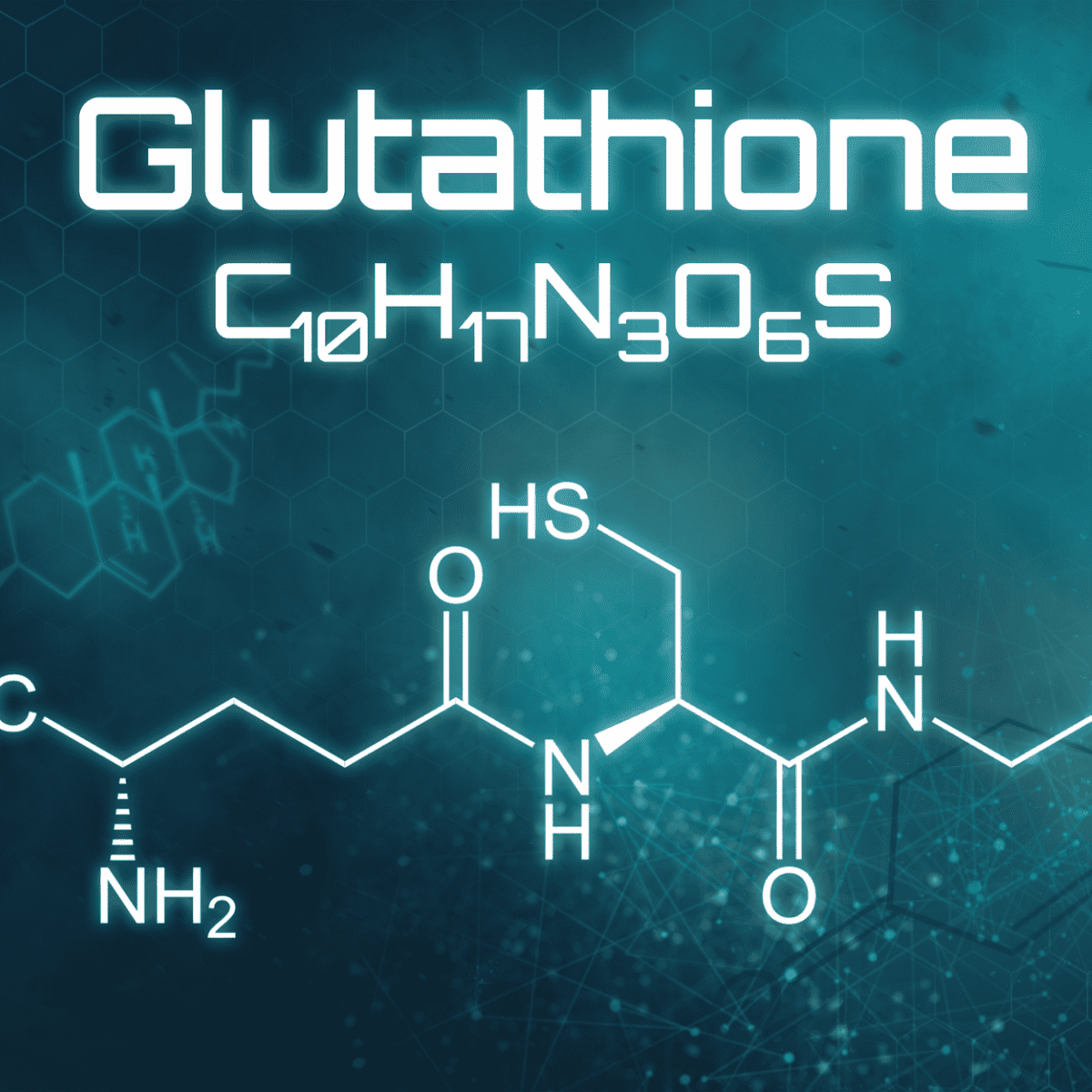
    Glutathione was first accurately described in 1935, but it wasn’t until the 1980s that research into the molecule’s function began to reveal its incredible abilities. At the molecular level, glutathione is a tripeptide, meaning it is composed of three amino acids – glutamate, cysteine ​​and glycine – linked by peptide bonds.

    These components combine to form a unique molecule that helps maintain cellular homeostasis or balance. It supports cell homeostasis largely by protecting against oxidative damage. This protective property makes glutathione exceptionally powerful.

    Many diseases and conditions are associated with decreased glutathione levels, including Alzheimer’s disease, cancer, chronic liver disease, cognitive disorders, diabetes, Parkinson’s disease, bone loss and more.1

    Since glutathione protects cells throughout the body, it makes sense that a deficiency could be linked to several health problems.

    Short content

    Glutathione is a molecule consisting of three amino acids: glutamate, cysteine ​​and glycine. Combined, they form a molecule that helps maintain cell homeostasis throughout the body. Low glutathione levels have been linked to a wide variety of diseases and conditions.

    Where glutathione comes from

    Maintaining optimal glutathione levels is essential for good health. Fortunately, every cell has the ability to produce glutathione in a cellular fluid called cytosol, provided it has access to the required precursor amino acids: glutamic acid (glutamate), cysteine ​​and glycine.

    The process occurs in two steps and requires special enzymes to complete. Glutathione is then pumped into the mitochondria, the organelles that create the energy that fuels every cell in our body. Glutathione protects the mitochondria against oxidative damage caused by radical oxygen species.

    Your body naturally knows how much glutathione is needed to ward off oxidative damage to your cells. But certain obstacles may prevent her from achieving that production.

    Without the building blocks of glutathione and the enzymes that enable its production, your body cannot maintain healthy levels of this powerful antioxidant.

    Short content

    Glutathione, synthesized from its constituent amino acids in a cellular fluid known as cytosol, is then transported to the mitochondria, the energy-producing organelles of the cell. Glutathione protects the mitochondria against oxidative damage.

    How to increase glutathione levels

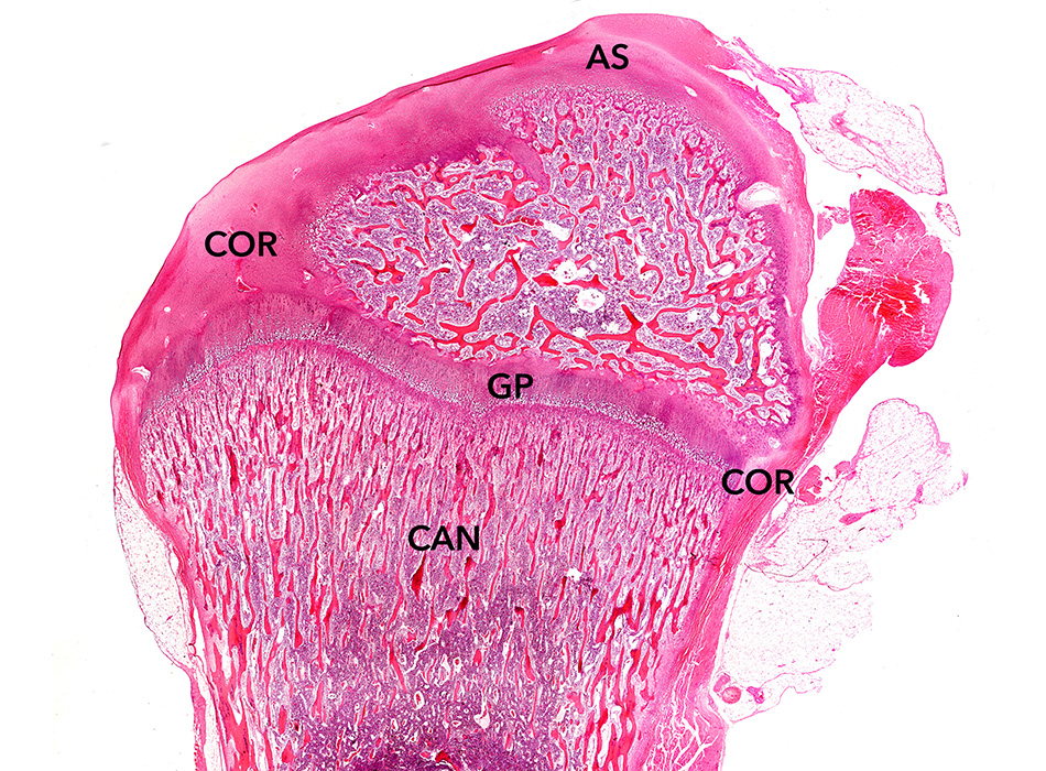
    In order for our cells to function optimally and prevent oxidative damage, it is imperative to ensure an ample supply of glutathione in our body. The antioxidant effect directly benefits the health and quality of our bones and our bone remodeling process.

    Recent research indicates that glutathione plays an even broader role by maintaining a balanced relationship between osteoclasts and osteoblast cells responsible for bone resorption and formation respectively – mainly by inhibiting osteoclast production. This action naturally increases bone mass.2

    These strategies can help us maintain healthy levels of the Master Antioxidant.

    • Eat sulfur-rich vegetables – Glutathione production requires sulfur, which is found in plant foods such as broccoli, Brussels sprouts, kale, cauliflower, watercress and mustard greens. Studies have linked a diet high in cruciferous vegetables to reduced oxidative stress and increased glutathione levels.
    • Eat glutathione-rich foods – Foods naturally rich in glutathione, such as spinach, avocados, asparagus and okra, all reduce oxidative stress. Our digestive system is not adept at absorbing glutathione from food, so the glutathione in these foods is not likely to get into our cells. However, because they reduce oxidative stress, they help keep glutathione levels high.
    • Increase vitamin C – Vitamin C has a similar effect as the glutathione-rich foods mentioned above. It’s a powerful antioxidant itself, so an abundance of vitamin C takes the pressure off glutathione, keeping levels robust. Additionally, studies have shown that vitamin C helps replenish glutathione molecules.3
    • Eat selenium-rich foods – Glutathione needs selenium to function. Include selenium-rich foods in your diet, such as chicken, fish, brown rice and Brazil nuts. Selenium is a basic supplement in the Osteoporosis Reversal Program.
    • Include foods high in cysteine – Cysteine, one of the three amino acids that make up glutathione, is found in foods such as whey protein, tuna, lean chicken, lentils, oatmeal, yogurt, carrots, shiitake mushrooms, almond butter and sunflower seeds. Using this building block allows your body to generate more of the master antioxidant.3
    • Turmeric extract curcumin Curcumin is an extract of the spice turmeric. Studies in animals have shown that curcumin has the ability to increase glutathione levels. Curcumin can be found in a supplement form.4
    • Take milk thistle – Milk thistle is a plant that contains a collection of compounds called silymarin. Silymarin is known for its antioxidant properties and studies have shown that it increases glutathione levels. 5
    • Get high-quality sleep – Glutathione, like all antioxidants, fights oxidative stress. Poor sleep quality can increase oxidative stress, making it harder for glutathione to be produced. Prioritize consistent, high-quality sleep to increase and maintain healthy glutathione levels.6
    • Regular exercise – Exercise helps our body maintain and increase antioxidant levels. Research has shown that combining cardio training with strength training has the most beneficial effect on glutathione levels. 7
    •  

    Short content

    Keep glutathione levels high to protect your bone remodeling process. See the list of strategies above, which includes dietary changes, supplements, sleep quality, and regular exercise.

    What this means for you

    You have the ability to equip your body with the essentials to protect itself from oxidative damage. Every cell in your body is ready and able to produce the Master Antioxidant if you provide the right materials and conditions.

    For tips on incorporating foods rich in sulfur, selenium, vitamin C and glutathione into your diet, try Bone Appétit, the Save Institute’s pH-balanced cookbook and meal planner. For help building a regular exercise routine you love, try SaveTrainer, the Save Institute’s online video training platform.

    You’re not the only one making healthy changes to improve your health. Find the support you need, in whatever form it takes, and celebrate that you have the wisdom to use the resources available to you!

    References

    1 https://www.ncbi.nlm.nih.gov/pmc/articles/PMC6770193/

    2 https://www.sciencedirect.com/science/article/pii/S0753332220304972

    3 https://www.ncbi.nlm.nih.gov/pubmed/12499341

    4 https://pubmed.ncbi.nlm.nih.gov/29484396/

    5 https://pubmed.ncbi.nlm.nih.gov/2353930/

    6 https://www.ncbi.nlm.nih.gov/pubmed/25945148

    7 https://pubmed.ncbi.nlm.nih.gov/17925621/


    Source link

  • Superior Polymers releases breakthrough medical-grade PEEK composite, combining carbon fiber, hydroxyapatite and polyether-ether ketone for medical applications

    Superior Polymers releases breakthrough medical-grade PEEK composite, combining carbon fiber, hydroxyapatite and polyether-ether ketone for medical applications


    First combination of three clinically proven biomaterials now available for the manufacture of medical devices such as spinal and cardiovascular implants

    FLOWOOD, Miss., Oct. 17, 2023 (GLOBE NEWSWIRE) — Superior Polymers, an innovator in advanced materials for medical applications, today announces the launch of Magnolia Trinity PEEK, which brings together three clinically proven biomaterials. Magnolia Trinity PEEK combines carbon fiber, hydroxyapatite and polyetheretherketone – three crucial materials needed in high-performance medical applications. Now Magnolia Trinity PEEK offers unparalleled versatility, biocompatibility and durability for medical devices such as orthopedic and cardiovascular implants or surgical instruments, catheter components and more.

    “Superior Polymers pushed the boundaries of materials science to create Magnolia Trinity PEEK,” said Bob Fruge, director of Business Development for Superior Polymers. “Bringing these three proven materials together in one application is unprecedented. This new composite material opens up new possibilities for the use of implantable polymers for engineers, designers and manufacturers. We are thrilled to bring Magnolia Trinity PEEK to our customers and the marketplace to ultimately improve patient outcomes.”

    Multiple studies have proven the positive effects of hydroxyapatite, carbon fiber and polyetheretherketone.i Carbon fiber is known for its exceptional mechanical properties, while hydroxyapatite is a natural mineral component of bone, known for its osteoconductive properties. By combining these materials, multifunctional composites can be created that can simultaneously provide structural support, promote bone growth and resist wear and tear.

    Magnolia Trinity PEEK is easily machinable and can be easily customized to specific medical device designs. This versatility allows for the creation of complex and patient-specific implants.

    Additional key features and benefits of Magnolia Trinity PEEK include:

    • Fatigue resistance: Carbon fiber reinforcement improves the fatigue resistance of PEEK.
    • Reduced wear and friction during joint replacement and prosthetics: High abrasion resistance and low friction properties due to the presence of carbon fiber are advantageous in applications involving hinged or sliding components, such as joint replacements and prosthetics.
    • Osteoconductive potential: Hydroxyapatite is a naturally occurring mineral found in bone and when combined with PEEK it improves the osteoconductive properties of the material.
    • Improved radiolucency: Carbon fibers improve radiolucency compared to pure PEEK, which is relatively radiopaque.
    • Better hemocompatibility: Higher crystallinity of the composite can cause less hemolysis and thrombin formation and reduce platelet adhesion, making it a promising material for cardiovascular applications.

    Magnolia Trinity PEEK is available now. Visit superior-polymers.com today to schedule a consultation to learn more and explore its potential applications with a materials expert. Representatives from Superior Polymers will attend the upcoming annual North American Spine Society (NASS) event in Los Angeles, CA, October 18-21, 2023. To schedule a meeting, email Bob Fruge at bob.fruge@superior-polymers.com.

    About superior polymers

    Superior Polymers is a leading innovator in advanced materials, committed to delivering cutting-edge solutions to industries around the world. With a strong focus on customer service, our team of experts strives to create materials that exceed expectations in performance, durability and versatility. More information at www.superior-polymers.com.

    Contact:

    Email address: bob.fruge@superior-polymers.com

    Phone number: 601-345-4515

    Company website: http://www.superior-polymers.com

    Social media links: https://www.linkedin.com/company/superior-polymers/

    PR contact
    Melany Joy Beck | melany.beck@512financial.com
    Cell: (737) 900-7986

    i https://www.ncbi.nlm.nih.gov/pmc/articles/PMC4987717/

    https://pubmed.ncbi.nlm.nih.gov/25780341/

    A photo accompanying this announcement is available at https://www.globenewswire.com/NewsRoom/AttachmentNg/c3d86fcc-172b-4d59-b90c-e983978048cf

    Source link

  • Mom’s Looking Forward Children’s Book Reviews

    Mom’s Looking Forward Children’s Book Reviews

    Screenshot%202023 09 28%20at%209.03.15%20AM

    Perhaps one of the most challenging aspects of motherhood with a chronic illness is helping your children understand what’s going on with your body and encouraging acceptance about how your illness affects them. How can you help them develop empathy for what you are experiencing, especially if your illness is invisible? Where is the line between being honest and worrying them? What’s the best way to address their concerns in child-friendly language? This is a topic that will probably need to be an ongoing conversation in your family – and sometimes reading a book together can help!

    Here’s a list of all the children’s books we’ve reviewed on our site to make it easier for you to browse all the options. All of these books can be considered tools to help children cope with having a parent with a chronic illness, and often they can be helpful even if the specific diagnosis mentioned is not yours. But please check out each individual review to find out if the book is right for your family!

    Review of: Aunt Barby’s invisible, endless Owie by Barbie Ingle and Tim Ingle

    Review of: How do you care for a very sick bear? by Vanessa Bayer

    Review of: How many marbles do you have? Helping children understand the limitations of people with chronic fatigue syndrome and fibromyalgia by Melinda Malott

    Review of: In my heart – A book full of feelings by Jo Witek

    Review of: Mommy has to stay in bed by Annette Rivlin-Gutman

    Review of: Mom Too bad by Rosana Sullivan

    Review of: Mom goes to the hospital by Josie Leon

    Review of: Mom, what’s wrong? Through Jessica Hensarling

    Review of: My special butterfly by Kelli Roseta

    Review of: Noah the Narwhal – A story of ups and downs by Judith Klausner

    Review of: Ravyn’s Doll – How to Explain Fibromyalgia to Your Child by Melissa Swanson

    Review of: Some days (board book) by Caitlyn PW Jones

    Review of: Some days: a story about love, ice cream and my mother’s chronic illness by Julie A. Stamm

    Review of: What does Super Jonny do when mom gets sick? by Simone Colwill

    Review of: When mom is sick by Ferne Sherkin-Langer

    Review of: When Pete’s father got sick by Kathleen Long Bostrom

    Review of: Why does mommy hurt? Helping children cope with the challenges of having a caregiver with chronic pain, fibromyalgia, or autoimmune diseases by Elizabeth M Christy

    Review of: Wonder Mom by Jennifer

    Source link

  • Bone Talk

    Bone Talk

    shutterstock 327287249 1

    Guidelines for guest blog posts

    Thank you for your interest in contributing to the Bone Talk blog! We periodically collaborate with health experts to demonstrate thought leadership and provide valuable resources and insights to our readers. The guidelines below describe the submission specifications.

    Blog post themes and topic suggestions

    • Nutrition for bone health

    • Exercise and physical activity

    • Safe movement

    • Diagnosis and treatment

    • Mental health

    • … and more! Do you have a suggestion? We’d love to hear it!

    • .Contact: Carina May, cmay(@)bonehealthandosteoporosis.org

    Quality specifications

    Pictures: Each message should have at least one main image that adds value to the information presented. Additional images may also be included in the message, but this is not required. We often use an image of the guest writer or we can use an agreed upon stock photo.

    • Originality: All content must be original. We do not accept syndicated or duplicate content.

    • Bio: Authors are invited to submit a short biography, photo and social links.

    Source link

  • 3 common myths about physical therapy

    3 common myths about physical therapy

    Physiotherapy is an essential aspect of post-operative recovery, injury rehabilitation and overall musculoskeletal health. However, despite its effectiveness, there are several misconceptions surrounding physical therapy that often prevent people from seeking this treatment.

    Learn the truth about physical therapy by understanding the truth of three common myths debunked by bone and joint specialists.

    Myth 1: Physical therapy is only for athletes

    A common misconception is that physical therapy is only for athletes recovering from sports-related injuries.

    The truth is that physical therapy is not limited to athletes. It benefits individuals of all ages and activity levels. Physical therapists are skilled in treating a variety of conditions, including chronic pain, postoperative rehabilitation, work-related injuries, and non-surgical orthopedic injuries.

    Whether you are an athlete, a sedentary person, or somewhere in between, physical therapy can be tailored to meet your specific needs.

    Myth 2: Physical therapy is painful

    Another prevailing myth is that physical therapy is painful and involves long-term discomfort to achieve results. On the contrary, the physiotherapists at Bone and Joint Specialists always put the patient’s comfort and safety first.

    Although some therapeutic exercises may cause mild discomfort because they target weakened or injured areas, physical therapists adjust the intensity of the exercises to avoid excessive pain.

    In addition, physical therapy aims to relieve pain and promote healing – not to worsen discomfort. The goal is to gradually improve strength, flexibility and range of motion. This actually helps patients with long-lasting pain relief.

    Myth 3: It is only for patients after surgery

    Another common misconception is that physical therapy is only necessary for patients recovering from surgery.

    While it is certainly important for postoperative recovery, physical therapy can benefit a wide range of people, including those with overuse injuries such as tendinitis or bursitis.

    It is beneficial for anyone looking to improve flexibility, muscle strength and movement mechanics. It is also an essential part of recovery for many patients at Bone and Joint Specialists, helping them reduce the risk of future injuries.

    Schedule a consultation today

    At Bone & Joint Specialists in Merrillville, IN, experienced physical therapists provide you with expert care and guidance throughout your therapeutic journey.

    Schedule a consultation with them by calling (219) 795-3360 and take the first step toward a healthier and less painful lifestyle.

    Source link