Welcome to our latest article on the many benefits of stair climbing. In this section, we will explore how climbing stairs is not just a simple task but also a fantastic exercise for improving your knee health and strength.
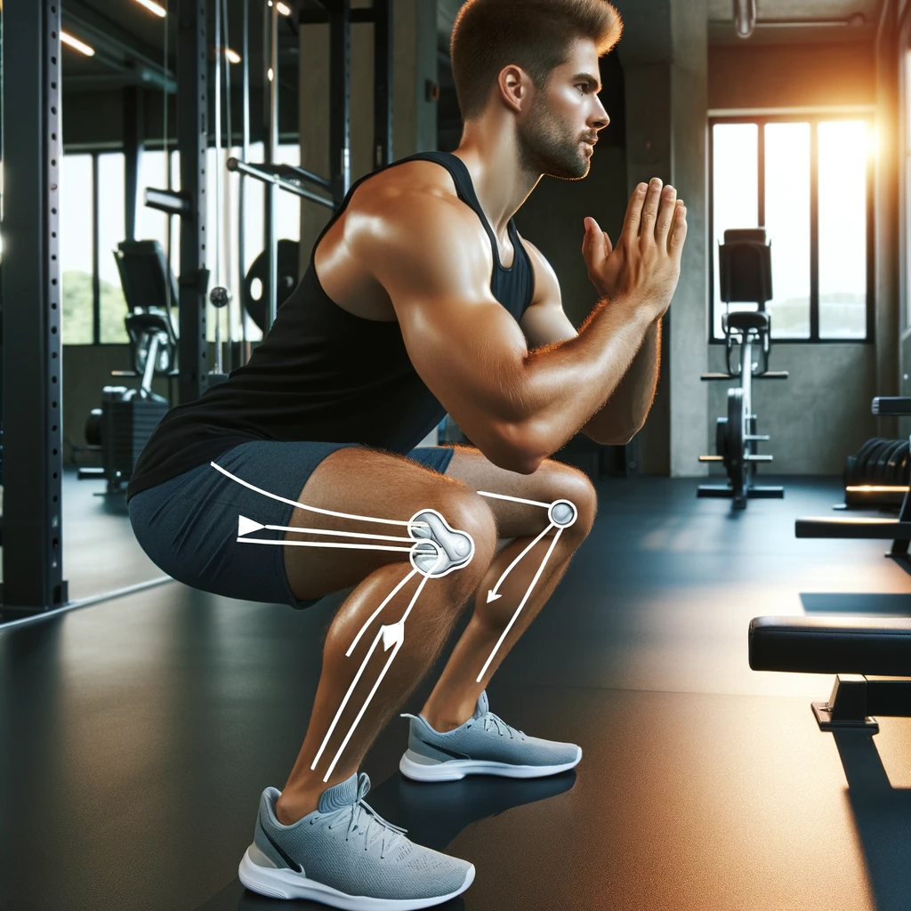
When you climb stairs, you engage the muscles around your knees, particularly the quadriceps and hamstrings, which play a crucial role in knee stability. By regularly incorporating stair climbing into your fitness routine, you can strengthen these muscles, leading to improved knee health and reduced risk of knee injuries.
Moreover, stair climbing is an effective calorie-burning activity that can aid in weight management. Excess weight puts additional pressure on your knees, increasing the risk of knee problems. By climbing stairs regularly, you can burn calories, reduce weight, and alleviate the stress on your knees.
However, it is important to note that stair climbing intensity and duration should be tailored to your specific needs and condition. We always recommend consulting with a professional or trainer to determine the appropriate approach to stair climbing for your knee health and strength goals.
Key Takeaways:
- Climbing stairs is an effective exercise for strengthening the muscles around the knees.
- Regular stair climbing can help improve knee health and reduce the risk of knee injuries.
- Stair climbing aids in weight management, reducing the pressure on your knees.
- Consult with a professional to determine the appropriate intensity and duration of stair climbing for your specific condition.
The Indirect Benefits of Stair Climbing
Stair climbing offers more than just direct benefits to knee strength. It also provides several indirect advantages for joint health. Let’s explore the various ways stair climbing contributes to overall joint well-being.
Benefit 1: Weight Management and Knee Arthritis Relief
One of the significant indirect benefits of stair climbing is the ability to manage weight effectively. By regularly engaging in this exercise, individuals can burn calories, shed excess pounds, and reduce the load on their joints, including the knees. This reduction in weight-bearing pressure can provide relief from knee arthritis symptoms, such as pain and inflammation.
“Stair climbing is a weight-bearing exercise that helps in managing weight, relieving pressure on the joints, and potentially reducing knee arthritis symptoms.” – [Source]
Benefit 2: Increased Bone Density and Joint Health
Stair climbing is a weight-bearing exercise that stimulates the growth and strengthening of bones. By subjecting the body to the force of gravity during stair climbing, individuals can increase their bone density. This is particularly crucial for maintaining joint health, as strong bones provide a stable foundation for the joints to function optimally.
Benefit 3: Improved Lower Limb Muscle Strength and Reduced Knee Pain
Research indicates that stair climbing is beneficial for individuals with osteoarthritis, a common form of joint degeneration that often affects the knees. The exercise can improve lower limb muscle strength and help alleviate knee pain associated with osteoarthritis.
“Stair climbing is beneficial for individuals with osteoarthritis, as it can improve lower limb muscle strength and reduce knee pain.” – [Source]
To summarize, stair climbing not only directly strengthens the knees but also provides indirect benefits for joint health. It aids in weight management and knee arthritis relief by reducing pressure on the joints. Furthermore, stair climbing supports bone density and helps individuals with osteoarthritis by improving lower limb muscle strength and alleviating knee pain.

| Indirect Benefits of Stair Climbing |
|---|
| Weight management and knee arthritis relief |
| Increased bone density and joint health |
| Improved lower limb muscle strength and reduced knee pain |
Precautions for Stair Climbing
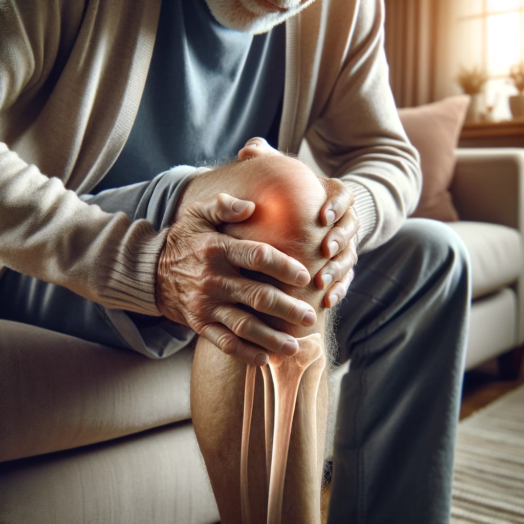
While stair climbing is generally safe and beneficial, it is important to take certain precautions, especially if you experience knee pain or are undergoing a knee rehabilitation program. Individuals with specific knee conditions, such as chondromalacia patella, may find that stair climbing exacerbates their pain and discomfort and may need to avoid it initially. To ensure the appropriateness of stair climbing for your specific condition, we strongly recommend consulting with a healthcare professional or a certified trainer.
During stair climbing, it is crucial to be mindful of any signs of strain or sharp pain in your knees. These may indicate underlying problems or issues that require immediate attention. If you experience such symptoms, it is important to cease the exercise and seek medical advice to prevent further injury or complications.
Stair climbing and knee rehabilitation programs require a personalized approach, taking into consideration your unique circumstances and medical history. Therefore, consulting with a healthcare professional or a certified trainer is essential to ensure the safe and effective integration of stair climbing into your exercise routine.
Quote:
“To prevent knee pain and injury during stair climbing, it is important to listen to your body and take necessary precautions. Consulting with a healthcare professional or a certified trainer can provide valuable guidance in designing an appropriate stair climbing regimen for your specific needs.” – Dr. Sarah Thompson, Orthopedic Specialist
| Precautions for Stair Climbing | Knee Pain or Rehabilitation Program |
|---|---|
| Avoid stair climbing if you have chondromalacia patella or other specific knee conditions | Consult with a healthcare professional or certified trainer to determine the appropriateness of stair climbing for your specific condition |
| Be aware of any signs of strain or sharp pain during stair climbing | Cease the exercise and seek medical advice if experiencing knee pain during or after stair climbing |
| Personalized approach required for stair climbing and knee rehabilitation | Consultation with a healthcare professional or certified trainer is essential to tailor the exercise program |

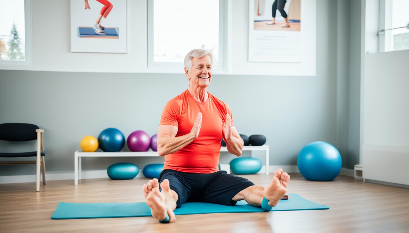
Exercises to Strengthen the Legs for Stair Climbing
In order to make stair climbing easier and more comfortable, it is beneficial to incorporate exercises that strengthen the legs. By targeting the muscles involved in stair climbing, you can improve muscle strength and balance, which are essential for successful and pain-free stair climbing.
According to the second source mentioned, the following exercises are highly effective in enhancing knee health and preparing the legs for stair climbing:
- Tandem Balance Passes: This exercise involves standing with one foot directly in front of the other and slowly moving the back foot in a forward and backward motion while maintaining balance. It helps improve stability and works the muscles in the calves and thighs.
- Step-Ups: Step-ups involve placing one foot on an elevated platform, such as a step or bench, and stepping onto it using the leg and glute muscles. This exercise targets the quads, hamstrings, and glutes, strengthening them for stair climbing.
- Lateral Banded Stepping: Using a resistance band around the ankles, perform sideways steps, maintaining tension in the band. This exercise targets the abductor muscles of the legs, which support proper knee alignment during stair climbing.
- Modified Lunges: Lunges are excellent for strengthening the quadriceps and glutes. Modify the movement by using a chair or wall for support, if needed, to reduce pressure on the knees.
- Alternating Single-Leg Stair Taps: Stand in front of a low step and step onto it with one foot, lifting the opposite knee. Alternate legs and repeat. This exercise mimics stair climbing and improves balance and coordination.
To ensure optimal results and minimize the risk of injury, it is crucial to perform these exercises consistently and under proper guidance. Start with lighter resistance or modifications if necessary, gradually increasing the intensity as strength and comfort levels improve.
| Exercise | Target Muscles | Benefit |
|---|---|---|
| Tandem Balance Passes | Calves, Thighs, Core | Improves stability and works the muscles involved in stair climbing. |
| Step-Ups | Quadriceps, Hamstrings, Glutes | Strengthens the major muscles involved in stair climbing. |
| Lateral Banded Stepping | Abductor Muscles | Targets the muscles that support proper knee alignment during stair climbing. |
| Modified Lunges | Quadriceps, Glutes | Strengthens the muscles involved in stair climbing while reducing knee strain. |
| Alternating Single-Leg Stair Taps | Calves, Thighs, Core | Improves balance, coordination, and mimics the movement of stair climbing. |
Remember to listen to your body and stop any exercise that causes pain or discomfort. If you have any underlying health conditions or concerns, it is always advisable to consult with a healthcare professional or a certified trainer before starting any new exercise routine.
Stair Climbing and Overall Health
Stair climbing is not only beneficial for knee health but also plays a significant role in promoting overall health and well-being. Research from the third source mentioned reveals that regular stair climbing is associated with improved cognitive function and a reduced risk of cognitive impairments such as mild cognitive impairment, Alzheimer’s disease, and vascular dementia. It’s an exercise that engages both the body and the mind, making it an excellent choice for cognitive health.
Moreover, stair climbing is an effective cardiovascular exercise that offers numerous benefits for heart health. Engaging in this activity helps improve cardiovascular fitness, increase stamina, and even lower the risk of mortality. It’s a natural form of aerobic exercise that strengthens the heart, promotes blood circulation, and enhances overall cardiovascular function.
Additionally, incorporating stair climbing into daily routines is a convenient and time-efficient way to improve overall health and vitality. It is an accessible exercise that can be easily performed in various settings, whether at home, work, or in public spaces. By simply taking the stairs instead of the elevator or escalator, individuals can reap the many health benefits associated with stair climbing.
| Benefits of Stair Climbing for Overall Health |
|---|
| Improved cognitive function |
| Reduced risk of cognitive impairments (mild cognitive impairment, Alzheimer’s disease, and vascular dementia) |
| Enhanced cardiovascular health |
| Increased stamina |
| Lowered risk of mortality |
| Easily incorporated into daily routines |
With its numerous benefits for cognitive health and cardiovascular well-being, stair climbing proves to be a holistic exercise that contributes to overall health. It’s a simple yet powerful activity that can positively impact multiple aspects of well-being, making it an ideal choice for individuals looking to improve their overall health and vitality.
Conclusion
In conclusion, climbing stairs offers numerous benefits for knee health and overall well-being. By engaging in this simple yet effective exercise, you can strengthen the muscles around the knee, leading to improved knee stability and reduced risk of injuries. Additionally, stair climbing aids in weight management by burning calories, which can alleviate the pressure on the knees caused by excess weight.
Furthermore, stair climbing provides indirect relief for knee arthritis symptoms. By managing weight and increasing bone density, this exercise can help alleviate knee arthritis pain and improve joint function. It’s important to note that individuals with certain knee conditions should consult with a healthcare professional or trainer to determine the appropriateness of stair climbing for their specific needs.
Moreover, stair climbing not only benefits knee health but also has positive effects on cognitive health and cardiovascular fitness. Regular stair climbing has been associated with improved cognitive function and a reduced risk of cognitive impairments and dementia. Additionally, as a cardiovascular exercise, stair climbing helps to improve heart health, increase stamina, and lower mortality risk.
Incorporating stair climbing into your daily routine is a practical and accessible way to improve knee health, overall fitness, and quality of life. So why wait? Start climbing those stairs and reap the numerous benefits it has to offer.
FAQ
Is climbing stairs good for knee health?
Yes, climbing stairs is beneficial for knee health. It strengthens the muscles around the knee, such as the quadriceps and hamstrings, which are essential for knee stability.
What are the knee health benefits of climbing stairs?
Climbing stairs helps to strengthen the muscles around the knee, improves knee stability, aids in weight management, and reduces the risk of excess pressure on the knees.
Can stair climbing improve knee strength?
Yes, stair climbing is an effective exercise for improving knee strength. It helps to strengthen the muscles around the knee, enhancing their ability to support the joint.
Does stair climbing help with knee arthritis?
Yes, stair climbing can provide relief for knee arthritis symptoms. It aids in weight management, reduces knee pain, and improves lower limb muscle strength.
Are there any precautions to consider before stair climbing?
Yes, individuals with certain knee conditions, such as chondromalacia patella, may experience increased pain with stair climbing and should consult with a healthcare professional or trainer. It is important to be aware of any signs of strain or sharp pain during stair climbing, as this may indicate underlying problems.
What exercises can I do to strengthen my legs for stair climbing?
Exercises such as tandem balance passes, step-ups, lateral banded stepping, modified lunges, and alternating single-leg stair taps can help improve leg muscle strength and balance, which are important for successful stair climbing.
Does stair climbing have benefits for overall health?
Yes, stair climbing has multiple benefits for overall health. It has been associated with improved cognitive function, reduced risks of cognitive impairment and dementia, improved heart health, increased stamina, and lower mortality risk.