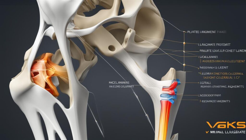
As we delve into the fascinating world of human anatomy, one area that captures our attention is the knee joint. This intricate joint, with its various structures, holds the distinction of being the largest and most complex joint in our body. Understanding the anatomy of the knee bones and how they fit together is crucial in comprehending knee problems and seeking appropriate treatment.
The knee consists of four bones: the femur (thigh bone), patella (kneecap), tibia (shin bone), and fibula (outer shin bone). Each bone plays a vital role in supporting our body, transferring forces between the hip and foot, and allowing smooth and efficient movement of the leg.
Key Takeaways:
- The knee joint is the largest and most complex joint in the human body.
- The knee consists of four bones: the femur, patella, tibia, and fibula.
- Understanding the knee bones structure is crucial in comprehending knee problems.
- The knee bones work together to support the body and allow efficient movement of the leg.
- Seeking appropriate treatment for knee conditions requires understanding the anatomy of the knee bones.
Knee Bone Anatomy: Structure and Function
The knee bones provide the structure to the knee joint and are composed of the femur, patella, tibia, and fibula. The femur is the thigh bone, while the patella is the kneecap. The tibia is the main shin bone, and the fibula is the outer shin bone. These bones work together to support the body, transfer forces between the hip and foot, and facilitate smooth and efficient movement of the leg.
The knee bones also encompass the tibiofemoral joint (between the thigh and shin bones) and the patellofemoral joint (between the kneecap and thigh bone). Understanding the anatomy and function of these knee bones is essential in diagnosing and treating knee conditions.
| Knee Bone | Common Name |
|---|---|
| Femur | Thigh bone |
| Patella | Kneecap |
| Tibia | Shin bone |
| Fibula | Outer shin bone |
Knee Joint Cartilage Anatomy: Types and Functions
The knee joint is supported by different types of cartilage, including articular cartilage and the knee meniscus. Articular cartilage is a thin layer that lines the surfaces of the knee joints, providing cushioning and reducing friction. The knee meniscus, on the other hand, is a thicker layer of cartilage that acts as a shock absorber, distributing forces and facilitating smooth movement of the knee bones.
These cartilages, along with the back of the patella, play crucial roles in knee joint function and can be susceptible to injuries such as tears or arthritis. Articular cartilage ensures that the surfaces of the knee bones glide smoothly over each other, preventing bone-on-bone contact and reducing wear and tear. The knee meniscus, with its C-shaped structure, helps with load-bearing, stability, and shock absorption. By distributing forces evenly, the meniscus prevents excessive pressure on specific areas of the knee joint.
Articular Cartilage
Articular cartilage covers the ends of the femur, tibia, and patella, allowing them to move smoothly against each other. This thin, flexible tissue is made up mostly of water, collagen fibers, and proteoglycans. Its unique composition enables it to withstand compression and abrasive forces during movement. Articular cartilage also plays a vital role in absorbing shock and dispersing the loads placed on the knee joint. However, it has a limited ability to repair itself, and damage can lead to pain, stiffness, and reduced joint function.
Knee Meniscus
The knee meniscus comprises two C-shaped pieces of cartilage: the medial meniscus on the inner side and the lateral meniscus on the outer side of the knee joint. These structures provide stability, cushioning, and load distribution to the knee joint. The menisci help to deepen the surface of the tibia, allowing a better fit with the rounded end of the femur. They also act as shock absorbers, dispersing forces and reducing the risk of injury to the underlying bone. However, the menisci can be susceptible to tears, which may impair their function and contribute to knee pain and instability.
A variety of factors can lead to knee cartilage injuries, such as traumatic incidents, repetitive stress, and degenerative conditions like osteoarthritis. These injuries can affect the articular cartilage, knee meniscus, or both. Treatment options for knee cartilage injuries range from conservative approaches, such as physical therapy and medication, to surgical interventions like arthroscopy and cartilage restoration procedures.
A comprehensive understanding of knee joint cartilage anatomy is essential for healthcare professionals and patients alike. By recognizing the types and functions of knee cartilage, individuals can make informed decisions regarding their knee health and work towards preserving or restoring the integrity of this vital joint structure.
| Cartilage Type | Location | Function |
|---|---|---|
| Articular cartilage | Covers the ends of the femur, tibia, and patella | Provides cushioning, reduces friction, and allows smooth joint movement |
| Knee meniscus | Located between the femur and tibia | Acts as a shock absorber, distributes forces, and improves stability |
Knee and Leg Muscle Anatomy: Roles and Importance
The knee and leg muscles play a vital role in the stability and movement of the knee joint. Understanding the anatomy of these muscles is key to maintaining healthy knee function. Let’s explore the main muscles involved and their importance in supporting the knee.
The Quadriceps
The quadriceps are a group of four muscles located on the front of the thigh. They include the rectus femoris, vastus lateralis, vastus medialis, and vastus intermedius. These muscles are responsible for extending the knee, allowing you to straighten your leg. They are crucial for activities such as walking, running, and jumping.
The Hamstrings
On the back of the thigh, you’ll find the hamstring muscles. This group consists of three muscles: the biceps femoris, semitendinosus, and semimembranosus. The hamstrings play an important role in knee flexion, bending the knee and bringing the heel towards the buttocks. They are engaged in activities like walking, running, kicking, and bending down.
The Glutes
The glutes, or gluteal muscles, are located in the buttock area. They consist of three muscles: the gluteus maximus, gluteus medius, and gluteus minimus. While primarily known for their role in hip movement, the glutes also contribute to knee stability. They help control side-to-side movements of the knee and provide stability during activities like squatting and lunging.
The Calves
The calf muscles, or gastrocnemius and soleus, are located on the back of the lower leg. Although they primarily act on the ankle joint, the calves also play a role in knee flexion and extension. They work together with the quadriceps and hamstrings to ensure smooth movement and stability of the knee during activities like running and jumping.
Weakness or tightness in these knee and leg muscles can lead to imbalances and increase the risk of knee pain and instability. To maintain knee health, it’s important to regularly engage in exercises that strengthen and stretch these muscles.

| Muscle Group | Main Muscles | Functions |
|---|---|---|
| Quadriceps | Rectus Femoris, Vastus Lateralis, Vastus Medialis, Vastus Intermedius | Extension of the knee, straightening the leg |
| Hamstrings | Biceps Femoris, Semitendinosus, Semimembranosus | Flexion of the knee, bending the leg |
| Glutes | Gluteus Maximus, Gluteus Medius, Gluteus Minimus | Control of side-to-side knee movements, stability during squatting and lunging |
| Calves | Gastrocnemius, Soleus | Assist in knee flexion and extension, contribute to overall leg stability |
Knee Ligament Anatomy: Stability and Function
The knee joint relies on its ligaments to provide stability and limit excessive movements that can lead to instability. Understanding the anatomy of the knee ligaments is essential for diagnosing and treating ligament injuries, such as sprains or tears, which can result in reduced joint function and instability.
The knee ligament anatomy consists of two sets of ligaments: the cruciate ligaments and the collateral ligaments. The cruciate ligaments, including the anterior cruciate ligament (ACL) and the posterior cruciate ligament (PCL), play a significant role in controlling the anterior and posterior movement of the knee, as well as rotational stability. On the other hand, the collateral ligaments, which consist of the medial collateral ligament (MCL) and the lateral collateral ligament (LCL), provide stability to the knee joint by limiting sideways movements.
Damage to these ligaments, resulting from sports injuries or accidents, can significantly impact knee stability and function. It is important to recognize the signs and symptoms of ligament injuries to initiate appropriate treatment promptly.
Cruciate Ligaments
The cruciate ligaments are located inside the knee joint and connect the femur (thigh bone) to the tibia (shin bone). They are named based on their attachment points. The anterior cruciate ligament (ACL) attaches from the anterior tibia to the posterior femur, while the posterior cruciate ligament (PCL) attaches from the posterior tibia to the anterior femur.
The ACL plays a crucial role in preventing the femur from sliding backward on the tibia, especially during activities that involve sudden stops or changes in direction. The PCL, on the other hand, prevents the femur from sliding forward on the tibia and stabilizes the knee when the knee is bent.
Collateral Ligaments
The collateral ligaments are located on the sides of the knee joint and provide stability by limiting sideways movements. The medial collateral ligament (MCL) is located on the inner side of the knee, connecting the femur to the tibia. It helps prevent the knee from bending inward. The lateral collateral ligament (LCL), on the other hand, is located on the outer side of the knee and connects the femur to the fibula. It helps prevent the knee from bending outward.
Together, the cruciate and collateral ligaments work in harmony to provide stability, control movement, and prevent excessive forces on the knee joint.

| Ligament | Location | Function |
|---|---|---|
| Anterior Cruciate Ligament (ACL) | Inside the knee joint, connecting the anterior tibia to the posterior femur | Prevents the femur from sliding backward on the tibia; stabilizes the knee during sudden stops or changes in direction |
| Posterior Cruciate Ligament (PCL) | Inside the knee joint, connecting the posterior tibia to the anterior femur | Prevents the femur from sliding forward on the tibia; stabilizes the knee when the knee is bent |
| Medial Collateral Ligament (MCL) | Located on the inner side of the knee, connecting the femur to the tibia | Prevents the knee from bending inward; provides stability against valgus (sideways) forces |
| Lateral Collateral Ligament (LCL) | Located on the outer side of the knee, connecting the femur to the fibula | Prevents the knee from bending outward; provides stability against varus (sideways) forces |
Proper understanding of the knee ligament anatomy is crucial for healthcare professionals in accurately diagnosing and effectively treating ligament injuries. By considering the location and function of each ligament and assessing the extent of ligament damage, appropriate treatment plans, including physical therapy, bracing, or surgical interventions, can be implemented to restore joint stability and function.
Conclusion: The Importance of Understanding Knee Bones Structure
Understanding the intricate anatomy of the knee bones structure is essential for comprehending the complexity of the knee joint and effectively diagnosing and treating various knee conditions. From the femur, patella, tibia, and fibula to the surrounding structures such as cartilage, muscles, ligaments, tendons, bursa, and joint capsule, each component plays a crucial role in the stability, function, and movement of the knee.
By gaining knowledge about the knee bones structure and their interconnections, individuals can make informed decisions about their knee health. Whether it’s seeking appropriate medical treatment for knee problems or taking preventive measures to maintain knee function, understanding the intricate anatomy of the knee bones provides a solid foundation for managing knee conditions.
From athletes striving to maintain peak performance to individuals dealing with knee pain or injury, knowledge of the knee bones structure empowers us to take control of our knee health. By working in partnership with healthcare professionals, we can ensure that our knees remain strong, stable, and capable of supporting us through all of life’s activities.
FAQ
What are the bones that make up the knee joint?
The knee joint is composed of the femur (thigh bone), patella (kneecap), tibia (shin bone), and fibula (outer shin bone).
What is the function of the knee bones?
The knee bones work together to support the body, transfer forces between the hip and foot, and allow smooth and efficient movement of the leg.
What types of cartilage are present in the knee joint?
The knee joint is supported by articular cartilage, which lines the surfaces of the knee joints, and the knee meniscus, which acts as a shock absorber.
Which muscles are involved in knee joint stability and movement?
The main muscles that control the leg are the quadriceps (front of the thigh), hamstrings (back of the thigh), glutes (buttock muscles), and calves (back of the lower leg).
What is the role of ligaments in the knee joint?
Ligaments in the knee provide stability and prevent excessive movements and instability. There are two sets of ligaments: the cruciate ligaments and the collateral ligaments.
Why is it important to understand the knee bones structure?
Understanding the knee bones structure is crucial in comprehending the complexity of the knee joint, diagnosing knee conditions, and seeking appropriate treatment.

Leave a Reply