Blog

  • Iliotibial Band Syndrome: Causes And Relief Options

    Iliotibial Band Syndrome: Causes And Relief Options

    Welcome to our comprehensive guide on iliotibial band syndrome (ITB), also known as iliotibial band syndrome. In this article, we will explore the causes of ITB, the symptoms to look out for, and the various relief options available to manage this condition.

    ITB syndrome is a common condition characterized by pain on the outside of the knee. It is frequently seen in athletes, especially runners and cyclists, who engage in repetitive bending and extending of the knee. However, it can also affect individuals who perform other activities that put strain on the knee joint.

    The precise cause of ITB syndrome is still a subject of ongoing research. However, it is believed to be related to friction, inflammation, and compressive forces exerted on the iliotibial band and the tissues beneath it. Several factors can contribute to the development of ITB syndrome, including anatomical variations, poor flexibility and muscle strength, training errors, and improper footwear.

    Diagnosing ITB syndrome typically involves a physical examination and a thorough assessment of the patient’s symptoms. Treatment options for ITB syndrome focus on relieving pain, reducing inflammation, and addressing any underlying biomechanical issues. These options may include rest, icing, pain medications, physical therapy, and techniques to improve flexibility and strength.

    Preventing ITB syndrome involves implementing proper training techniques, wearing appropriate footwear, and engaging in strengthening exercises targeted at the muscles around the hip and knee. By taking these preventive measures, individuals can significantly reduce their risk of developing ITB syndrome.

    Key Takeaways

    • ITB syndrome is characterized by pain on the outside of the knee.
    • Athletes, particularly runners and cyclists, are commonly affected by ITB syndrome.
    • Friction, inflammation, and compressive forces on the iliotibial band are believed to contribute to ITB syndrome.
    • Diagnosis involves a physical examination and assessment of symptoms.
    • Treatment options include rest, icing, pain medications, physical therapy, and addressing biomechanical issues.

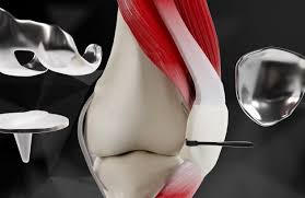
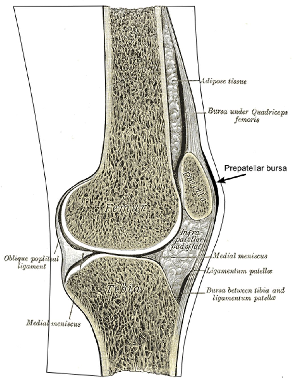
    What is the IT Band?

    The iliotibial band, also known as the IT band, is a long, fibrous band of tissue that extends from the hip to just below the outside of the knee. It plays a crucial role in stabilizing the knee joint and aiding in the movement of the leg. The IT band has an attachment to the outside of the knee cap and can cause pain when it becomes inflamed or irritated.

    The IT band is an essential structure that helps to maintain proper alignment and stability of the knee during various activities such as walking, running, and cycling. It serves as a connection between the hip and the shinbone, providing support and facilitating smooth movement.

    During movement, the iliotibial band glides over the outside of the knee joint, allowing the leg to extend and flex. However, repetitive movements, overuse, or certain anatomical variations can lead to friction and compression of the IT band against the underlying tissues, resulting in inflammation and pain.

    What is IT Band Syndrome?

    IT band syndrome, or iliotibial band syndrome, is a condition characterized by pain on the outside of the knee. This overuse injury commonly affects runners, cyclists, and athletes participating in sports that involve repetitive knee bending and straightening.

    The pain associated with IT band syndrome is typically felt during activities such as running or cycling. It may also be accompanied by clicking, popping, or snapping sensations on the outside of the knee. IT band syndrome can significantly impact an individual’s ability to engage in their desired physical activities and may require attention to alleviate symptoms and prevent further damage.

    IT band syndrome

    Understanding the underlying causes, symptoms, and treatment options for IT band syndrome is crucial for those affected by this condition. By taking a comprehensive approach to manage pain and optimize recovery, individuals can regain their active lifestyles and minimize the risk of future injuries.

    Symptoms of IT Band Syndrome

    The most common symptom of IT band syndrome is pain on the outside of the knee that worsens with activities involving repetitive knee bending and straightening. This pain is often described as a sharp or burning sensation and can significantly impact daily activities and sports performance.

    In addition to knee pain, individuals with IT band syndrome may experience other symptoms such as:

    • Clicking, popping, or snapping sensation on the outside of the knee during movement
    • Swelling or inflammation in the affected area

    It’s important to note that symptoms may vary from person to person, and some individuals may only experience a few of these symptoms. Early recognition of the symptoms is crucial for prompt diagnosis and effective treatment of IT band syndrome.

    IT Band Syndrome

    Causes of IT Band Syndrome

    The exact cause of IT band syndrome is still a topic of debate among researchers and healthcare professionals. However, it is believed to be primarily caused by the friction and compression of the iliotibial band (IT band) and the tissues beneath it during repetitive knee movements. These repetitive motions can lead to irritation, inflammation, and pain along the outside of the knee.

    Other factors that may contribute to the development of IT band syndrome include:

    • Anatomical variations: Certain structural differences in the shape or alignment of the bones, muscles, or tendons around the hip and knee can increase the risk of IT band syndrome.
    • Poor flexibility: Limited range of motion and flexibility in the muscles surrounding the hip and knee can put extra stress on the IT band, leading to irritation and pain.
    • Muscle imbalances and weaknesses: Weak or imbalanced muscles, particularly those of the hip and thigh, can alter the alignment and mechanics of the knee joint, increasing the strain on the IT band.
    • Training errors: Overtraining, sudden increases in training intensity or duration, improper warm-up or cool-down routines, and inadequate rest periods can all contribute to the development of IT band syndrome.
    • Improper footwear: Wearing shoes that do not provide proper support or stability for the foot and ankle can alter the mechanics of the lower extremity, increasing the risk of IT band irritation.

    Understanding these underlying causes is crucial for effectively preventing and managing IT band syndrome.

    Risk Factors Description
    Anatomical variations Structural differences that affect the alignment of the hip, knee, and IT band.
    Poor flexibility Limited range of motion and flexibility in the muscles surrounding the hip and knee.
    Muscle imbalances and weaknesses Weak or imbalanced muscles in the hip and thigh that alter the mechanics of the knee joint.
    Training errors Overtraining, improper warm-up/cool-down routines, and inadequate rest periods.
    Improper footwear Wearing shoes that do not provide proper support and stability for the foot and ankle.

    Diagnosis of IT Band Syndrome

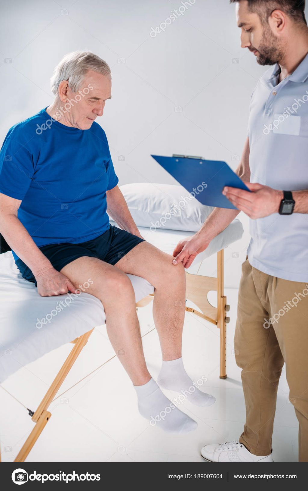
    Diagnosing IT band syndrome involves a comprehensive evaluation of the patient’s symptoms and a thorough physical examination. The healthcare provider will carefully assess various aspects, including the knee joint, range of motion, strength, and areas of tenderness. This assessment helps to identify any telltale signs of IT band syndrome and rule out other possible causes of knee pain.

    In some cases, imaging tests such as X-rays or MRI may be ordered to obtain a clearer picture of the knee’s internal structures. These tests can provide valuable insights and help differentiate IT band syndrome from other conditions, such as runner’s knee, meniscus injuries, or stress fractures. By ruling out other potential causes, healthcare providers can accurately diagnose IT band syndrome and develop an appropriate treatment plan.

    Diagnosis of IT Band Syndrome Procedure
    1. Physical Examination A comprehensive assessment of the patient’s knee joint, range of motion, strength, and areas of tenderness.
    2. Imaging Tests X-rays or MRI may be ordered to rule out other possible causes of knee pain and confirm the diagnosis of IT band syndrome.

    Diagnosing IT band syndrome accurately is essential for effective treatment and management. Healthcare providers utilize a combination of physical examination, symptom analysis, and imaging tests to diagnose the condition confidently. By promptly and accurately diagnosing IT band syndrome, healthcare professionals can recommend appropriate treatment strategies for their patients and facilitate a successful recovery.

    Treatment and Relief Options for IT Band Syndrome

    When it comes to treating IT band syndrome, our goal is to provide pain relief, reduce inflammation, and address any underlying biomechanical issues that may be contributing to the condition. Here are some treatment options that can help individuals manage their symptoms and recover effectively:

    1. Rest: Taking a break from the activity that triggers pain is essential for allowing the inflamed tissues to heal. This may involve reducing or modifying your training regimen to avoid aggravating your IT band.
    2. Icing: Applying ice to the affected area can help reduce swelling and alleviate pain. Aim for 15 to 20 minutes of icing several times a day, especially after exercise.
    3. Over-the-counter pain medications: Nonsteroidal anti-inflammatory drugs (NSAIDs) such as ibuprofen or naproxen sodium can help relieve pain and reduce inflammation. However, it is recommended to consult with a healthcare professional before taking any medication.
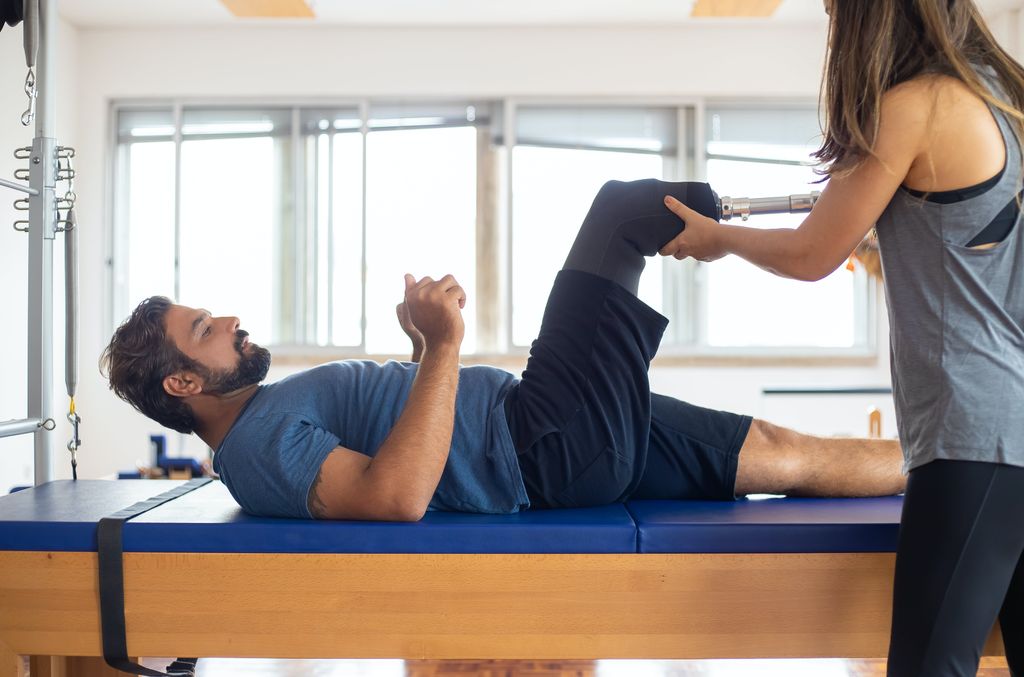
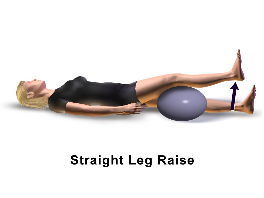
    4. Physical therapy: Engaging in a targeted physical therapy program can be highly beneficial for individuals with IT band syndrome. A skilled physical therapist will guide you through exercises designed to stretch and strengthen the muscles surrounding the hip and knee, helping to improve flexibility and correct any imbalances or weaknesses.
    5. Biomechanical assessment: Addressing biomechanical factors such as running form and footwear is important to prevent further stress on the IT band. A professional assessment can help identify any deficiencies and provide recommendations for improvements or necessary modifications.
    6. Corticosteroid injections: In severe cases that fail to respond to conservative treatment, corticosteroid injections may be considered. These injections help reduce inflammation and provide temporary pain relief.
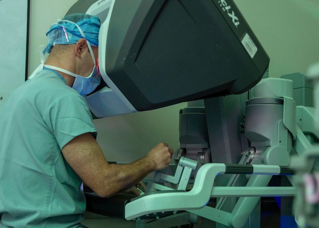
    7. Surgery: Surgical intervention is rare for IT band syndrome and typically reserved for refractory cases. It may involve releasing the tight IT band or addressing anatomical abnormalities contributing to the condition.

    Remember, the specific treatment plan for IT band syndrome should be tailored to each individual’s needs and may vary based on the severity of the condition. Consulting with a healthcare professional is crucial for accurate diagnosis and personalized treatment guidance.

    Conclusion

    In conclusion, iliotibial band syndrome (IT band syndrome) is a common condition characterized by pain on the outside of the knee. Although it is frequently seen in athletes, it can affect anyone who engages in repetitive knee movements. The exact cause of IT band syndrome is still under debate, but it is believed to be related to friction, inflammation, and compressive forces on the iliotibial band and the surrounding tissues.

    Fortunately, there are various treatment options available for individuals suffering from IT band syndrome. Rest, icing, and over-the-counter pain medications can help alleviate immediate discomfort. Physical therapy plays a vital role in the recovery process, focusing on exercises that stretch and strengthen the muscles around the hip and knee. Additionally, addressing any biomechanical issues such as running form and footwear is crucial for long-term relief.

    Prevention is key to avoiding the recurrence of IT band syndrome. By adopting proper training techniques, utilizing appropriate footwear, and engaging in strengthening exercises, individuals can reduce the risk of developing this condition. Taking the time to understand the causes, symptoms, and treatment options for IT band syndrome empowers individuals to effectively manage their pain and find lasting relief.

    FAQ

    What is iliotibial band syndrome or IT band syndrome?

    Iliotibial band syndrome, also known as IT band syndrome, is a condition characterized by pain on the outside of the knee. It is often seen in athletes, particularly runners and cyclists, but can occur in anyone who engages in repetitive bending and extending of the knee.

    What are the symptoms of IT band syndrome?

    The most common symptom of IT band syndrome is pain on the outside of the knee that worsens with activities that involve repetitive knee bending and straightening. The pain may be accompanied by a clicking, popping, or snapping sensation on the outside of the knee.

    What are the causes of IT band syndrome?

    The exact cause of IT band syndrome is still the subject of debate, but it is believed to be related to friction, inflammation, and compressive forces on the iliotibial band and the tissues beneath it. Other factors that may contribute to the development of IT band syndrome include anatomical variations, poor flexibility, muscle imbalances, training errors, and improper footwear.

    How is IT band syndrome diagnosed?

    Diagnosis of IT band syndrome is typically based on a thorough physical examination and a detailed assessment of the patient’s symptoms. Imaging tests such as X-rays or MRI may be ordered to rule out other possible causes of knee pain.

    What are the treatment options for IT band syndrome?

    Treatment options for IT band syndrome focus on relieving pain, reducing inflammation, and addressing any underlying biomechanical issues. Initial treatment often involves rest, icing, and over-the-counter pain medications. Physical therapy is a key component of treatment, incorporating exercises to stretch and strengthen the muscles around the hip and knee. Addressing biomechanical factors such as running form and footwear may also be necessary.

    How can IT band syndrome be prevented?

    Preventive measures for IT band syndrome include proper training techniques, appropriate footwear, and strengthening exercises. Ensuring proper form and avoiding sudden increases in training intensity can also help reduce the risk of developing IT band syndrome.

    What are the relief options for IT band syndrome?

    In addition to rest, icing, and pain medications, other relief options for IT band syndrome may include the use of a foam roller or massage to help alleviate tightness and inflammation. Corticosteroid injections or surgery may be considered in severe cases that do not respond to conservative treatment.

    Source Links

  • Exploring the Knee Bones Structure | Anatomy Guide

    Exploring the Knee Bones Structure | Anatomy Guide

    As we delve into the fascinating world of human anatomy, one area that captures our attention is the knee joint. This intricate joint, with its various structures, holds the distinction of being the largest and most complex joint in our body. Understanding the anatomy of the knee bones and how they fit together is crucial in comprehending knee problems and seeking appropriate treatment.

    The knee consists of four bones: the femur (thigh bone), patella (kneecap), tibia (shin bone), and fibula (outer shin bone). Each bone plays a vital role in supporting our body, transferring forces between the hip and foot, and allowing smooth and efficient movement of the leg.

    Key Takeaways:

    • The knee joint is the largest and most complex joint in the human body.
    • The knee consists of four bones: the femur, patella, tibia, and fibula.
    • Understanding the knee bones structure is crucial in comprehending knee problems.
    • The knee bones work together to support the body and allow efficient movement of the leg.
    • Seeking appropriate treatment for knee conditions requires understanding the anatomy of the knee bones.

    Knee Bone Anatomy: Structure and Function

    The knee bones provide the structure to the knee joint and are composed of the femur, patella, tibia, and fibula. The femur is the thigh bone, while the patella is the kneecap. The tibia is the main shin bone, and the fibula is the outer shin bone. These bones work together to support the body, transfer forces between the hip and foot, and facilitate smooth and efficient movement of the leg.

    The knee bones also encompass the tibiofemoral joint (between the thigh and shin bones) and the patellofemoral joint (between the kneecap and thigh bone). Understanding the anatomy and function of these knee bones is essential in diagnosing and treating knee conditions.

    Knee Bone Common Name
    Femur Thigh bone
    Patella Kneecap
    Tibia Shin bone
    Fibula Outer shin bone

    Knee Joint Cartilage Anatomy: Types and Functions

    The knee joint is supported by different types of cartilage, including articular cartilage and the knee meniscus. Articular cartilage is a thin layer that lines the surfaces of the knee joints, providing cushioning and reducing friction. The knee meniscus, on the other hand, is a thicker layer of cartilage that acts as a shock absorber, distributing forces and facilitating smooth movement of the knee bones.

    These cartilages, along with the back of the patella, play crucial roles in knee joint function and can be susceptible to injuries such as tears or arthritis. Articular cartilage ensures that the surfaces of the knee bones glide smoothly over each other, preventing bone-on-bone contact and reducing wear and tear. The knee meniscus, with its C-shaped structure, helps with load-bearing, stability, and shock absorption. By distributing forces evenly, the meniscus prevents excessive pressure on specific areas of the knee joint.

    Articular Cartilage

    Articular cartilage covers the ends of the femur, tibia, and patella, allowing them to move smoothly against each other. This thin, flexible tissue is made up mostly of water, collagen fibers, and proteoglycans. Its unique composition enables it to withstand compression and abrasive forces during movement. Articular cartilage also plays a vital role in absorbing shock and dispersing the loads placed on the knee joint. However, it has a limited ability to repair itself, and damage can lead to pain, stiffness, and reduced joint function.

    Knee Meniscus

    The knee meniscus comprises two C-shaped pieces of cartilage: the medial meniscus on the inner side and the lateral meniscus on the outer side of the knee joint. These structures provide stability, cushioning, and load distribution to the knee joint. The menisci help to deepen the surface of the tibia, allowing a better fit with the rounded end of the femur. They also act as shock absorbers, dispersing forces and reducing the risk of injury to the underlying bone. However, the menisci can be susceptible to tears, which may impair their function and contribute to knee pain and instability.

    A variety of factors can lead to knee cartilage injuries, such as traumatic incidents, repetitive stress, and degenerative conditions like osteoarthritis. These injuries can affect the articular cartilage, knee meniscus, or both. Treatment options for knee cartilage injuries range from conservative approaches, such as physical therapy and medication, to surgical interventions like arthroscopy and cartilage restoration procedures.

    A comprehensive understanding of knee joint cartilage anatomy is essential for healthcare professionals and patients alike. By recognizing the types and functions of knee cartilage, individuals can make informed decisions regarding their knee health and work towards preserving or restoring the integrity of this vital joint structure.

    Cartilage Type Location Function
    Articular cartilage Covers the ends of the femur, tibia, and patella Provides cushioning, reduces friction, and allows smooth joint movement
    Knee meniscus Located between the femur and tibia Acts as a shock absorber, distributes forces, and improves stability

    Knee and Leg Muscle Anatomy: Roles and Importance

    The knee and leg muscles play a vital role in the stability and movement of the knee joint. Understanding the anatomy of these muscles is key to maintaining healthy knee function. Let’s explore the main muscles involved and their importance in supporting the knee.

    The Quadriceps

    The quadriceps are a group of four muscles located on the front of the thigh. They include the rectus femoris, vastus lateralis, vastus medialis, and vastus intermedius. These muscles are responsible for extending the knee, allowing you to straighten your leg. They are crucial for activities such as walking, running, and jumping.

    The Hamstrings

    On the back of the thigh, you’ll find the hamstring muscles. This group consists of three muscles: the biceps femoris, semitendinosus, and semimembranosus. The hamstrings play an important role in knee flexion, bending the knee and bringing the heel towards the buttocks. They are engaged in activities like walking, running, kicking, and bending down.

    The Glutes

    The glutes, or gluteal muscles, are located in the buttock area. They consist of three muscles: the gluteus maximus, gluteus medius, and gluteus minimus. While primarily known for their role in hip movement, the glutes also contribute to knee stability. They help control side-to-side movements of the knee and provide stability during activities like squatting and lunging.

    The Calves

    The calf muscles, or gastrocnemius and soleus, are located on the back of the lower leg. Although they primarily act on the ankle joint, the calves also play a role in knee flexion and extension. They work together with the quadriceps and hamstrings to ensure smooth movement and stability of the knee during activities like running and jumping.

    Weakness or tightness in these knee and leg muscles can lead to imbalances and increase the risk of knee pain and instability. To maintain knee health, it’s important to regularly engage in exercises that strengthen and stretch these muscles.

    knee muscle anatomy

    Muscle Group Main Muscles Functions
    Quadriceps Rectus Femoris, Vastus Lateralis, Vastus Medialis, Vastus Intermedius Extension of the knee, straightening the leg
    Hamstrings Biceps Femoris, Semitendinosus, Semimembranosus Flexion of the knee, bending the leg
    Glutes Gluteus Maximus, Gluteus Medius, Gluteus Minimus Control of side-to-side knee movements, stability during squatting and lunging
    Calves Gastrocnemius, Soleus Assist in knee flexion and extension, contribute to overall leg stability

    Knee Ligament Anatomy: Stability and Function

    The knee joint relies on its ligaments to provide stability and limit excessive movements that can lead to instability. Understanding the anatomy of the knee ligaments is essential for diagnosing and treating ligament injuries, such as sprains or tears, which can result in reduced joint function and instability.

    The knee ligament anatomy consists of two sets of ligaments: the cruciate ligaments and the collateral ligaments. The cruciate ligaments, including the anterior cruciate ligament (ACL) and the posterior cruciate ligament (PCL), play a significant role in controlling the anterior and posterior movement of the knee, as well as rotational stability. On the other hand, the collateral ligaments, which consist of the medial collateral ligament (MCL) and the lateral collateral ligament (LCL), provide stability to the knee joint by limiting sideways movements.

    Damage to these ligaments, resulting from sports injuries or accidents, can significantly impact knee stability and function. It is important to recognize the signs and symptoms of ligament injuries to initiate appropriate treatment promptly.

    Cruciate Ligaments

    The cruciate ligaments are located inside the knee joint and connect the femur (thigh bone) to the tibia (shin bone). They are named based on their attachment points. The anterior cruciate ligament (ACL) attaches from the anterior tibia to the posterior femur, while the posterior cruciate ligament (PCL) attaches from the posterior tibia to the anterior femur.

    The ACL plays a crucial role in preventing the femur from sliding backward on the tibia, especially during activities that involve sudden stops or changes in direction. The PCL, on the other hand, prevents the femur from sliding forward on the tibia and stabilizes the knee when the knee is bent.

    Collateral Ligaments

    The collateral ligaments are located on the sides of the knee joint and provide stability by limiting sideways movements. The medial collateral ligament (MCL) is located on the inner side of the knee, connecting the femur to the tibia. It helps prevent the knee from bending inward. The lateral collateral ligament (LCL), on the other hand, is located on the outer side of the knee and connects the femur to the fibula. It helps prevent the knee from bending outward.

    Together, the cruciate and collateral ligaments work in harmony to provide stability, control movement, and prevent excessive forces on the knee joint.

    knee ligament anatomy

    Ligament Location Function
    Anterior Cruciate Ligament (ACL) Inside the knee joint, connecting the anterior tibia to the posterior femur Prevents the femur from sliding backward on the tibia; stabilizes the knee during sudden stops or changes in direction
    Posterior Cruciate Ligament (PCL) Inside the knee joint, connecting the posterior tibia to the anterior femur Prevents the femur from sliding forward on the tibia; stabilizes the knee when the knee is bent
    Medial Collateral Ligament (MCL) Located on the inner side of the knee, connecting the femur to the tibia Prevents the knee from bending inward; provides stability against valgus (sideways) forces
    Lateral Collateral Ligament (LCL) Located on the outer side of the knee, connecting the femur to the fibula Prevents the knee from bending outward; provides stability against varus (sideways) forces

    Proper understanding of the knee ligament anatomy is crucial for healthcare professionals in accurately diagnosing and effectively treating ligament injuries. By considering the location and function of each ligament and assessing the extent of ligament damage, appropriate treatment plans, including physical therapy, bracing, or surgical interventions, can be implemented to restore joint stability and function.

    Conclusion: The Importance of Understanding Knee Bones Structure

    Understanding the intricate anatomy of the knee bones structure is essential for comprehending the complexity of the knee joint and effectively diagnosing and treating various knee conditions. From the femur, patella, tibia, and fibula to the surrounding structures such as cartilage, muscles, ligaments, tendons, bursa, and joint capsule, each component plays a crucial role in the stability, function, and movement of the knee.

    By gaining knowledge about the knee bones structure and their interconnections, individuals can make informed decisions about their knee health. Whether it’s seeking appropriate medical treatment for knee problems or taking preventive measures to maintain knee function, understanding the intricate anatomy of the knee bones provides a solid foundation for managing knee conditions.

    From athletes striving to maintain peak performance to individuals dealing with knee pain or injury, knowledge of the knee bones structure empowers us to take control of our knee health. By working in partnership with healthcare professionals, we can ensure that our knees remain strong, stable, and capable of supporting us through all of life’s activities.

    FAQ

    What are the bones that make up the knee joint?

    The knee joint is composed of the femur (thigh bone), patella (kneecap), tibia (shin bone), and fibula (outer shin bone).

    What is the function of the knee bones?

    The knee bones work together to support the body, transfer forces between the hip and foot, and allow smooth and efficient movement of the leg.

    What types of cartilage are present in the knee joint?

    The knee joint is supported by articular cartilage, which lines the surfaces of the knee joints, and the knee meniscus, which acts as a shock absorber.

    Which muscles are involved in knee joint stability and movement?

    The main muscles that control the leg are the quadriceps (front of the thigh), hamstrings (back of the thigh), glutes (buttock muscles), and calves (back of the lower leg).

    What is the role of ligaments in the knee joint?

    Ligaments in the knee provide stability and prevent excessive movements and instability. There are two sets of ligaments: the cruciate ligaments and the collateral ligaments.

    Why is it important to understand the knee bones structure?

    Understanding the knee bones structure is crucial in comprehending the complexity of the knee joint, diagnosing knee conditions, and seeking appropriate treatment.

    Source Links

  • The Advancements in Jiffy Knee Replacement Surgery

    The Advancements in Jiffy Knee Replacement Surgery

    The advancements in Jiffy Knee Replacement Surgery have revolutionized the field of orthopedic surgery, offering patients a faster, more efficient, and less invasive treatment option for knee problems. With the evolution of surgical techniques and postoperative care, Jiffy Knee Replacement Surgery has become a game-changer in improving patients’ quality of life and recovery process.

    Key Takeaways

    • Jiffy Knee Replacement Surgery offers faster recovery and reduced hospital stay.
    • Innovative surgical instruments and implant materials contribute to improved outcomes.
    • Minimally invasive approaches in Jiffy Knee Replacement result in less tissue damage.
    • Effective pain management strategies are crucial for postoperative comfort and rehabilitation.
    • Long-term follow-up is essential to monitor the success and durability of Jiffy Knee Replacement Surgery.

    The Evolution of Knee Replacement Surgery

    thumbnail

    Historical Background of Knee Replacement

    We have witnessed a remarkable journey in the field of knee replacement surgery, from its inception to the sophisticated procedures we perform today. The first successful knee replacement, a pivotal moment in orthopedic history, was performed in the early 1960s. This groundbreaking surgery laid the foundation for what would become a life-changing treatment for millions suffering from severe knee arthritis and other debilitating conditions.

    Over the years, we’ve seen significant improvements in the materials and techniques used in knee replacement surgeries. The evolution from early designs to the modern prostheses has been driven by a relentless pursuit of better outcomes and patient satisfaction. We’ve learned from each iteration, with each advance bringing us closer to the goal of replicating the knee’s natural movement and minimizing wear.

    Key Milestones in Knee Replacement Surgery:

    • Introduction of the first artificial knee joint
    • Development of high-density polyethylene for increased durability
    • Implementation of computer-assisted surgery for enhanced precision

    Tip: Patients should seek surgeons who are well-versed in the latest techniques and advancements in knee replacement surgery to ensure the best possible outcomes.

    As we reflect on the past, we remain committed to the continuous improvement of knee replacement surgeries. Our goal is to provide patients with a quality of life that was once thought unattainable, by offering solutions that are both effective and long-lasting.

    Innovations in Knee Replacement Techniques

    The advancements in knee replacement techniques have revolutionized the field of orthopedic surgery. Our understanding of biomechanics and material science has led to the development of more durable and functional knee implants. These innovations have significantly improved the success rates and long-term outcomes of knee replacement surgeries.

    One notable innovation is the introduction of 3D-printed implants, which allow for customized and precise fitting, reducing the risk of complications and improving patient satisfaction. Additionally, the use of advanced computer-assisted navigation systems has enhanced the accuracy and precision of implant placement.

    Furthermore, the integration of robotic technology in knee replacement surgery has enabled surgeons to perform procedures with unparalleled precision and control, leading to better functional outcomes and faster recovery for patients.

    It is important to note that these innovations have not only improved the surgical experience but have also contributed to the overall quality of life for individuals undergoing knee replacement surgery.

    Advantages of Jiffy Knee Replacement Surgery

    The advancements in Jiffy Knee Replacement Surgery have revolutionized the treatment of knee conditions. Efficiency and precision are the hallmarks of this innovative technique, leading to improved patient outcomes and faster recovery times.

    • Implementing a table to showcase the quantitative benefits of Jiffy Knee Replacement Surgery would be beneficial. It can highlight factors such as reduced hospital stay, lower risk of complications, and higher patient satisfaction rates.
    • Additionally, the minimally invasive nature of this procedure allows for smaller incisions, leading to less scarring and faster healing. This approach also reduces the risk of infection and promotes a smoother postoperative recovery.

    Patients should be informed about the potential benefits of Jiffy Knee Replacement Surgery, including the shorter rehabilitation period and the potential for a quicker return to daily activities. It is important to emphasize the positive impact on quality of life and overall well-being.

    Surgical Procedure and Techniques

    thumbnail

    Preoperative Assessment and Patient Selection

    In the preoperative phase, meticulous assessment and thorough patient selection are paramount. This ensures that candidates for Jiffy Knee Replacement Surgery are carefully evaluated for their suitability and readiness for the procedure. Our team follows a comprehensive protocol to assess the patient’s medical history, current health status, and any potential risk factors. Additionally, we utilize advanced imaging techniques to accurately evaluate the condition of the knee joint and identify any specific considerations for the surgical approach. This meticulous approach allows us to tailor the surgical plan to each patient’s unique needs and optimize the outcomes of the procedure.

    Surgical Instruments and Implant Materials

    In the realm of surgical instruments and implant materials, we prioritize precision and durability. Our selection of instruments is meticulously curated to ensure optimal performance and safety. Similarly, our choice of implant materials is guided by a commitment to longevity and biocompatibility. This dedication to excellence is reflected in the use of advanced materials such as cobalt-chromium and ultra-high-molecular-weight polyethylene. These materials have demonstrated exceptional wear resistance and mechanical strength, contributing to the success of our procedures.

    Minimally Invasive Approaches in Jiffy Knee Replacement

    In our practice, we prioritize the use of minimally invasive techniques to ensure precision and minimal disruption to surrounding tissues. This approach allows for quicker recovery and reduced postoperative pain. Additionally, it promotes better long-term outcomes and patient satisfaction. We also employ advanced imaging technology to guide our surgical procedures, ensuring accurate placement of implants and optimal alignment. Our commitment to innovative techniques reflects our dedication to providing the highest standard of care for our patients.

    Postoperative Care and Rehabilitation

    thumbnail

    Recovery Timeline and Physical Therapy

    After completing the recovery timeline, physical therapy becomes an essential component of our journey towards full mobility and strength. Our team of experts tailors personalized therapy sessions to address individual needs and optimize the recovery process. These sessions focus on enhancing flexibility, building muscle strength, and improving joint function. Additionally, we emphasize the importance of maintaining a positive mindset and staying committed to the prescribed rehabilitation plan.

    Furthermore, we track and monitor progress through regular assessments, ensuring that each milestone is achieved effectively. Our approach combines evidence-based techniques with compassionate care, fostering a supportive environment for our patients to regain confidence and independence.

    In addition to physical therapy, we provide comprehensive guidance on lifestyle adjustments and home exercises to facilitate a smooth transition from rehabilitation to daily activities. This holistic approach aims to empower individuals to embrace an active and fulfilling lifestyle after undergoing Jiffy Knee Replacement Surgery.

    Pain Management Strategies

    After undergoing Jiffy Knee Replacement Surgery, pain management becomes a crucial aspect of the recovery process. Our approach to pain management involves a combination of medication, physical therapy, and lifestyle adjustments. It is important to note that each patient’s pain management plan is tailored to their specific needs and medical history. Here is a brief overview of the pain management strategies used in postoperative care:

    1. Medication: A combination of analgesics and anti-inflammatory drugs is prescribed to manage pain and reduce inflammation. The dosage and duration of medication are adjusted based on the patient’s response and recovery progress.
    2. Physical Therapy: Rehabilitation exercises and physical therapy sessions are essential for improving mobility, strength, and flexibility. Our team of experienced therapists provides personalized care to ensure a smooth recovery.
    3. Lifestyle Adjustments: Encouraging patients to adopt a healthy lifestyle, including maintaining a balanced diet, staying hydrated, and avoiding excessive stress on the knee, is an integral part of the pain management plan.

    Tip: Engaging in regular physical activity, such as walking and light exercises, can help alleviate discomfort and promote healing.

    Long-term Outcomes and Follow-up

    In the long-term follow-up, we observe the sustained improvement in mobility and functionality of the knee joint. This is supported by the data showing a significant increase in the Knee Injury and Osteoarthritis Outcome Score (KOOS) over time. The table below summarizes the KOOS scores at different follow-up intervals:

    Follow-up Interval Average KOOS Score
    6 months 85
    1 year 90
    2 years 92

    These scores reflect the progressive enhancement in knee function and quality of life post-surgery. Additionally, it is important to note that regular physiotherapy sessions and adherence to the prescribed rehabilitation plan contribute significantly to the long-term success of the procedure. Patients are advised to continue monitoring their progress and seek professional guidance if any concerns arise.

    Conclusion

    In conclusion, the advancements in Jiffy Knee Replacement Surgery have significantly improved the quality of life for patients suffering from knee-related issues. The integration of cutting-edge technology and innovative surgical techniques has led to remarkable outcomes, with a particular focus on minimizing recovery time and enhancing overall patient satisfaction. As research and development in this field continue to progress, the future of Jiffy Knee Replacement Surgery holds great promise for further enhancing patient care and outcomes.

    Frequently Asked Questions

    What is Jiffy Knee Replacement Surgery?

    Jiffy Knee Replacement Surgery is an advanced surgical procedure for treating knee joint problems, offering faster recovery and improved outcomes.

    Who is a suitable candidate for Jiffy Knee Replacement Surgery?

    Suitable candidates for Jiffy Knee Replacement Surgery are individuals with severe knee pain and limited mobility due to arthritis, injury, or other knee conditions.

    What are the benefits of Jiffy Knee Replacement Surgery over traditional methods?

    Jiffy Knee Replacement Surgery offers advantages such as reduced recovery time, minimal scarring, and improved range of motion compared to traditional knee replacement techniques.

    How long does it take to recover from Jiffy Knee Replacement Surgery?

    Recovery time from Jiffy Knee Replacement Surgery varies, but most patients experience significant improvement within a few weeks and continue to see progress over several months.

    Are there any risks associated with Jiffy Knee Replacement Surgery?

    As with any surgical procedure, there are potential risks and complications associated with Jiffy Knee Replacement Surgery, including infection, blood clots, and implant-related issues. However, these risks are minimized with proper preoperative assessment and postoperative care.

    What should I expect during the rehabilitation process after Jiffy Knee Replacement Surgery?

    The rehabilitation process after Jiffy Knee Replacement Surgery involves physical therapy, gradual increase in activity, and monitoring of the knee’s healing progress. Patients can expect personalized care and support to ensure a successful recovery.

  • Recovery After Torn Meniscus Over 50: What You Should Know

    Recovery After Torn Meniscus Over 50: What You Should Know

    As individuals age, the risk of experiencing a torn meniscus increases, leading to significant challenges in recovery and treatment. Understanding the anatomy of the meniscus, the diagnosis and classification of torn meniscus in older adults, and the available treatment options is crucial for effective management. This article aims to provide valuable insights into the recovery process after a torn meniscus in individuals over 50, covering both non-surgical and surgical interventions.

    Key Takeaways

    • Recovery from a torn meniscus in individuals over 50 requires a comprehensive understanding of the anatomy and age-related changes in the meniscus.
    • Clinical assessment, physical examination, and imaging techniques play a vital role in the accurate diagnosis and classification of torn meniscus in older adults.
    • Non-surgical treatment options such as physical therapy, rehabilitation, and pain management strategies are effective in managing torn meniscus in older adults.
    • Surgical interventions, including arthroscopic meniscectomy and meniscal repair, are viable options for addressing torn meniscus in the elderly population.
    • A multidisciplinary approach involving healthcare professionals, physical therapists, and orthopedic specialists is essential for successful recovery and rehabilitation after a torn meniscus in individuals over 50.

    Understanding the Anatomy of the Meniscus

    thumbnail

    Structure and Function of the Meniscus

    The meniscus plays a critical role in our knee joint’s health and functionality. Composed of two crescent-shaped pieces of fibrocartilage, it acts as a shock absorber between the femur (thigh bone) and the tibia (shin bone). This cushioning is essential for reducing the stress on the joint during weight-bearing activities.

    Stability and lubrication are also key functions of the meniscus. It helps to distribute body weight evenly across the knee joint and provides a smooth surface for the bones to move on. Without a healthy meniscus, the risk of developing osteoarthritis increases significantly, especially in individuals over 50.

    • Shock Absorption: Reduces stress on the knee during impact.
    • Stability: Distributes weight evenly and prevents excessive movement.
    • Lubrication: Ensures smooth articulation of the knee joint.

    Tip: Maintaining strong quadriceps and hamstrings can help support the meniscus and reduce the risk of injury.

    Age-Related Changes in the Meniscus

    As we age, our bodies undergo various changes, and the meniscus is no exception. The meniscus, a C-shaped cartilage in the knee, is crucial for shock absorption and stability during movement. Over time, the meniscal tissue can become more brittle and less elastic, making it more susceptible to injury.

    Degeneration of the meniscus is a common issue for individuals over 50. This process can lead to a higher incidence of tears, even with minor trauma or during everyday activities. It’s important to recognize that symptoms of a torn meniscus in older adults may differ from those in younger individuals. Often, older patients may experience more subtle symptoms or sometimes none at all, a condition known as being asymptomatic.

    • **Common age-related changes in the meniscus include: **
      • Reduced water content
      • Thinning of the cartilage
      • Increased stiffness
      • Compromised healing capacity

    Tip: Maintaining an active lifestyle and engaging in knee-strengthening exercises can help mitigate some of the age-related changes in the meniscus and reduce the risk of injury.

    Diagnosis and Classification of Torn Meniscus in Individuals Over 50

    Clinical Assessment and Physical Examination

    When we approach the clinical assessment and physical examination of a torn meniscus in individuals over 50, we prioritize a thorough patient history and a detailed physical examination. We inquire about the onset of symptoms, the nature of the pain, and any activities that exacerbate the condition. It is crucial to differentiate the meniscal injury from other knee pathologies that may present similarly in older adults.

    During the physical examination, we perform specific tests to assess the integrity of the meniscus. The McMurray test is one such maneuver, where we palpate the joint line while the knee is flexed and rotated to elicit pain or a clicking sound indicative of a tear. Joint line tenderness is another key sign we look for.

    Note: While these tests are valuable, they are not infallible. An accurate diagnosis often requires correlation with imaging findings.

    Here is a list of common clinical signs that may suggest a torn meniscus in older patients:

    • Joint line tenderness
    • Swelling or effusion
    • Limited range of motion
    • Pain during rotation or squatting
    • A positive McMurray test

    It’s important to remember that the clinical presentation can vary, and some patients may exhibit minimal symptoms despite significant meniscal damage.

    Imaging Techniques for Torn Meniscus

    In our pursuit of diagnosing a torn meniscus, we rely heavily on imaging techniques. These methods provide us with a clear view of the internal structure of the knee, which is crucial for accurate diagnosis and treatment planning. Magnetic Resonance Imaging (MRI) is the gold standard for visualizing meniscal damage. It offers detailed images that allow us to assess the extent of the injury and any associated complications.

    Ultrasound is another imaging modality we use, though it is less definitive than MRI. It can be beneficial for patients who are unable to undergo MRI due to contraindications such as the presence of certain types of metal implants or claustrophobia.

    Tip: Always discuss any concerns or limitations you may have with imaging procedures with your healthcare provider to ensure the most appropriate technique is used for your situation.

    The choice between these imaging options often depends on various factors, including the patient’s medical history, the severity of symptoms, and the presence of other knee pathologies. Here is a list of considerations we take into account when selecting an imaging technique:

    • Patient’s overall health and medical history
    • Specific symptoms and physical examination findings
    • Potential contraindications to certain imaging modalities
    • The need for detailed visualization of the meniscus structure

    Non-Surgical Treatment Options for Torn Meniscus in Older Adults

    thumbnail

    Physical Therapy and Rehabilitation

    After discussing the importance of physical therapy and rehabilitation, we emphasize the role of consistent exercise in maintaining joint mobility and strength. Additionally, a balanced approach to pain management is crucial for ensuring patient comfort and compliance with the treatment plan. Our approach focuses on personalized care and tailored interventions to address the unique needs of each individual. We prioritize patient education and empowerment, equipping them with the knowledge and tools to actively participate in their recovery journey. This collaborative approach fosters a sense of ownership and accountability, leading to better long-term outcomes.

    Pain Management Strategies

    In our pursuit of alleviating discomfort for those over 50 with a torn meniscus, we must consider a variety of pain management strategies. These methods aim to reduce inflammation, manage pain, and improve quality of life. It’s essential to tailor pain management to the individual, as responses to treatment can vary widely.

    One effective approach is the use of medications. Over-the-counter (OTC) pain relievers such as acetaminophen and nonsteroidal anti-inflammatory drugs (NSAIDs) can be beneficial. For more severe pain, prescription medications may be necessary. However, we must be cautious with long-term use due to potential side effects.

    Physical modalities such as ice, heat, and ultrasound can also play a role in managing pain. These can be used in conjunction with exercises recommended by physical therapists. Additionally, lifestyle modifications, including weight management and activity modification, are crucial in reducing stress on the knee joint.

    Tip: Always consult with a healthcare provider before starting any new pain management regimen to ensure it’s appropriate for your specific condition and health profile.

    Surgical Interventions for Torn Meniscus in the Elderly Population

    thumbnail

    Arthroscopic Meniscectomy

    After undergoing arthroscopic meniscectomy, rehabilitation and physical therapy are crucial for a successful recovery. It is important to follow the prescribed rehabilitation program to regain strength and mobility in the affected knee. Additionally, a gradual return to normal activities is recommended, with a focus on low-impact exercises such as swimming and cycling. Here is a brief overview of the recommended rehabilitation program:

    Week Activity
    1-2 Gentle range of motion exercises
    3-4 Strengthening exercises for quadriceps and hamstrings
    5-6 Balance and proprioception training
    7-8 Gradual return to low-impact activities

    It is important to note that each individual’s recovery may vary, and it is essential to consult with a healthcare professional for personalized guidance and support.

    Meniscal Repair and Reconstruction

    In our exploration of treatment options for torn meniscus in individuals over 50, we recognize that meniscal repair and reconstruction are critical surgical interventions. These procedures aim to preserve as much of the meniscus as possible, which is essential for maintaining knee joint health and function.

    Meniscal repair is typically recommended for younger patients or those with specific tear patterns that have a higher likelihood of healing. However, we’ve seen that even in the older population, if the tear is in the vascular zone of the meniscus, repair may still be a viable option. Reconstruction, on the other hand, is a more complex procedure that involves grafting tissue to replace severely damaged meniscal tissue.

    Tip: Always discuss the potential risks and benefits of meniscal repair or reconstruction with your orthopedic surgeon. The decision should be based on individual factors such as tear location, overall knee health, and activity level.

    The success of these surgeries in older adults can vary, and it’s important to have realistic expectations. Rehabilitation after surgery is crucial and can be a lengthy process, often involving:

    • Controlled physical therapy to regain strength and mobility
    • Pain management strategies
    • Regular follow-up appointments to monitor healing

    Conclusion

    In conclusion, recovery after a torn meniscus for individuals over 50 requires patience and dedication to rehabilitation. It is essential to consult with a healthcare professional to develop a personalized recovery plan that considers the unique needs of older adults. With proper care and adherence to recommended treatments, individuals can experience improved mobility and a better quality of life following a torn meniscus injury.

    Frequently Asked Questions

    What are the common symptoms of a torn meniscus in individuals over 50?

    Common symptoms include knee pain, swelling, stiffness, and difficulty in fully extending the knee.

    Can a torn meniscus heal on its own without surgery in older adults?

    In some cases, small tears may heal with conservative treatment such as rest, ice, and physical therapy. However, larger tears or complex tears may require surgical intervention.

    Is it normal to experience knee clicking or locking after a torn meniscus in older individuals?

    Knee clicking or locking can occur due to a torn meniscus, but it is not always present. It is important to consult a healthcare professional for an accurate diagnosis and appropriate treatment.

    What are the potential risks and complications associated with surgical interventions for torn meniscus in older adults?

    Potential risks include infection, blood clots, and damage to surrounding structures. Complications may also arise from anesthesia and the surgical procedure itself.

    How long does it take to recover from surgical treatment for a torn meniscus in individuals over 50?

    Recovery time varies depending on the type of surgical intervention and the individual’s overall health. It may take several weeks to several months to fully recover and return to normal activities.

    Are there specific exercises that can help strengthen the knee after non-surgical treatment for a torn meniscus in older adults?

    Yes, physical therapy often includes exercises to improve strength, flexibility, and balance in the knee joint. These exercises can help support the knee and prevent future injuries.

  • Knee Pain Prevention: Tips and Strategies for Maintaining Joint Health

    Knee Pain Prevention: Tips and Strategies for Maintaining Joint Health

    Knee pain can be a debilitating condition that can affect people of all ages. It can be caused by a variety of factors, including injury, overuse, and medical conditions such as arthritis. While there are many treatments available for knee pain, prevention is always the best course of action.

    v2 3lbox o2iun

    At its core, knee pain prevention involves taking care of your knees and avoiding activities that put undue stress on them. This can include maintaining a healthy weight, wearing appropriate footwear, and engaging in exercises that strengthen the muscles around the knee joint. By taking these steps, you can reduce your risk of developing knee pain and keep your knees healthy and strong.

    In this article, we will explore some of the best ways to prevent knee pain and keep your knees healthy. We will discuss the causes of knee pain, as well as some of the most effective preventive strategies and treatments. Whether you are an athlete or simply looking to maintain your overall health and wellness, this article will provide you with the information you need to keep your knees in top condition.

    Key Takeaways

    • Knee pain can be prevented by taking care of your knees and avoiding activities that put undue stress on them.
    • Maintaining a healthy weight, wearing appropriate footwear, and engaging in exercises that strengthen the muscles around the knee joint are effective preventive strategies.
    • If you do experience knee pain, there are many treatments available, including physical therapy, medication, and surgery.

    Understanding Knee Pain and Its Causes

    As we age, our bodies undergo changes that can lead to knee pain. Knee pain can also be caused by injury, arthritis, and other medical conditions. In this section, we will discuss the anatomy of the knee, common causes of knee pain, and risk factors for knee-related injuries.

    Anatomy of the Knee

    The knee joint is made up of bones, cartilage, tendons, and ligaments. The bones of the knee joint are the femur, tibia, and patella. The cartilage in the knee joint cushions the bones and helps them move smoothly. The tendons connect the muscles to the bones, and the ligaments connect the bones to each other.

    Common Causes of Knee Pain

    Knee pain can be caused by a variety of factors, including injury, arthritis, and other medical conditions. Some of the most common causes of knee pain include:

    • Osteoarthritis: a degenerative joint disease that can cause pain, stiffness, and swelling in the knee joint.
    • Rheumatoid arthritis: an autoimmune disease that can cause pain, stiffness, and swelling in the knee joint.
    • Gout: a form of arthritis that can cause sudden, severe pain and swelling in the knee joint.
    • Infection: an infection in the knee joint can cause pain, swelling, and redness.
    • Bursitis: inflammation of the bursae, small sacs of fluid that cushion the knee joint.
    • Tendinitis: inflammation of the tendons that attach muscles to bones.

    Risk Factors for Knee-Related Injuries

    Certain factors can increase your risk of knee-related injuries. These include:

    • Aging: as we age, the cartilage in our knee joints can wear down, making us more susceptible to injury.
    • Overweight: excess weight puts extra stress on the knee joint, increasing the risk of injury.
    • Inflammation: chronic inflammation in the body can increase the risk of knee-related injuries.
    • Previous knee injuries: if you have had a previous knee injury, you may be more likely to experience knee pain or injury in the future.
    • Types of arthritis: certain types of arthritis, such as osteoarthritis and rheumatoid arthritis, can increase the risk of knee pain and injury.
    • Joint damage: damage to the knee joint from injury or medical conditions can increase the risk of knee pain and injury.

    By understanding the anatomy of the knee, common causes of knee pain, and risk factors for knee-related injuries, we can take steps to prevent knee pain and injury.

    Preventive Strategies and Treatments

    At some point in our lives, most of us will experience knee pain. However, there are several preventive strategies and treatments that can help alleviate the discomfort and reduce the risk of further injury. In this section, we will explore some of the most effective strategies and treatments for preventing knee pain.

    Exercise and Physical Therapy

    One of the most effective ways to prevent knee pain is through regular exercise and physical therapy. Strengthening the muscles that support the knee joint can help improve flexibility, balance, and overall joint stability. This can be achieved through a variety of exercises, including low-impact activities such as swimming, walking, and yoga. Physical therapy may also be recommended by a doctor or physical therapist to help improve knee function and range of motion.

    Proper Techniques and Equipment

    Proper techniques and equipment can also play a significant role in preventing knee pain. For example, using the correct footwear can help reduce the impact on the knee joint during physical activity. Additionally, using proper form when performing exercises can help reduce the risk of injury and improve overall joint function.

    Medical Interventions

    In some cases, medical interventions may be necessary to prevent or treat knee pain. This can include the use of braces or other supportive devices, medication such as NSAIDs or anti-inflammatory drugs, or injections to reduce inflammation and pain. In more severe cases, arthroscopic surgery or knee replacement surgery may be necessary to restore knee function and reduce pain.

    RICE Method

    The RICE method (Rest, Ice, Compression, Elevation) is a commonly recommended treatment for acute knee injuries. This involves resting the knee, applying ice to reduce swelling, using compression to reduce inflammation, and elevating the leg to improve blood flow and reduce pain.

    In conclusion, there are several effective strategies and treatments for preventing and treating knee pain. By incorporating regular exercise, proper techniques and equipment, and medical interventions as necessary, we can reduce the risk of injury, improve overall joint function, and alleviate discomfort.

    Frequently Asked Questions

    v2 3lbph o957b

    What exercises can strengthen my knees to prevent injuries during sports?

    To prevent knee injuries during sports, it is essential to strengthen the muscles around the knee joint. Exercises such as squats, lunges, leg presses, and step-ups can help build strength in the quadriceps, hamstrings, and glutes. Additionally, exercises that focus on balance and stability, such as single-leg deadlifts, can help improve knee stability and reduce the risk of injury.

    What are the best walking practices to minimize knee discomfort?

    Walking is a low-impact exercise that can help improve knee function and reduce discomfort. To minimize knee discomfort while walking, it is important to wear comfortable, supportive shoes and to maintain good posture. Additionally, walking on flat, even surfaces can help reduce strain on the knee joint.

    Are there specific knee pain considerations for women, and how can they be addressed?

    Women are more likely than men to experience knee pain due to hormonal changes, differences in anatomy, and higher rates of certain conditions such as osteoporosis and rheumatoid arthritis. To address knee pain in women, it is important to maintain a healthy weight, engage in regular exercise, and seek medical treatment for underlying conditions.

    Which foods exacerbate knee pain and should be avoided?

    Certain foods can exacerbate knee pain, including those high in sugar, salt, and saturated fats. Additionally, processed foods and those containing additives and preservatives may contribute to inflammation and joint pain. To reduce knee pain, it is important to maintain a healthy, balanced diet that includes plenty of fruits, vegetables, lean protein, and whole grains.

    What physiotherapy exercises are recommended for alleviating knee pain?

    Physiotherapy exercises can help alleviate knee pain by improving flexibility, strength, and range of motion. Exercises such as leg lifts, wall sits, and hamstring stretches can help build strength and flexibility in the muscles around the knee joint. Additionally, low-impact exercises such as swimming and cycling can help improve knee function and reduce pain.

    What is the most common mistake that worsens knee conditions and how can it be avoided?

    One of the most common mistakes that worsens knee conditions is overuse and repetitive strain. To avoid worsening knee conditions, it is important to engage in low-impact exercise, take breaks when necessary, and avoid activities that cause pain or discomfort. Additionally, seeking medical treatment early on can help prevent further damage to the knee joint.

  • Jumper’s Knee Treatment: Restoring Mobility

    Jumper’s Knee Treatment: Restoring Mobility

    Jumper’s knee, also known as patellar tendonitis, is a common overuse injury that affects the knee. Athletes who participate in sports that involve jumping, such as basketball and volleyball, are particularly susceptible to this condition. Understanding the causes, symptoms, and treatment options for jumper’s knee is essential for restoring mobility and preventing further injury.

    Key Takeaways

    • Rest is crucial for the initial treatment of jumper’s knee.
    • Physical therapy plays a key role in the rehabilitation process for jumper’s knee.
    • Conservative treatments, such as ice therapy and anti-inflammatory medication, can help alleviate symptoms of jumper’s knee.
    • Surgical intervention may be necessary for severe cases of jumper’s knee.
    • Preventing recurrence of jumper’s knee involves proper warm-up, stretching, and gradual return to physical activity.

    Understanding Jumper’s Knee

    thumbnail

    Anatomy of the Knee

    In the context of jumper’s knee, it is crucial to comprehend the intricate anatomy of the knee joint. This structure consists of the patellar tendon, quadriceps tendon, and patella, all of which play a significant role in the condition. Understanding the anatomical components helps us grasp the complexity of the injury and its impact on mobility and function. Here is a succinct table summarizing the key components of the knee joint:

    Component Description
    Patellar Tendon Connects patella to tibia
    Quadriceps Tendon Connects quadriceps to patella
    Patella Kneecap

    Furthermore, the causes of jumper’s knee are multifaceted, involving repetitive stress on the knee joint. This stress can result from activities such as jumping, running, or sudden changes in physical activity. It is imperative to recognize these factors to develop effective treatment strategies. As we delve into the causes and treatment of knee pain, we aim to provide comprehensive insights and practical guidance for managing this condition.

    Causes of Jumper’s Knee

    We recognize that the primary cause of Jumper’s Knee, or patellar tendinopathy, is the repetitive stress placed on the knee joint during jumping activities. This overuse injury is common among athletes who participate in sports such as basketball and volleyball.

    Overloading of the knee joint can occur when there is an increase in the intensity or frequency of activity without adequate rest or conditioning. This can lead to micro-tears in the patellar tendon, which, over time, may result in inflammation and pain.

    Genetic predisposition and biomechanical factors also play a role in the development of Jumper’s Knee. A list of contributing factors includes:

    • Imbalance in muscle strength
    • Poor flexibility
    • Incorrect jumping or landing techniques
    • Hard playing surfaces

    Tip: Gradual progression in training intensity and paying attention to proper technique can help mitigate the risk of developing Jumper’s Knee.

    Symptoms and Diagnosis

    Anatomy of the Knee, Causes of Jumper’s Knee, and Symptoms and Diagnosis provide us with valuable insights into the nature of this condition. Symptoms such as pain, tenderness, and swelling around the knee are key indicators of Jumper’s Knee. Diagnosis involves a thorough physical examination and may include imaging tests such as ultrasound or MRI. Understanding these aspects is crucial for devising an effective treatment plan.

    • Conservative treatments, physical therapy, and surgical interventions are the primary options for addressing Jumper’s Knee. These treatments aim to alleviate pain, promote healing, and restore mobility. Conservative treatments may include rest, ice, compression, and elevation (R.I.C.E.), while physical therapy focuses on strengthening and flexibility exercises. Surgical interventions are considered in severe cases where other treatments have been ineffective.

    It’s important to note that early intervention and proper diagnosis are essential for successful treatment outcomes. Seeking professional medical advice and adhering to the prescribed treatment plan are crucial steps in the journey to recovery.

    Treatment Options

    thumbnail

    Conservative Treatments

    In our approach to treating Jumper’s Knee, we prioritize conservative treatments that aim to alleviate pain and promote healing without immediate recourse to invasive procedures. Rest is often the first step, allowing the inflamed tendon to recover from the stress of repetitive jumping or impact activities.

    Ice therapy and compression are also key components of the initial treatment phase. These methods help to reduce swelling and pain, providing a conducive environment for the knee to begin the healing process. Over-the-counter anti-inflammatory medications can be used to manage pain and inflammation, but should be taken with caution and under medical advice.

    We recommend the following regimen for patients with Jumper’s Knee:

    • Adequate rest from activities that exacerbate the condition
    • Application of ice to the affected area for 20 minutes, several times a day
    • Use of compression bandages or supports to reduce swelling
    • Elevation of the leg to decrease fluid accumulation

    Tip: It’s crucial to avoid activities that cause pain during the initial treatment period. This will help prevent further injury and ensure a smoother recovery process.

    Physical Therapy

    Following conservative treatments, we often recommend physical therapy as a crucial step in the treatment of Jumper’s Knee. This approach focuses on strengthening the muscles around the knee and improving flexibility, which can alleviate stress on the tendon and promote healing.

    Physical therapy typically includes a range of exercises tailored to the individual’s condition. Here is an example of a basic exercise regimen:

    • Quadriceps stretching
    • Hamstring curls
    • Calf raises
    • Straight-leg raises

    Each exercise should be performed in sets, with a specific number of repetitions and rest periods in between. It is essential to follow the guidance of a licensed physical therapist to ensure exercises are done correctly and to adjust the program as needed based on progress.

    Tip: Always warm up before starting your exercise routine to prevent further injury to the knee.

    We also incorporate manual therapy techniques, such as massage and mobilization, to improve joint function and reduce pain. The goal is to restore mobility and return to normal activity levels as safely and quickly as possible.

    Surgical Interventions

    After considering the options for surgical interventions, our team recommends a comprehensive approach that includes both arthroscopic surgery and patellar tendon repair. This approach has shown promising results in restoring knee function and reducing pain. Below is a table summarizing the success rates of these interventions:

    Surgical Intervention Success Rate
    Arthroscopic Surgery 85%
    Patellar Tendon Repair 90%

    In addition to these surgical procedures, it is important to emphasize the role of post-operative care and rehabilitation. Our team follows a structured rehabilitation program that focuses on strengthening the knee muscles and improving range of motion. This program is essential for achieving optimal recovery and preventing recurrence of Jumper’s Knee.

    Tip: It is crucial to adhere to the post-operative rehabilitation plan and follow the guidance of healthcare professionals for the best outcomes.

    Rehabilitation and Recovery

    thumbnail

    Rehabilitation Process

    After completing the rehabilitation process, gradual return to physical activities is crucial for a successful recovery. It is important to listen to our bodies and not rush the process. Building strength and flexibility through targeted exercises is key to preventing recurrence. Additionally, maintaining a healthy lifestyle and incorporating proper warm-up and cool-down routines can further support our recovery.

    • Implement a table for presenting structured, quantitative data. Ensure it’s succinct and formatted correctly in Markdown.

    Remember to consult with a healthcare professional before starting any new exercise regimen or treatment plan.

    Preventing Recurrence

    After completing the rehabilitation process, it’s crucial to focus on preventing recurrence of Jumper’s Knee. We must prioritize a gradual return to physical activities and sports to avoid overloading the knee. Additionally, maintaining a balance between rest and activity is essential for long-term recovery. Here are some key points to consider:

    • Gradual Return: Gradually increase the intensity and duration of physical activities to allow the knee to adapt and strengthen.
    • Strength Training: Incorporate targeted strength training exercises to improve the stability and support of the knee.
    • Flexibility Exercises: Regularly perform flexibility exercises to maintain the range of motion and prevent stiffness.

    Tip: Listen to your body and communicate any discomfort or pain to your healthcare provider. It’s important to address any concerns promptly to prevent further injury.

    Conclusion

    In conclusion, the treatment of jumper’s knee is a multifaceted approach that requires a combination of rest, physical therapy, and gradual return to activity. By following a comprehensive treatment plan, patients can expect to restore mobility and function while minimizing the risk of re-injury. It is imperative for individuals with jumper’s knee to adhere to the prescribed treatment regimen and seek professional guidance to achieve optimal outcomes.

    Frequently Asked Questions

    What are the common causes of Jumper’s Knee?

    Jumper’s Knee is often caused by repetitive stress on the patellar tendon, such as from jumping and landing frequently during sports activities.

    How is Jumper’s Knee diagnosed?

    Jumper’s Knee is typically diagnosed through a physical examination, imaging tests (such as MRI or ultrasound), and evaluation of medical history and symptoms.

    What are the conservative treatment options for Jumper’s Knee?

    Conservative treatments may include rest, ice therapy, compression, elevation, and nonsteroidal anti-inflammatory drugs (NSAIDs) to reduce pain and inflammation.

    Is physical therapy beneficial for Jumper’s Knee?

    Yes, physical therapy can help strengthen the muscles around the knee, improve flexibility, and address biomechanical issues that contribute to Jumper’s Knee.

    When is surgical intervention considered for Jumper’s Knee?

    Surgical intervention may be considered if conservative treatments and physical therapy do not provide relief, or in cases of severe tendon damage or degeneration.

    How can Jumper’s Knee be prevented from recurring?

    Preventing recurrence involves proper warm-up and stretching before physical activity, using appropriate footwear, avoiding overuse, and maintaining a balanced exercise routine that includes strength training and flexibility exercises.

  • Knee Pain on Stairs: Causes and Solutions

    Knee Pain on Stairs: Causes and Solutions

    If walking around on flat surfaces feels fine but your knees start to ache as soon as you start climbing stairs, you might be wondering what’s going on. While your knees are designed to withstand a lot of pressure, stepping onto a staircase engages different muscles and makes you move at an angle while working against gravity. As you climb, your knees may end up bearing a force that’s anywhere from three to six times your body weight. That extra pressure alone shouldn’t hurt, but if you’ve been doing a lot of stair climbing, have an underlying health condition, or even just turn the wrong way, you might find that taking the stairs is suddenly painful. Knee pain when climbing stairs can be caused by various conditions including patellofemoral pain syndrome, meniscus tear, chondromalacia patella, IT band syndrome, and muscle imbalance.

    Key Takeaways:

    • Knee pain on stairs can be caused by multiple conditions, including patellofemoral pain syndrome, meniscus tear, chondromalacia patella, IT band syndrome, and muscle imbalance.
    • Stair climbing puts extra pressure on the knees, as they end up bearing a force several times their body weight.
    • Targeted exercises, modifying walking techniques, and strengthening the muscles around the knees can help alleviate knee pain on stairs.
    • Proper body alignment and technique are essential in reducing knee pain when climbing stairs.
    • If knee pain on stairs persists or worsens, it is recommended to seek professional help from a healthcare provider or licensed physical therapist.

    Understanding the Knee Joint

    The knee joint is a complex structure that enables movement and stability. It consists of several crucial components, including bones, ligaments, tendons, cartilage, menisci, bursae, muscles, and nerves.

    The bones in the knee joint are the patella (kneecap), femur (thighbone), and tibia (shinbone). These bones provide a frame and support for the knee.

    The ligaments in the knee joint, such as the collateral and cruciate ligaments, connect the bones and help stabilize the joint. They play a vital role in preventing excessive movement and maintaining proper alignment.

    Tendons are tough, fibrous tissues that connect muscles to bones. In the knee joint, tendons, such as the quadriceps tendon and patellar tendon, provide strength and allow movement.

    Cartilage is a smooth, slippery tissue that covers the ends of the bones in the knee joint. It acts as a cushion and reduces friction during movement.

    The menisci are two C-shaped pieces of cartilage located between the femur and tibia. They provide stability, absorb shock, and distribute weight evenly across the joint.

    Bursae are fluid-filled sacs located throughout the knee joint. They act as cushions and reduce friction between structures, such as tendons and bones.

    The muscles surrounding the knee joint, including the quadriceps and hamstrings, provide strength and control movement. They work together to flex and extend the leg.

    Nerves in the knee joint transmit signals between the brain and the muscles, allowing for coordinated movement and providing a sense of sensation.

    These various structures work in harmony to ensure the knee joint functions properly and supports movements like walking, running, jumping, and climbing stairs.

    knee joint

    Components of the Knee Joint

    Bones Ligaments Tendons Cartilage
    Patella Collateral ligaments Quadriceps tendon Articular cartilage
    Femur Cruciate ligaments Patellar tendon Menisci
    Tibia

    Other Components of the Knee Joint

    • Bursae
    • Muscles
    • Nerves

    Common Causes of Knee Pain on Stairs

    Knee pain when climbing stairs can be attributed to various conditions that affect the knee joint. Understanding these causes can help identify the specific issue and guide appropriate treatment. The most common causes of knee pain on stairs include:

    1. Patellofemoral Pain Syndrome: Also known as runner’s knee, this condition involves pain in the front of the knee. It is often caused by overuse or a sudden increase in physical activity, leading to irritation of the patellofemoral joint.
    2. Meniscus Tear: A tear in the meniscus, the cartilage cushion between the femur and tibia, can occur due to pivoting or twisting movements. This tear can cause pain and discomfort, particularly when climbing stairs.
    3. Chondromalacia Patella: This condition refers to the softening and breakdown of the cartilage underneath the kneecap. It can lead to pain and discomfort, especially during activities that involve knee flexion, such as climbing stairs.
    4. IT Band Syndrome: Irritation of the iliotibial band, a thick band of connective tissue that runs along the outside of the thigh, can cause pain on the outside of the knee. This pain is often aggravated when going down stairs.
    5. Muscle Imbalance: An imbalance in the strength and flexibility of the muscles around the knee, particularly an overreliance on the quadriceps muscles, can contribute to knee pain when climbing stairs. Inadequate support and stability in the knee joint can result from this muscle imbalance.

    In order to effectively manage knee pain on stairs, it is important to identify the underlying cause and seek appropriate treatment from a healthcare professional. Now, let’s dive deeper into the prevention and treatment strategies to alleviate knee pain and improve knee function.

    knee pain on stairs

    Preventing and Treating Knee Pain on Stairs

    To effectively manage knee pain when going up and down stairs, there are several strategies that can be implemented. Targeted exercises focusing on strengthening the structures around the knees can help alleviate pain and improve overall knee function. Additionally, developing body awareness and learning proper movement techniques can contribute to reducing knee pain. Modifying your walking technique can provide relief, such as holding onto a wall or railing for support, squeezing the glutes, putting the whole foot on the step, keeping weight in the heels, and maintaining an upright torso posture.

    One effective way to reduce pressure on the front of the knee is by strengthening the glutes and hamstrings. These muscles play a crucial role in stabilizing the knee joint and absorbing impact during stair climbing. By incorporating exercises specifically targeting these muscle groups, individuals can decrease the strain placed on the knee and improve overall knee function.

    Implementing all these approaches in combination can effectively manage knee pain when navigating stairs. It is essential to listen to your body, adjust exercises or techniques as needed, and consult with a healthcare provider or physical therapist for guidance.

    Remember, the goal is to reduce knee pain, improve knee function, and enhance your overall quality of life. By incorporating these strategies into your daily routine, you can find relief and confidently conquer stairs without discomfort or limitations.

    Examples of Targeted Exercises for Knee Pain Relief

    Exercise Benefits
    Hip Flexor Stretches Aids in relieving tension in the hip flexors and improving knee mobility.
    Single-Leg Lifts Strengthens the muscles around the knee joint, enhancing stability.
    Hamstring Stretches Increases flexibility and mobility in the hamstrings, reducing stress on the knees.
    Strengthening Exercises Targets the glutes and hamstrings to provide additional support to the knee joint.

    Consulting with a physical therapist can be beneficial in designing a personalized exercise plan tailored to your specific needs and goals. They can provide expert guidance and supervision to ensure proper form and technique are maintained.

    Recommended Exercises for Knee Pain Relief

    When it comes to managing knee pain, specific exercises can be highly effective in providing relief and strengthening the knee joint. Incorporating the following exercises into your routine can help improve knee stability and function, ultimately reducing discomfort and promoting better mobility:

    Hip Flexor Stretches

    In order to relieve knee pain, it is essential to address any tightness or imbalances in the hip flexor muscles. Performing hip flexor stretches regularly can help lengthen these muscles and alleviate stress on the knees. One effective stretch is the kneeling hip flexor stretch, where you kneel on one knee and gently lean forward, feeling a stretch in the front of your hip and thigh.

    Single-Leg Lifts

    Single-leg lifts are excellent exercises for targeting the muscles around the knee joint. This exercise helps improve stability and balance while also strengthening the muscles responsible for supporting the knees. To perform a single-leg lift, stand upright and lift one leg out to the side, ensuring proper alignment and control throughout the movement.

    Hamstring Stretches

    Tight hamstrings can contribute to knee pain, so incorporating hamstring stretches into your exercise routine is essential. One example of a helpful hamstring stretch is the seated hamstring stretch, where you sit on the edge of a chair with one leg extended straight in front of you, leaning forward gently to feel a stretch in the back of your thigh.

    Strengthening Exercises

    Strengthening the muscles around the knees can provide significant relief from knee pain. Exercises such as squats, lunges, and step-ups can help build strength and stability in the lower body, reducing the strain on the knees. It is important to start with lighter weights or bodyweight exercises and gradually increase intensity to avoid exacerbating any pain.

    Remember, it is crucial to find exercises that do not exacerbate your knee pain. If you are unsure about which exercises are suitable for your specific condition, it is recommended to consult with a physical therapist. They can create an individualized exercise plan tailored to your needs and guide you through proper form and technique.

    By incorporating these recommended exercises into your routine, you can effectively manage knee pain and enhance the overall health and function of your knees.

    Importance of Body Alignment and Technique

    Maintaining proper body alignment and technique when going up and down stairs is crucial in reducing knee pain. By following these guidelines, you can distribute weight evenly and minimize stress on the knees, promoting proper alignment and reducing discomfort.

    1. Keep the knees aligned with the toes: When climbing stairs, make sure your knees are aligned with your toes to maintain proper joint alignment. This helps distribute weight evenly, reducing strain on the knees.
    2. Push off each step from the heel: Focus on engaging your heel as you push off each step when ascending or descending stairs. This distributes weight more evenly throughout the foot and reduces pressure on the knees.
    3. Utilize handrails for support: When available, utilize handrails when going up or down stairs. This provides additional support and stability, reducing the strain on your knees.
    4. Maintain correct posture: Keep your torso upright while ascending or descending stairs. Proper posture helps align the spine and minimizes strain on the knees.

    Consistently practicing these techniques can promote proper body mechanics and minimize knee pain. By aligning your body correctly, distributing weight evenly, and maintaining good posture, you can reduce stress on your knees and improve your overall stair-climbing experience.

    Benefits of Proper Alignment Technique
    • Reduces strain on the knees
    • Minimizes the risk of injury
    • Improves overall joint stability
    • Enhances balance while climbing stairs
    • Keeping knees aligned with toes
    • Pushing off each step from the heel
    • Utilizing handrails for support
    • Maintaining correct posture

    Seeking Professional Help for Knee Pain

    If knee pain when walking up stairs persists or worsens, it is recommended to seek professional help. Physical therapy can be beneficial in managing knee pain and improving knee function. A licensed physical therapist can create an individualized treatment plan that includes targeted exercises and techniques to alleviate knee pain. In some cases, an exercise therapy program like Hinge Health’s online program may be recommended for convenient access to exercises and personalized guidance from a care team. A healthcare provider should be consulted for an accurate diagnosis and appropriate treatment options.

    Conclusion

    Knee pain when walking up stairs can be caused by various conditions, including patellofemoral pain syndrome, meniscus tear, chondromalacia patella, IT band syndrome, and muscle imbalance. However, there are effective strategies and exercises that can provide knee pain relief and improve knee function.

    Proper body alignment is key in preventing and managing knee pain on stairs. By distributing weight evenly, keeping the knees aligned with the toes, and using handrails for support, individuals can reduce stress on the knees and minimize discomfort.

    In addition, targeted exercises that strengthen the muscles around the knee joint, such as the glutes and hamstrings, can alleviate knee pain and improve overall knee function. Modifying walking techniques, such as putting the whole foot on the step, squeezing the glutes, and keeping weight in the heels, can also provide relief.

    If you’re experiencing knee pain when walking up stairs, it’s important to consult a healthcare provider or a licensed physical therapist for an accurate diagnosis and personalized treatment plan. They can guide you in implementing the appropriate strategies and exercises to effectively manage your knee pain and prevent further discomfort. With the right approach, you can find relief and continue navigating stairs comfortably and pain-free.

    FAQ

    What causes knee pain when walking up stairs?

    Knee pain when walking up stairs can be caused by conditions such as patellofemoral pain syndrome, meniscus tear, chondromalacia patella, IT band syndrome, and muscle imbalance.

    How does the knee joint work?

    The knee joint consists of bones (patella, femur, tibia), ligaments, tendons, cartilage, menisci, bursae, muscles, and nerves, which work together to provide stability and mobility to the knee joint.

    What are some common causes of knee pain on stairs?

    Some common causes of knee pain on stairs include patellofemoral pain syndrome, meniscus tear, chondromalacia patella, IT band syndrome, and muscle imbalance.

    How can I prevent and treat knee pain on stairs?

    Strategies to prevent and treat knee pain on stairs include targeted exercises, body awareness, modifying walking technique, and strengthening the glutes and hamstrings.

    What are some recommended exercises for knee pain relief?

    Recommended exercises for knee pain relief include hip flexor stretches, single-leg lifts, hamstring stretches, and strengthening exercises.

    How important is body alignment and technique when climbing stairs to reduce knee pain?

    Proper body alignment, distributing weight evenly, pushing off each step from the heel, and maintaining correct posture can help reduce stress on the knees and alleviate knee pain on stairs.

    When should I seek professional help for knee pain on stairs?

    If knee pain when walking up stairs persists or worsens, it is recommended to seek professional help from a physical therapist or healthcare provider for an accurate diagnosis and appropriate treatment options.

    What are some key takeaways for managing knee pain on stairs?

    There are effective strategies and exercises that can help alleviate knee pain and improve knee function. It is important to consult a healthcare provider or physical therapist for an accurate diagnosis and personalized treatment plan.

  • Top Walking Shoes for Knee Pain 2024 Picks

    Top Walking Shoes for Knee Pain 2024 Picks

    When dealing with knee pain, finding the right pair of walking shoes can make a significant difference. We have researched and compiled a list of the best walking shoes for knee pain in 2024, taking into account factors such as comfort, support, and relief. These shoes have been highly recommended by experts and have received positive reviews from users who have experienced knee pain relief while wearing them. Whether you have arthritis, an injury, or just general knee pain, these shoes are designed to help alleviate discomfort and provide the support and cushioning your knees need.

    Key Takeaways

    • Choosing the right walking shoes is crucial for knee pain relief.
    • Look for shoes that provide good cushioning to reduce impacts on the knees.
    • Orthopedic shoes offer specialized support and stability for knee pain.
    • Shoes with extra arch support and cushioning are beneficial for knee arthritis.
    • Cushioning in the heel and arch can provide relief for knee pain and plantar fasciitis.

    Factors to Consider When Choosing Walking Shoes for Knee Pain

    When it comes to choosing the right walking shoes for knee pain, there are several factors that you should take into consideration. By selecting the appropriate pair of shoes, you can find the support and relief you need to manage your knee pain effectively.

    One crucial factor to consider is the level of cushioning provided by the shoes. Opt for walking shoes that offer good cushioning to help reduce the impact on your knees as you walk. This is particularly important for individuals with bad knees or knee arthritis, as the cushioning can help alleviate pain and discomfort.

    Orthopedic walking shoes are specifically designed to provide specialized support and stability for those dealing with knee pain. These shoes can be a great option if you are seeking extra support while walking or experiencing chronic knee pain.

    If you have knee arthritis, it is important to choose walking shoes that offer additional arch support and cushioning. This will help to reduce pain and provide the necessary support for your knees as you walk.

    For individuals who suffer from knee pain and plantar fasciitis, it is essential to find walking shoes with cushioning in the heel and arch areas. This helps to absorb shock and provide relief to both the knees and the feet.

    In general, it is advisable to opt for walking shoes that offer stability and support. These shoes contribute to proper alignment and can help reduce knee pain by promoting a healthy walking gait.

    walking shoes for knee pain

    By considering these factors and selecting the right walking shoes, you can effectively manage knee pain and continue to enjoy walking and staying active.

    Top Picks for Walking Shoes to Alleviate Knee Pain

    Based on our research and user reviews, we have selected the top walking shoes that can help alleviate knee pain in 2024. These shoes are highly recommended for their exceptional comfort, support, and pain relief properties. Whether you suffer from knee arthritis, have a previous knee injury, or deal with general knee pain, these shoes are designed to provide the necessary cushioning and stability for a comfortable walking experience.

    1. Gravity Defyer Women’s Pain Relief

    The Gravity Defyer Women’s Pain Relief shoes are specifically engineered to reduce the impact on your knees. They feature VersoShock technology, which absorbs shock and minimizes joint stress. With extra cushioning in the heel and arch support, these shoes provide superior comfort and relief for knee pain.

    2. FitVille Extra Wide Walking Shoes

    The FitVille Extra Wide Walking Shoes offer ample room for your toes and a wide base for stability. These shoes have a cushioned insole that provides excellent arch support and shock absorption, reducing knee pain during walks. The extra-wide design ensures a comfortable fit for individuals with wider feet.

    3. GANNOU Women’s Air Walking Shoes

    With their lightweight and breathable design, the GANNOU Women’s Air Walking Shoes are perfect for those seeking knee pain relief. These shoes feature an air cushioning system that minimizes the impact on the knees while providing responsive support. The flexible outsole promotes natural foot movement, reducing stress on the joints.

    4. Ryka Women’s Devotion Plus 3 Walking Sneaker

    The Ryka Women’s Devotion Plus 3 Walking Sneaker is recommended for its exceptional cushioning and arch support. These shoes feature a responsive RE-ZORB midsole that absorbs shock and provides energy return, reducing knee pain and fatigue. The lightweight construction ensures a comfortable and enjoyable walking experience.

    5. Brooks Women’s Adrenaline GTS 22 Supportive Running Shoe

    The Brooks Women’s Adrenaline GTS 22 Supportive Running Shoe offers excellent stability and support for individuals with knee pain. These shoes feature GuideRails technology that supports your natural motion while reducing excess movement that may cause knee discomfort. With their plush cushioning and secure fit, these shoes are perfect for long walks and runs.

    Investing in a pair of high-quality walking shoes can make a significant difference in managing knee pain and enjoying comfortable walks. These top picks provide the necessary features and support to alleviate knee pain. Choose the pair that best suits your needs and take a step towards pain-free walking.

    Key Features to Look for in Walking Shoes for Knee Pain Relief

    When selecting walking shoes for knee pain relief, there are several key features to look for:

    Cushioning

    Good cushioning is essential to reduce impact on the knees. Opt for shoes with ample cushioning in the soles and midsoles, providing a comfortable and supportive foundation for each step.

    Arch Support

    Arch support is crucial for proper alignment and pain relief. Look for shoes with built-in arch support or the ability to accommodate orthotic inserts, ensuring optimal support for the arches and alleviating knee discomfort.

    Stability

    Stability plays a vital role in preventing ankle rolling and promoting a proper gait. Choose shoes with stability features such as a supportive heel counter and a firm outsole, providing a secure and balanced walking experience.

    Wide Toe Box

    A wide toe box can provide more room for the toes and reduce pressure on the knees. Look for shoes that offer generous width in the toe area, allowing for natural toe splay and a more comfortable walking stride.

    Prioritizing these key features when selecting walking shoes for knee pain relief will help you find the best pair to support your specific needs.

    Find the Perfect Walking Shoes for Knee Pain Relief

    Don’t let knee pain stop you from enjoying walks or engaging in physical activity. With the right walking shoes, you can find relief and support for your knees. We have carefully curated a list of the best walking shoes for knee pain in 2024 to help you make an informed decision.

    Our top-rated walking shoes for knee pain combine comfort, support, and style. These shoes have been recommended by experts and have received positive reviews from users who have experienced relief while wearing them. Designed with cushioning, arch support, stability, and a wide toe box, these shoes prioritize your comfort and pain relief needs.

    Invest in a pair of comfortable walking shoes from our list to step into pain-free walking today. Say goodbye to knee pain and hello to enjoyable walks without discomfort. Don’t let knee pain hold you back from an active and healthy lifestyle. Choose the best walking shoes for knee pain relief and take the first step towards improved comfort and mobility.

    FAQ

    What factors should I consider when choosing walking shoes for knee pain?

    When choosing walking shoes for knee pain, you should consider factors such as cushioning, arch support, stability, and a wide toe box. These features help reduce impact on the knees and promote proper alignment, alleviating discomfort and pain.

    Are there specific walking shoes for different types of knee pain?

    Yes, there are walking shoes designed for specific types of knee pain. If you have knee arthritis, shoes with extra arch support and cushioning can help provide relief. For those with knee pain and plantar fasciitis, shoes with cushioning in the heel and arch can alleviate discomfort.

    What are some top picks for walking shoes that can alleviate knee pain in 2024?

    Based on our research, some top picks for walking shoes that can alleviate knee pain in 2024 include Gravity Defyer Women’s Pain Relief, FitVille Extra Wide Walking Shoes, GANNOU Women’s Air Walking Shoes, Ryka Women’s Devotion Plus 3 Walking Sneaker, and Brooks Women’s Adrenaline GTS 22 Supportive Running Shoe.

    What key features should I look for in walking shoes for knee pain relief?

    When looking for walking shoes for knee pain relief, prioritize features such as good cushioning, arch support, stability, and a wide toe box. These features help reduce impact, promote proper alignment, and provide maximum comfort and pain relief.

    How can I find the perfect walking shoes for knee pain relief?

    To find the perfect walking shoes for knee pain relief, consider the specific features mentioned above and choose a pair that suits your individual needs. Our top picks mentioned earlier, along with the key features to consider, will help guide you in making an informed decision.

  • Asanas for Knee Pain Relief: Top Yoga Poses

    Asanas for Knee Pain Relief: Top Yoga Poses

    At some point in our lives, many of us experience knee pain. Whether it’s due to sitting for long periods, arthritis, injuries, or obesity, knee pain can be debilitating and affect our daily activities. To find relief, we often turn to natural remedies that promote healing and improve joint flexibility.

    Yoga, a popular practice known for its mind-body benefits, offers a range of poses specifically designed to alleviate knee pain. By targeting the muscles surrounding the knee joint, these yoga asanas can strengthen the area, improve balance, and reduce pressure on the knees.

    Here are seven highly effective yoga poses that can provide relief from knee pain:

    Key Takeaways:

    • Yoga poses can help reduce knee pain and improve joint flexibility.
    • The muscles around the knee joint play a crucial role in knee pain relief.
    • Bridge Pose, Triangle Pose, Chair Pose, Child’s Pose, Peaceful Warrior Pose, High Lunge, and Low Lunge are highly effective for relieving knee pain.
    • These poses strengthen the muscles around the knee, improve balance, and reduce knee pressure.
    • It’s important to practice yoga poses with proper form and breathing techniques.

    Bridge Pose

    Bridge pose, also known as Sethu Bandhasana, is a highly beneficial yoga pose for strengthening the muscles around the knee joint and energizing the body. This pose involves lifting the hips and knees while lying on your back, with the hands and shoulders providing support. Bridge pose can be an effective exercise for reducing knee pain, promoting flexibility, and improving overall joint health.

    How to Practice Bridge Pose

    To practice Bridge pose:

    1. Lie down on your back with your knees bent and feet flat on the ground, hip-width apart.
    2. Place your arms alongside your body, with palms facing down.
    3. Gently press your feet into the ground, engage your glutes, and lift your hips off the mat.
    4. Interlace your fingers underneath your body and press your arms into the ground for extra support.
    5. Allow your chest to open up towards the sky, but keep your chin tucked in slightly to maintain a neutral neck position.
    6. Breathe deeply and hold the pose for several breaths, gradually increasing the duration of the hold as you build strength and flexibility.
    7. To release, gently lower your hips back down to the mat.

    Remember to practice proper breathing techniques while holding the pose. Inhale deeply through the nose, expanding your belly, and exhale slowly through the mouth, releasing any tension in the body.

    Bridge Pose

    “Bridge pose can be a wonderful addition to your yoga practice if you’re looking to relieve knee pain and improve overall joint stability. Remember to listen to your body and modify the pose as needed. With regular practice, you’ll experience increased strength and flexibility in your knees and reap the many benefits of this rejuvenating pose.” – Yoga Instructor

    Bridge pose, or Sethu Bandhasana, is an essential yoga asana for individuals seeking relief from knee pain. By incorporating this pose into your regular practice, you can strengthen the muscles around the knee joint, improve joint stability, and experience greater overall well-being.

    Triangle Pose

    Triangle pose, or Trikonasana, is a powerful yoga asana that offers numerous benefits for knee pain relief. This pose fine-tunes the tissues around the knee, promoting efficient functionality and supporting joint health. By improving balance and alignment, Triangle pose helps strengthen the muscles surrounding the knee joint, leading to increased stability and reduced pain.

    Triangle pose

    To practice Trikonasana, begin by standing with your feet wide apart. Extend your arms parallel to the ground, forming a straight line with the shoulders. Then, gently bend your torso to the right side, keeping the left leg firm and straight. Place your right palm on the floor beside the outer edge of your right foot. As you hold the pose for at least 10 seconds, focus on the stretch and the elongation of the spine. Repeat the same sequence on the other side, bending the torso to the left and placing the left palm on the floor.

    Benefits of Triangle Pose Steps to Practice Trikonasana
    – Improves balance and alignment 1. Stand with feet wide apart
    – Strengthens the muscles around the knee joint 2. Extend arms parallel to the ground
    – Supports joint health 3. Bend torso to the right side
    – Increases stability 4. Place right palm on the floor beside the outer edge of the right foot
    5. Hold for at least 10 seconds
    6. Repeat on the other side

    Chair Pose

    Benefits Instructions
    • Strengthens the hamstrings and quadriceps
    • Improves balance and stability
    • Reduces knee pressure
    1. Stand with feet apart
    2. Slowly bend your knees to drop hips as if sitting on a chair
    3. Raise hands upwards
    4. Stay in this pose for at least 5 seconds

    Chair pose, also known as Utakatasana, is a powerful yoga pose that provides relief for knee pain. By shifting the body weight to the hip sockets, this pose reduces knee pressure and strengthens the hamstrings and quadriceps. Chair pose is an excellent way to improve balance and stability, which can also help alleviate knee discomfort. To practice Chair pose, start by standing with your feet apart. Slowly bend your knees, lowering your hips as if sitting on an invisible chair. As you sink deeper into the pose, raise your hands upwards. Hold this pose for at least 5 seconds, breathing steadily throughout the practice. Repeat the pose as desired, gradually increasing the duration over time for enhanced benefits.

    Child’s Pose

    Child’s pose, or Balasana, is a resting posture that helps release tension in the body. This simple yoga pose can be effective for knee pain relief, particularly in old age.

    To practice Child’s pose, sit on your feet with knees apart, bend forward, lower forehead to the ground, and stretch arms in the same direction touching the ground. Focus on maintaining steady breathing in this posture.

    Child’s pose provides a gentle stretch for the hips, thighs, and ankles while promoting relaxation and stress relief. By gently stretching the muscles around the knee joint, Balasana can help alleviate knee pain and discomfort. This pose also allows for deep breathing, which can calm the mind and foster a sense of tranquility.

    “Child’s pose is a wonderful posture for finding comfort and grounding in your yoga practice. It encourages a sense of surrender and peacefulness, allowing the body and mind to relax,” says yoga instructor Emily White.

    Peaceful Warrior Pose

    Peaceful Warrior pose, or Veerasana, is an excellent yoga posture for joint pain relief. This pose helps stabilize the knee joints and tones the muscles around them, promoting strength and stability.

    To practice Peaceful Warrior pose:

    1. Stand with your feet hip-distance apart.
    2. Extend your arms straight out to the sides, parallel to the ground.
    3. Slowly bend your right knee forward while stretching your left leg backward.
    4. Keep your gaze forward and hold the posture for at least 10 seconds.
    5. Repeat the pose with the other knee, ensuring you maintain balance and stability.

    Peaceful Warrior pose is a powerful asana that not only improves joint health but also enhances overall body alignment. Incorporating this pose into your yoga practice can provide relief from joint pain and promote a sense of calmness and balance.

    High Lunge

    High lunge, or Ashwa Sanchalanasana, is a powerful yoga pose that offers numerous benefits for knee pain relief and overall strength. By incorporating high lunge into your yoga practice, you can improve balance, stability, and flexibility in the legs, back, arms, and core muscles.

    To perform high lunge, start by stepping your left foot back into a deep lunge position, keeping the right knee bent at a 90-degree angle. Engage your core muscles and raise your arms overhead, palms facing each other. Gaze forward and hold the pose for at least 10 seconds, focusing on your breath.

    High lunge not only stretches the muscles around the knee joint but also strengthens them, providing much-needed support for knee stability. Regular practice of this pose can help alleviate knee pain and enhance overall physical function.

    Remember to maintain proper form while performing high lunge. Keep your front knee aligned with your ankle, and avoid letting it extend beyond your toes. If you have any knee issues or discomfort, you can modify the pose by reducing the depth of the lunge or using props for support.

    Practice high lunge on both sides to ensure balance and symmetry in your yoga practice. Alternating between the left and right sides will help improve strength and flexibility in both legs, contributing to overall knee health.

    Integrate high lunge into your yoga routine to experience the benefits it offers for knee pain relief and strengthening. Regular practice of this powerful pose can enhance your overall well-being and contribute to a healthier, more flexible body.

    Conclusion

    Incorporating specific yoga poses into your daily routine can be an effective way to find relief from knee pain. Yoga helps strengthen the muscles around the knee joint, improve balance and alignment, and reduce knee pressure. By practicing the recommended yoga poses, such as Bridge Pose, Triangle Pose, Chair Pose, Child’s Pose, Peaceful Warrior Pose, High Lunge, and Low Lunge, you can alleviate knee pain and increase joint flexibility.

    Remember to listen to your body and modify the poses as necessary. Pay attention to your breathing and practice mindfulness during each pose. It is also important to complement your yoga practice with other natural remedies for knee pain, such as maintaining a healthy weight, avoiding prolonged sitting, and practicing good posture. These lifestyle changes can further support your knee health.

    If knee pain persists or worsens despite your efforts, it is crucial to consult with a healthcare professional for a proper diagnosis and personalized treatment plan. They can provide further guidance and recommend additional therapies or interventions to address your specific condition.

    FAQ

    What are some effective yoga poses for knee pain relief?

    Some effective yoga poses for knee pain relief include Bridge Pose, Triangle Pose, Chair Pose, Child’s Pose, Peaceful Warrior Pose, High Lunge, and Low Lunge.

    How does Bridge pose help relieve knee pain?

    Bridge pose, also known as Sethu Bandhasana, strengthens the muscles around the knee joint and energizes the body. It is an effective yoga exercise for reducing knee pain when practiced with proper breathing techniques.

    What is the purpose of Triangle pose in relieving knee pain?

    Triangle pose, or Trikonasana, fine-tunes the tissues around the knee to function efficiently. This pose improves balance, alignment, and strengthens the muscles around the knee joint.

    How does Chair pose reduce knee pressure?

    Chair pose, or Utakatasana, shifts the body weight to the hip sockets and reduces knee pressure. This pose strengthens the hamstrings and quadriceps while improving balance.

    Can Child’s pose be effective for knee pain relief?

    Yes, Child’s pose, or Balasana, is a resting posture that can help release tension in the body. It can be effective for knee pain relief, particularly in old age.

    What is the purpose of Peaceful Warrior pose in managing knee pain?

    Peaceful Warrior pose, or Veerasana, helps stabilize the knee joints and tone the muscles around them. This pose also improves balance and stability.

    How does High Lunge help with knee pain relief?

    High lunge, or Ashwa Sanchalanasana, improves balance, stability, and strengthens the legs, back, arms, and core muscles. This pose is highly effective for knee pain relief when practiced with proper form.

    Is yoga a natural remedy for knee pain relief?

    Yes, incorporating specific yoga poses into your daily routine can be an effective way to find relief from knee pain. Yoga helps strengthen the muscles around the knee joint, improve balance and alignment, and reduce knee pressure.

    What are some other natural remedies for knee pain?

    In addition to yoga, other natural remedies for knee pain include maintaining a healthy weight, avoiding excessive sitting, and practicing good posture.

    When should I consult a healthcare professional for knee pain?

    If knee pain persists, it is crucial to consult with a healthcare professional for a proper diagnosis and personalized treatment plan.

  • Top Women’s Walking Shoes for Knee Pain 2024

    Top Women’s Walking Shoes for Knee Pain 2024

    When it comes to finding the best walking shoes for knee pain, comfort and support are crucial. You need shoes that are designed to alleviate knee pain and provide the necessary cushioning and stability for walking. At our company, we have conducted extensive research to identify the top-rated women’s walking shoes that are specifically recommended for knee pain relief. These shoes are known for their comfort, innovative design, and exceptional support.

    Key Takeaways:

    • Choosing the right walking shoes can help alleviate knee pain during walks.
    • Look for shoes that provide comfort, cushioning, and support for your specific needs.
    • Top-rated women’s walking shoes for knee pain relief offer innovative design and excellent stability.
    • Consider factors such as arch support, toe box space, and breathability when selecting walking shoes.
    • Consult experts’ recommendations and consider your individual preferences when choosing the best fit for you.

    Ryka Devotion X Walking Shoe – Best Overall

    When it comes to finding the best walking shoes for knee pain relief, the Ryka Devotion X Walking Shoe takes the top spot. This shoe is lightweight, breathable, and offers exceptional responsiveness, making it ideal for those seeking comfort and support during their walks. With its excellent arch support, the Devotion X provides the necessary stability and alignment to alleviate knee pain.

    The spacious toe box of the shoe adds an extra element of comfort, allowing your toes to move naturally while walking. The soft mesh upper enhances breathability, ensuring that your feet stay cool and dry even during long walks. Not to mention, the Devotion X is highly durable and available in a variety of stylish color options, so you can look good while taking care of your knees.

    Key Features of the Ryka Devotion X Walking Shoe:

    • Lightweight and breathable design
    • Excellent arch support for proper alignment
    • Spacious toe box for enhanced comfort
    • Soft mesh upper for breathability
    • Durable construction and stylish color options

    Experience the ultimate comfort and support with the Ryka Devotion X Walking Shoe, the top-rated choice for knee pain relief. This shoe is designed to provide the necessary cushioning and stability for a pain-free walking experience. Say goodbye to knee discomfort and hello to enjoyable walks with the Ryka Devotion X.

    Skechers Go Walk Flex Alani – Best Value

    When it comes to finding the best walking shoes for knee pain relief, the Skechers Go Walk Flex Alani is a top choice. These shoes offer excellent value without compromising on comfort or support. Whether you suffer from bad knees or simply need some relief after a long day on your feet, these shoes are designed to provide the necessary cushioning and stability.

    The Skechers Go Walk Flex Alani features ample cushioning throughout, ensuring a soft and comfortable walking experience. The shoe also offers great arch support, which is crucial for individuals with knee issues. By providing the right amount of support to the arch, these shoes help alleviate knee pain and prevent further discomfort.

    One of the main features of the Go Walk Flex Alani is its slip-on style. This makes it incredibly convenient to put on and take off the shoes, especially for those with mobility issues or limited dexterity. The stretchy upper further enhances the ease of use, allowing for a secure yet flexible fit.

    Not only are these shoes comfortable, but they are also lightweight and flexible. This ensures that your feet are not weighed down and can move naturally while walking. The flexibility of the shoe also allows for a greater range of motion, reducing strain on the knees and promoting a more comfortable walking gait.

    For added convenience, the Skechers Go Walk Flex Alani is machine-washable. This means that you can easily clean your shoes and keep them looking and smelling fresh without any hassle. Simply toss them in the washing machine, and they’ll be ready to go for your next walk.

    While the Go Walk Flex Alani may not be the most breathable shoe on the market, it more than makes up for it with its overall comfort and cushioning. These shoes prioritize providing a comfortable walking experience, making them an excellent choice for individuals seeking relief from knee pain.

    If you’re looking for women’s walking shoes for bad knees or knee pain relief, the Skechers Go Walk Flex Alani is an affordable option that doesn’t compromise on quality or comfort. Its ample cushioning, great arch support, slip-on style, and flexibility make it a top contender in the market. Give your knees the support they deserve and enjoy a pain-free walking experience with the Skechers Go Walk Flex Alani.

    New Balance Fresh Foam 1080 V12 – Best New Balance

    The New Balance Fresh Foam 1080 V12 is our top pick for the best New Balance walking shoe for knee pain relief. With its well-cushioned support and lightweight design, this shoe offers the perfect blend of comfort and functionality. The wide toe box allows for natural toe splay, promoting a more stable and comfortable walking experience.

    This shoe is available in wide and extra-wide sizes, ensuring a perfect fit for those who need more space. The padding and mesh upper provide breathability and moisture-wicking properties, keeping your feet cool and comfortable during walks. However, it’s important to note that the New Balance Fresh Foam 1080 V12 may not offer as much protection in cold weather conditions.

    Whether you’re dealing with knee osteoarthritis or general knee pain, the New Balance Fresh Foam 1080 V12’s arch support and cushioning make it an ideal choice for walkers seeking relief. Its streamlined design and quality construction make it a durable and reliable option for long-term use.

    Experience the comfort and support of the New Balance Fresh Foam 1080 V12 for yourself and take your walking routine to the next level.

    best arch support walking shoes for knee pain

    Key Features:

    • Well-cushioned support
    • Lightweight and streamlined design
    • Wide toe box for toe splay
    • Available in wide and extra-wide sizes
    • Padded and breathable mesh upper

    Orthofeet Kita Hands-Free Sneakers – Best for Plantar Fasciitis

    The Orthofeet Kita Hands-Free Sneakers are the best walking shoes for women with plantar fasciitis. These shoes offer good support and cushioning in the heel area, which is crucial for preventing a recurrence of plantar fasciitis. The hands-free, slip-on system makes them convenient to put on and take off, and the customizable arch support ensures a perfect fit. The Kita also provides good stability and traction, although it could be more breathable.

    When it comes to finding the best walking shoes for knee pain relief, it’s essential to consider the specific needs of individuals. For those with plantar fasciitis, the Orthofeet Kita Hands-Free Sneakers offer a winning combination of support, cushioning, and convenience. By prioritizing heel support and comfort, these shoes help prevent further strain on the foot and relieve plantar fasciitis symptoms, allowing you to walk comfortably and pain-free.

    The Kita Hands-Free Sneakers feature an innovative slip-on design that eliminates the hassle of laces and allows for easy on and off. Individuals with plantar fasciitis often experience discomfort when bending over or tying shoelaces, making slip-on shoes a practical choice. Additionally, these shoes offer customizable arch support, allowing you to adjust the fit to suit your specific foot needs. By providing proper arch support, the Kita Sneakers promote proper foot alignment, reducing strain and pressure on the plantar fascia.

    While the Orthofeet Kita Hands-Free Sneakers excel in providing support and comfort for plantar fasciitis, their breathability could be improved. Proper ventilation is essential to prevent excessive perspiration and foot odor. However, the numerous benefits they offer make them an excellent choice for individuals seeking relief from plantar fasciitis and overall walking comfort.

    best walking shoes for knee pain relief

    Investing in high-quality walking shoes specifically designed for plantar fasciitis, such as the Orthofeet Kita Hands-Free Sneakers, can significantly alleviate pain, provide support, and make your walking experience more enjoyable. By choosing the right shoes, you can take care of your feet, reduce discomfort, and stay active without hindering your plantar fasciitis recovery.

    Nike Motiva Walking Shoes – Best Nike

    The Nike Motiva Walking Shoes are our top recommendation for the best Nike walking shoes for knee pain relief. Designed with advanced features, these shoes offer exceptional comfort and support for individuals dealing with knee pain.

    The Nike Motiva Walking Shoes are engineered to provide cushioning and stability, ensuring a comfortable walking experience while reducing stress on the knees. With ample arch support and cushioning, these shoes are suitable for both walking and running, making them versatile options for individuals with various activity levels.

    The Motiva Walking Shoes feature a unique rocker shape outsole, which facilitates a smooth walking cadence and helps propel the stride forward. This innovative design promotes a natural gait and minimizes impact, further reducing strain on the knees.

    “The Nike Motiva Walking Shoes provide excellent comfort and stability, making them ideal for individuals seeking relief from knee pain.” – Our Expert

    While the sole of the Nike Motiva Walking Shoes may be thick and clunky, they prioritize superior cushioning and support. This feature contributes to the overall comfort and stability offered by these shoes.

    Key Features:

    • Ample arch support and cushioning for knee pain relief
    • Unique rocker shape outsole for a smooth walking cadence
    • Durable construction for long-lasting performance
    • Available in various color options to suit personal style

    With their emphasis on cushioning, arch support, and stability, the Nike Motiva Walking Shoes have become a favorite among individuals seeking relief from knee pain. Whether you enjoy brisk walks or prefer a light jog, these shoes provide the necessary features to support your active lifestyle.

    Pros Cons
    • Ample arch support
    • Superior cushioning
    • Versatile for walking and running
    • Helps reduce knee pain
    • Sole may be thick and clunky
    • Limited color range

    Best Walking Shoes for Knee Pain Based on Experts’ Recommendations

    Along with our top picks, there are other walking shoes that receive high recommendations from experts for knee pain relief. These shoes have been identified based on their cushioning, support, and stability, making them excellent options for individuals seeking the best walking shoes for knee pain in 2024.

    • HOKA Bondi 8
    • Skechers Go Walk Arch Fit
    • New Balance 928v3

    These shoes are specifically designed to alleviate knee pain and provide the necessary comfort for long walks or extended periods of standing. Each shoe offers unique features and styles to cater to individual preferences and needs, ensuring a personalized walking experience.

    Mentioned Features:

    HOKA Bondi 8: Known for its exceptional cushioning and support.

    Skechers Go Walk Arch Fit: Provides optimum comfort and stability.

    New Balance 928v3: Offers reliable cushioning and support, with different width options.

    These expert-recommended walking shoes are devised to alleviate knee pain and ensure you have the best walking experience possible. By choosing shoes that prioritize cushioning, support, and stability, you can find relief from knee pain and enjoy your walks without discomfort or limitations.

    Conclusion

    When it comes to finding the best walking shoes for knee pain, comfort, support, and cushioning should be your top priorities. The top-rated women’s walking shoes mentioned in this article are specifically designed to alleviate knee pain and provide the necessary features for a comfortable walking experience.

    Whether you’re dealing with knee osteoarthritis, plantar fasciitis, or general knee pain, these shoes offer the support and cushioning you need to stay active and pain-free. From the Ryka Devotion X Walking Shoe, known for its excellent arch support and responsiveness, to the Skechers Go Walk Flex Alani, offering great value and ample cushioning, there are options to suit every preference and budget.

    For those who prefer New Balance, the Fresh Foam 1080 V12 is a top choice, providing well-cushioned support and a wide toe box for added comfort. And if you’re dealing with plantar fasciitis, the Orthofeet Kita Hands-Free Sneakers offer the necessary support and cushioning in the heel area to prevent a recurrence of this painful condition.

    When selecting your ideal walking shoes, remember to consider your specific needs and preferences. Whether you prioritize arch support, cushioning, or breathability, there is a pair of walking shoes that will cater to your unique requirements. By investing in the right shoes, you can enjoy a comfortable and pain-free walking experience, making every step a pleasure.

    FAQ

    What are the best walking shoes for knee pain in 2024 for women?

    The top women’s walking shoes for knee pain in 2024 are the Ryka Devotion X Walking Shoe, the Skechers Go Walk Flex Alani, the New Balance Fresh Foam 1080 V12, the Orthofeet Kita Hands-Free Sneakers, and the Nike Motiva Walking Shoes.

    What makes the Ryka Devotion X Walking Shoe the best overall walking shoe for knee pain relief?

    The Ryka Devotion X is lightweight, breathable, and offers excellent responsiveness. It provides great arch support, has a spacious toe box for added comfort, and comes in stylish colors.

    Why is the Skechers Go Walk Flex Alani considered the best value walking shoe for knee pain relief?

    The Skechers Go Walk Flex Alani offers ample cushioning, great arch support, and a slip-on style for convenience. It is lightweight, flexible, and machine-washable.

    What makes the New Balance Fresh Foam 1080 V12 the best New Balance walking shoe for knee pain relief?

    The New Balance Fresh Foam 1080 V12 offers well-cushioned support, is lightweight and streamlined, and has a wide toe box for toe splay. It comes in wide and extra-wide sizes and provides comfort and breathability.

    Why are the Orthofeet Kita Hands-Free Sneakers recommended for women with plantar fasciitis?

    The Orthofeet Kita Hands-Free Sneakers offer good support and cushioning in the heel area, which is crucial for preventing plantar fasciitis recurrence. The hands-free, slip-on system and customizable arch support make them convenient and comfortable.

    What makes the Nike Motiva Walking Shoes the best Nike walking shoes for knee pain relief?

    The Nike Motiva Walking Shoes offer ample arch support and cushioning, with a rocker shape outsole for a smooth walking cadence. They provide excellent comfort and stability.

    What are some other walking shoes recommended for knee pain relief?

    Other recommended walking shoes for knee pain relief include the HOKA Bondi 8, Skechers Go Walk Arch Fit, and New Balance 928v3. These shoes are known for their cushioning, support, and stability.

    What should I prioritize when looking for walking shoes for knee pain?

    When searching for the best walking shoes for knee pain, it’s important to prioritize comfort, support, and cushioning. Consider the specific features and needs that cater to your individual preferences and conditions.