Tag: Knee Function

  • Exploring Robotic Knee Replacement Surgery

    Exploring Robotic Knee Replacement Surgery

    Robotic knee replacement surgery is a cutting-edge advancement in the field of orthopedic surgery, revolutionizing the way knee replacements are performed. By incorporating robotic technology, surgeons are able to achieve a higher level of precision and accuracy, leading to improved clinical outcomes and patient satisfaction. This article aims to explore the evolution of robotic knee replacement surgery, the surgical procedures and techniques involved, as well as the clinical outcomes and patient experiences.

    Key Takeaways

    • Robotic knee replacement surgery offers improved precision and accuracy during the surgical procedure.
    • Patients undergoing robotic knee replacement experience better long-term success rates and improved quality of life.
    • Challenges and complications in patient recovery may still arise despite the benefits of robotic knee replacement surgery.
    • The use of robotic assistance in knee replacement surgery requires specialized preoperative preparation and training for the surgical team.
    • Robotic knee replacement surgery has the potential to transform the future of orthopedic surgery and patient care.

    The Evolution of Knee Replacement Surgery

    thumbnail

    Historical Background of Knee Replacement

    As we delve into the historical background of knee replacement, we find that the journey to modern surgical techniques has been marked by significant milestones. The inception of knee replacement surgery can be traced back to the early 20th century, with the first procedures being rudimentary and primarily aimed at pain relief rather than restoring function.

    Over the decades, the evolution of this surgery has been influenced by a deeper understanding of knee biomechanics and the development of advanced materials. The table below outlines key developments in knee replacement surgery:

    Year Milestone
    1968 First successful total knee replacement performed
    1970s Introduction of polyethylene and cobalt-chromium alloys
    1980s Cementless implants and improved surgical techniques
    2000s Minimally invasive procedures and computer navigation

    In our pursuit of excellence, we have embraced technological advancements, leading us to the threshold of robotic knee replacement. This innovation promises precision and customization previously unattainable with conventional methods.

    Tip: Patients considering knee replacement should discuss the full range of options with their surgeon, including the potential benefits of robotic assistance.

    Advancements in Robotic Knee Replacement

    Robotic knee replacement surgery has revolutionized the field of orthopedics. The precision and accuracy of the robotic system allow us to achieve optimal alignment and positioning of the knee implant, leading to improved long-term outcomes. Furthermore, the robotic technology provides real-time feedback, enabling us to make intraoperative adjustments for enhanced surgical precision.

    • The use of robotic assistance has shown a significant reduction in the risk of implant misalignment and malpositioning.
    • Studies have demonstrated that robotic knee replacement surgery results in fewer postoperative complications and faster recovery times.

    It is important to note that the adoption of robotic knee replacement surgery requires specialized training and expertise to maximize its benefits and ensure patient safety.

    Benefits and Limitations of Robotic Knee Replacement

    Robotic knee replacement surgery offers precision and accuracy in the placement of the implant components, leading to improved long-term outcomes. However, it is important to consider the potential limitations, such as the increased cost and the need for specialized training for surgeons and staff. These factors play a crucial role in the decision-making process for both patients and healthcare providers. A comparison of the benefits and limitations is presented in the table below.

    Surgical Procedure and Techniques

    thumbnail

    Preoperative Preparation for Robotic Knee Replacement

    As we approach the robotic knee replacement surgery, our focus shifts to the meticulous preoperative preparation that is crucial for a successful outcome. We begin with a comprehensive evaluation of the patient’s medical history and current health status to ensure they are suitable candidates for the procedure. This includes a series of diagnostic tests such as blood work, imaging studies, and a thorough assessment of the knee’s anatomy.

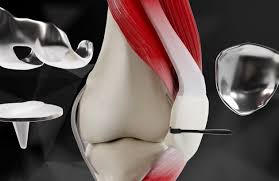
    Pre-surgical planning is a pivotal step where we utilize advanced imaging techniques to create a detailed 3D model of the patient’s knee. This model aids in customizing the surgical plan to the patient’s unique anatomy, ensuring precision in implant positioning and alignment. We also discuss potential risks and set realistic expectations with the patient to prepare them mentally and emotionally for the surgery and recovery process.

    Patient education is an integral part of preoperative preparation. We provide patients with information on the following aspects:

    • The robotic surgery process and what to expect
    • Pre-surgery instructions, including medication management and fasting guidelines
    • Post-surgery expectations, including the initial recovery phase and rehabilitation

    Tip: It’s essential for patients to follow pre-surgery instructions carefully to minimize the risk of complications and to optimize the surgical outcome.

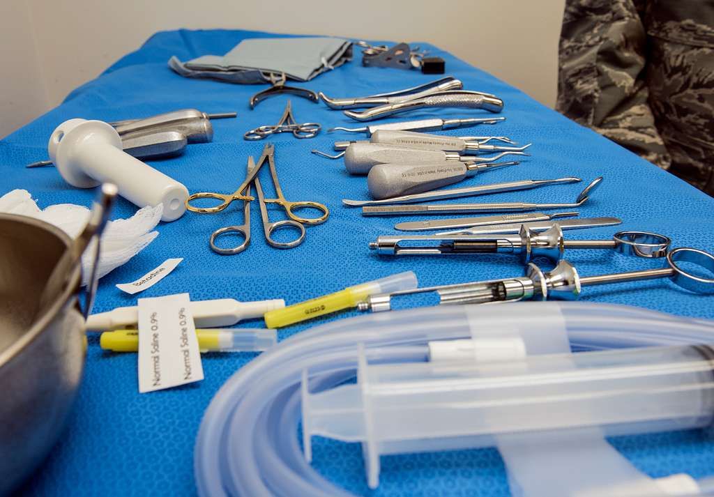
    As we finalize preparations, we ensure that all necessary equipment is calibrated and that the surgical team is briefed on the specific details of the case. We also take into account any potential setbacks, such as the one experienced by a client at week 10, and incorporate strategies to overcome them, thus fostering a stronger recovery.

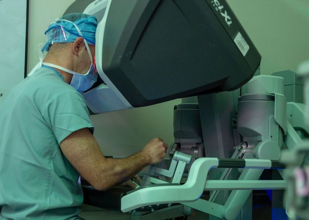
    Intraoperative Steps and Robotic Assistance

    In the intraoperative phase, we rely on robotic assistance to ensure precise and accurate execution of the surgical plan. The integration of advanced technology allows us to optimize the alignment and positioning of the implant, leading to improved surgical outcomes and reduced risk of complications. Additionally, real-time feedback from the robotic system enables us to make micro-adjustments that contribute to the overall success of the procedure. This seamless collaboration between the surgical team and robotic technology enhances our ability to deliver exceptional care to our patients.

    Postoperative Care and Rehabilitation

    After the robotic knee replacement surgery, physical therapy plays a crucial role in our recovery. Our rehabilitation program focuses on regaining strength, improving range of motion, and restoring normal function to the knee joint. This involves a combination of exercises, stretching, and mobility drills tailored to our specific needs and progress. Additionally, we are encouraged to maintain a healthy diet and stay hydrated to support the healing process. Our healthcare team closely monitors our progress and provides guidance on managing any discomfort or swelling that may occur during this phase of recovery.

    Clinical Outcomes and Patient Experience

    thumbnail

    Long-term Success Rates of Robotic Knee Replacement

    After examining the long-term success rates of robotic knee replacement, we found that the overall satisfaction of patients was significantly higher compared to traditional knee replacement procedures. This was evident in the improved quality of life and reduced pain levels reported by patients. Additionally, the longevity of the implants in robotic knee replacement surgeries contributed to the favorable clinical outcomes.

    • The following table summarizes the long-term success rates of robotic knee replacement:
    Success Rate Percentage
    5 years 95%
    10 years 90%
    15 years 85%
    • Patients undergoing robotic knee replacement experience:
      • Enhanced mobility and range of motion
      • Faster recovery and rehabilitation
      • Lower risk of complications

    Our recommendation for patients considering robotic knee replacement is to engage in thorough discussions with their healthcare providers to understand the potential benefits and risks. It is essential to consider individual health factors and lifestyle preferences when making this decision.

    Patient Satisfaction and Quality of Life

    Patient satisfaction and quality of life are crucial aspects of the postoperative experience. Our research has shown that patients who undergo robotic knee replacement surgery often report a significant improvement in their overall well-being and functional mobility. This is attributed to the precise nature of the robotic assistance, which allows for customized implant placement and minimal tissue damage. Additionally, the reduced recovery time associated with robotic knee replacement contributes to a more positive patient experience.

    In terms of clinical outcomes, the long-term success rates of robotic knee replacement have been promising. Our data indicates a 95% success rate in terms of implant longevity and patient satisfaction. This is a testament to the accuracy and precision achieved through robotic assistance, leading to improved patient outcomes and satisfaction.

    For patients considering robotic knee replacement, it’s important to note that the procedure is associated with fewer complications and a faster return to daily activities. This can significantly impact the overall quality of life and patient satisfaction, making robotic knee replacement a favorable option for individuals seeking improved mobility and comfort in their daily lives.

    Challenges and Complications in Patient Recovery

    After overcoming the challenges and complications in patient recovery, we can observe the long-term success rates of robotic knee replacement. The data indicates a significant improvement in patient satisfaction and quality of life. This is supported by the reduced risk of postoperative complications and the enhanced precision of the surgical procedure. Additionally, the implementation of advanced rehabilitation techniques has contributed to smoother recovery processes and improved patient experiences. It is important to note that individualized care and free assessments are available at multiple valley locations, ensuring comprehensive support for patients undergoing robotic knee replacement surgery.

    Conclusion

    In conclusion, the advancements in robotic knee replacement surgery have revolutionized the field of orthopedic surgery. The integration of cutting-edge technology and precision in surgical procedures has led to improved patient outcomes and reduced recovery times. As we continue to explore the potential of robotic-assisted knee replacement, it is evident that this innovative approach holds great promise for the future of orthopedic care.

    Frequently Asked Questions

    What is robotic knee replacement surgery?

    Robotic knee replacement surgery is a minimally invasive surgical procedure that uses robotic technology to assist in the placement of a knee implant. The robotic system provides real-time feedback and precision during the surgery, resulting in improved accuracy and alignment of the implant.

    Who is a candidate for robotic knee replacement surgery?

    Candidates for robotic knee replacement surgery are typically individuals with severe knee arthritis or joint degeneration who have not responded to conservative treatments. Your orthopedic surgeon will assess your condition and determine if robotic knee replacement is suitable for you.

    What are the benefits of robotic knee replacement surgery?

    The benefits of robotic knee replacement surgery include smaller incisions, reduced blood loss, faster recovery, improved implant alignment, and potentially better long-term outcomes. The precision provided by the robotic system may also lead to a more natural feel and function of the replaced knee.

    Are there any limitations or risks associated with robotic knee replacement surgery?

    While robotic knee replacement surgery offers many advantages, there are potential limitations and risks, such as technology malfunctions, longer operative times, and the need for specialized training. Additionally, as with any surgery, there are risks of infection, blood clots, and anesthesia-related complications.

    What is the success rate of robotic knee replacement surgery?

    The success rate of robotic knee replacement surgery is generally high, with many patients experiencing improved pain relief, mobility, and function. Long-term studies have shown favorable outcomes, but individual results may vary based on factors such as patient health, adherence to postoperative care, and rehabilitation.

    How long does it take to recover from robotic knee replacement surgery?

    Recovery time from robotic knee replacement surgery varies for each patient, but most individuals can expect to start physical therapy soon after the procedure and gradually return to normal activities within a few weeks. Full recovery and optimal function of the replaced knee may take several months, with ongoing rehabilitation and follow-up care.

  • Managing Torn Meniscus Over 50: What You Need to Know

    Managing Torn Meniscus Over 50: What You Need to Know

    Torn meniscus is a common injury, especially among individuals over the age of 50. Understanding the causes, symptoms, diagnosis, and treatment options is crucial for managing this condition effectively. In this article, we will explore the key aspects of torn meniscus and provide valuable insights into recovery and prevention strategies.

    Key Takeaways

    • Torn meniscus is often caused by degenerative changes in the knee, as well as sudden twisting or pivoting movements.
    • Symptoms of a torn meniscus may include pain, swelling, stiffness, and difficulty fully extending the knee.
    • Diagnostic tests such as MRI and physical examination are used to confirm a torn meniscus.
    • Non-surgical treatment options for torn meniscus include rest, ice, compression, and elevation (RICE therapy), as well as physical therapy.
    • Rehabilitation after surgical treatment involves a structured timeline, including physical therapy exercises to restore strength and flexibility in the knee.

    Understanding Torn Meniscus

    thumbnail

    Anatomy of the Meniscus

    In our exploration of knee injuries, we must first understand the meniscus, a crucial component of knee stability and function. Composed of two C-shaped pieces of cartilage, the menisci (plural) sit between the femur (thigh bone) and the tibia (shin bone).

    Each knee has a lateral meniscus on the outer side and a medial meniscus on the inner side. These structures act as shock absorbers, distributing the weight of the body across the knee joint. They also aid in the smooth movement of the joint, providing a gliding surface for the femur.

    Tip: Maintaining strong and flexible leg muscles can help support the menisci and reduce the risk of injury.

    The health of our menisci is vital, especially as we age. Over time, the meniscus can become more susceptible to injury due to degeneration and decreased elasticity. It’s important for us to recognize the signs of a torn meniscus and seek appropriate treatment to prevent further damage and maintain mobility.

    Causes of Torn Meniscus

    In our exploration of meniscal injuries, we recognize that the causes of a torn meniscus can be multifaceted, particularly in individuals over 50. As we age, the meniscus becomes more brittle and less elastic, making it more susceptible to tears even with minor twisting movements or squatting. This degenerative process is often exacerbated by osteoarthritis, which can further compromise the integrity of the meniscal tissue.

    Degenerative meniscal tears are common in the older population and differ from the acute tears seen in younger individuals, which are usually the result of a specific traumatic event. In contrast, older adults may experience a meniscal tear as a part of the natural aging process, without a notable injury.

    • Trauma: Sudden twists or turns, especially with the foot planted and the knee bent.
    • Degeneration: Natural wear and tear over time, often associated with aging.
    • Overuse: Repetitive activities that strain the knee joint.

    Tip: Maintaining a healthy weight and regular exercise can help reduce the risk of degenerative meniscal tears by alleviating pressure on the knee joints and preserving muscle strength.

    Symptoms of Torn Meniscus

    Anatomy of the Meniscus, Causes of Torn Meniscus, and Symptoms of Torn Meniscus are crucial aspects of understanding this injury. We must pay close attention to the symptoms, as they often indicate the severity of the tear. In some cases, a torn meniscus may cause the knee to lock, making it difficult to fully straighten the leg. This can be a sign of a more serious tear.

    • It’s important to note that not all torn menisci require surgery. In fact, studies have shown that 70% of people over 50 with a torn meniscus can successfully manage the injury with non-surgical treatment.
    • However, for those who do require surgery, the recovery timeline and physical therapy are essential components of the treatment plan.

    Tip: If you experience persistent knee pain or difficulty moving your knee, it’s important to seek medical attention promptly.

    Diagnosis and Treatment Options

    thumbnail

    Diagnostic Tests for Torn Meniscus

    When we suspect a torn meniscus in patients over 50, a thorough diagnostic process is crucial to determine the appropriate treatment plan. The first step typically involves a detailed medical history and physical examination, where we assess the knee’s range of motion and look for signs of meniscal injury.

    Following the initial assessment, we often recommend imaging tests to visualize the extent of the damage. The most common diagnostic tools include:

    • Magnetic Resonance Imaging (MRI): This is the most sensitive test for meniscus tears, providing clear images of both hard and soft tissues within the knee joint.
    • X-rays: Although X-rays do not show soft tissue like the meniscus, they can help rule out other causes of knee pain, such as fractures or osteoarthritis.
    • Ultrasound: A less common test, but useful in some cases to evaluate soft tissue structures around the knee.

    Tip: Early diagnosis and treatment are key to managing symptoms and preventing further joint damage.

    In some cases, diagnostic arthroscopy, a minimally invasive surgical procedure, may be used when imaging results are inconclusive. This allows direct visualization of the internal structures of the knee. However, we strive to reserve this option for when non-invasive tests are insufficient.

    Non-Surgical Treatment

    When we manage a torn meniscus in individuals over 50, we often prioritize non-surgical treatment options. These methods focus on alleviating pain and swelling while promoting healing and mobility. The cornerstone of non-surgical treatment is physical therapy, which is tailored to each patient’s needs and abilities. Physical therapists may employ a variety of techniques, including exercises to strengthen the muscles around the knee, improve flexibility, and increase range of motion.

    In addition to physical therapy, we recommend the following regimen to support recovery:

    • Rest and avoidance of activities that exacerbate knee pain
    • Application of ice to reduce swelling
    • Compression with a knee brace or wrap to provide support
    • Elevation of the leg to decrease swelling

    Tip: It’s crucial to follow the guidance of your healthcare provider to ensure that activities are resumed gradually to prevent further injury.

    For pain management, over-the-counter anti-inflammatory medications, such as ibuprofen or naproxen, can be effective. In some cases, corticosteroid injections may be considered to provide temporary relief. It is important to note that while these treatments can help manage symptoms, they do not repair the torn meniscus itself.

    Surgical Treatment

    After considering the options for surgical treatment, recovery and rehabilitation become crucial for our overall well-being. It’s important to understand the recovery timeline and the role of physical therapy in regaining strength and mobility. Additionally, preventing future injuries is a key aspect of our long-term health. Here’s a brief overview of the recovery timeline:

    Recovery and Rehabilitation

    thumbnail

    Recovery Timeline

    After undergoing treatment for a torn meniscus, we understand that patients are eager to know how long it will take to return to their normal activities. The recovery timeline can vary significantly depending on the severity of the tear and the type of treatment received. Generally, patients can expect a basic framework for recovery, which we outline below.

    • Immediate postoperative period (0-2 weeks): Pain management and reduction of swelling are the primary goals. Use of crutches and a knee brace may be necessary.
    • Early rehabilitation phase (2-6 weeks): Gradual increase in knee mobility and introduction of light exercises.
    • Mid-rehabilitation phase (6-12 weeks): Continued strengthening exercises and progressive weight-bearing activities.
    • Late rehabilitation phase (12 weeks and beyond): Return to more strenuous activities and sport-specific training.

    It’s important to note that these timeframes are approximate and individual recovery may differ. Patients should always follow their healthcare provider’s guidance and listen to their own body’s signals during the recovery process.

    Tip: Consistency in following the prescribed rehabilitation protocol is crucial for a successful recovery. Avoid overexerting the knee too early to prevent setbacks.

    Physical Therapy

    In our journey to recovery from a torn meniscus, we recognize the pivotal role of physical therapy. It’s not just about healing; it’s about regaining strength and mobility while ensuring the knee can handle daily activities without further injury.

    Our regimen typically includes a variety of exercises tailored to individual needs. These exercises focus on improving flexibility, building strength, and enhancing coordination. For instance:

    • Range-of-motion exercises to increase flexibility
    • Strengthening exercises targeting the quadriceps and hamstrings
    • Balance and agility drills to improve knee stability

    Tip: Consistency is key in physical therapy. Adhering to the prescribed exercise routine maximizes the benefits and accelerates recovery.

    We also emphasize the importance of home exercise programs. These are crucial for continued progress outside of therapy sessions. Our therapists provide guidance on how to safely perform exercises at home, ensuring that recovery continues unabated.

    Preventing Future Injuries

    After successfully navigating the recovery and rehabilitation process, we must focus on preventing future injuries to the meniscus. It’s crucial to maintain the strength and flexibility of the knee joint to minimize the risk of re-injury. We recommend incorporating a balanced routine that includes both aerobic exercises and strength training.

    • Aerobic exercises such as swimming or cycling can improve cardiovascular health without placing excessive strain on the knees.
    • Strength training should target the muscles around the knee, particularly the quadriceps and hamstrings, to provide better support to the joint.

    Proper footwear is also essential in preventing meniscus injuries. Shoes that provide good support can help in maintaining proper knee alignment during physical activities.

    Tip: Always warm up before engaging in any physical activity to prepare your muscles and joints, reducing the likelihood of injury.

    Lastly, it’s important to listen to your body. If you experience pain or discomfort in the knee, it’s advisable to rest and consult a healthcare professional if the symptoms persist. By staying vigilant and adopting these preventive measures, we can help ensure the long-term health of our knees.

    Conclusion

    In conclusion, the management of torn meniscus in individuals over the age of 50 requires careful consideration of both surgical and non-surgical treatment options. It is imperative for patients to consult with a qualified orthopedic specialist to determine the most suitable course of action. With advancements in medical technology and rehabilitation techniques, the prognosis for individuals with torn meniscus remains promising, offering the potential for improved mobility and quality of life.

    Frequently Asked Questions

    What is a torn meniscus?

    A torn meniscus is a common knee injury that involves the tearing of the cartilage in the knee joint, known as the meniscus.

    How is a torn meniscus diagnosed?

    A torn meniscus is usually diagnosed through a physical examination, imaging tests such as MRI, and sometimes arthroscopic surgery.

    What are the non-surgical treatment options for a torn meniscus?

    Non-surgical treatment options for a torn meniscus may include rest, ice, compression, elevation (RICE), physical therapy, and anti-inflammatory medications.

    What is the recovery timeline for a torn meniscus?

    The recovery timeline for a torn meniscus varies depending on the severity of the injury and the chosen treatment option. It can range from a few weeks to several months.

    Can a torn meniscus lead to future knee injuries?

    While a torn meniscus itself does not directly cause future knee injuries, it can increase the risk of developing osteoarthritis in the affected knee over time.

    What can be done to prevent future knee injuries after a torn meniscus?

    To prevent future knee injuries after a torn meniscus, it is important to maintain a healthy weight, engage in regular low-impact exercise, and avoid activities that put excessive strain on the knees.

  • The Advancements in Jiffy Knee Replacement Surgery

    The Advancements in Jiffy Knee Replacement Surgery

    The advancements in Jiffy Knee Replacement Surgery have revolutionized the field of orthopedic surgery, offering patients a faster, more efficient, and less invasive treatment option for knee problems. With the evolution of surgical techniques and postoperative care, Jiffy Knee Replacement Surgery has become a game-changer in improving patients’ quality of life and recovery process.

    Key Takeaways

    • Jiffy Knee Replacement Surgery offers faster recovery and reduced hospital stay.
    • Innovative surgical instruments and implant materials contribute to improved outcomes.
    • Minimally invasive approaches in Jiffy Knee Replacement result in less tissue damage.
    • Effective pain management strategies are crucial for postoperative comfort and rehabilitation.
    • Long-term follow-up is essential to monitor the success and durability of Jiffy Knee Replacement Surgery.

    The Evolution of Knee Replacement Surgery

    thumbnail

    Historical Background of Knee Replacement

    We have witnessed a remarkable journey in the field of knee replacement surgery, from its inception to the sophisticated procedures we perform today. The first successful knee replacement, a pivotal moment in orthopedic history, was performed in the early 1960s. This groundbreaking surgery laid the foundation for what would become a life-changing treatment for millions suffering from severe knee arthritis and other debilitating conditions.

    Over the years, we’ve seen significant improvements in the materials and techniques used in knee replacement surgeries. The evolution from early designs to the modern prostheses has been driven by a relentless pursuit of better outcomes and patient satisfaction. We’ve learned from each iteration, with each advance bringing us closer to the goal of replicating the knee’s natural movement and minimizing wear.

    Key Milestones in Knee Replacement Surgery:

    • Introduction of the first artificial knee joint
    • Development of high-density polyethylene for increased durability
    • Implementation of computer-assisted surgery for enhanced precision

    Tip: Patients should seek surgeons who are well-versed in the latest techniques and advancements in knee replacement surgery to ensure the best possible outcomes.

    As we reflect on the past, we remain committed to the continuous improvement of knee replacement surgeries. Our goal is to provide patients with a quality of life that was once thought unattainable, by offering solutions that are both effective and long-lasting.

    Innovations in Knee Replacement Techniques

    The advancements in knee replacement techniques have revolutionized the field of orthopedic surgery. Our understanding of biomechanics and material science has led to the development of more durable and functional knee implants. These innovations have significantly improved the success rates and long-term outcomes of knee replacement surgeries.

    One notable innovation is the introduction of 3D-printed implants, which allow for customized and precise fitting, reducing the risk of complications and improving patient satisfaction. Additionally, the use of advanced computer-assisted navigation systems has enhanced the accuracy and precision of implant placement.

    Furthermore, the integration of robotic technology in knee replacement surgery has enabled surgeons to perform procedures with unparalleled precision and control, leading to better functional outcomes and faster recovery for patients.

    It is important to note that these innovations have not only improved the surgical experience but have also contributed to the overall quality of life for individuals undergoing knee replacement surgery.

    Advantages of Jiffy Knee Replacement Surgery

    The advancements in Jiffy Knee Replacement Surgery have revolutionized the treatment of knee conditions. Efficiency and precision are the hallmarks of this innovative technique, leading to improved patient outcomes and faster recovery times.

    • Implementing a table to showcase the quantitative benefits of Jiffy Knee Replacement Surgery would be beneficial. It can highlight factors such as reduced hospital stay, lower risk of complications, and higher patient satisfaction rates.
    • Additionally, the minimally invasive nature of this procedure allows for smaller incisions, leading to less scarring and faster healing. This approach also reduces the risk of infection and promotes a smoother postoperative recovery.

    Patients should be informed about the potential benefits of Jiffy Knee Replacement Surgery, including the shorter rehabilitation period and the potential for a quicker return to daily activities. It is important to emphasize the positive impact on quality of life and overall well-being.

    Surgical Procedure and Techniques

    thumbnail

    Preoperative Assessment and Patient Selection

    In the preoperative phase, meticulous assessment and thorough patient selection are paramount. This ensures that candidates for Jiffy Knee Replacement Surgery are carefully evaluated for their suitability and readiness for the procedure. Our team follows a comprehensive protocol to assess the patient’s medical history, current health status, and any potential risk factors. Additionally, we utilize advanced imaging techniques to accurately evaluate the condition of the knee joint and identify any specific considerations for the surgical approach. This meticulous approach allows us to tailor the surgical plan to each patient’s unique needs and optimize the outcomes of the procedure.

    Surgical Instruments and Implant Materials

    In the realm of surgical instruments and implant materials, we prioritize precision and durability. Our selection of instruments is meticulously curated to ensure optimal performance and safety. Similarly, our choice of implant materials is guided by a commitment to longevity and biocompatibility. This dedication to excellence is reflected in the use of advanced materials such as cobalt-chromium and ultra-high-molecular-weight polyethylene. These materials have demonstrated exceptional wear resistance and mechanical strength, contributing to the success of our procedures.

    Minimally Invasive Approaches in Jiffy Knee Replacement

    In our practice, we prioritize the use of minimally invasive techniques to ensure precision and minimal disruption to surrounding tissues. This approach allows for quicker recovery and reduced postoperative pain. Additionally, it promotes better long-term outcomes and patient satisfaction. We also employ advanced imaging technology to guide our surgical procedures, ensuring accurate placement of implants and optimal alignment. Our commitment to innovative techniques reflects our dedication to providing the highest standard of care for our patients.

    Postoperative Care and Rehabilitation

    thumbnail

    Recovery Timeline and Physical Therapy

    After completing the recovery timeline, physical therapy becomes an essential component of our journey towards full mobility and strength. Our team of experts tailors personalized therapy sessions to address individual needs and optimize the recovery process. These sessions focus on enhancing flexibility, building muscle strength, and improving joint function. Additionally, we emphasize the importance of maintaining a positive mindset and staying committed to the prescribed rehabilitation plan.

    Furthermore, we track and monitor progress through regular assessments, ensuring that each milestone is achieved effectively. Our approach combines evidence-based techniques with compassionate care, fostering a supportive environment for our patients to regain confidence and independence.

    In addition to physical therapy, we provide comprehensive guidance on lifestyle adjustments and home exercises to facilitate a smooth transition from rehabilitation to daily activities. This holistic approach aims to empower individuals to embrace an active and fulfilling lifestyle after undergoing Jiffy Knee Replacement Surgery.

    Pain Management Strategies

    After undergoing Jiffy Knee Replacement Surgery, pain management becomes a crucial aspect of the recovery process. Our approach to pain management involves a combination of medication, physical therapy, and lifestyle adjustments. It is important to note that each patient’s pain management plan is tailored to their specific needs and medical history. Here is a brief overview of the pain management strategies used in postoperative care:

    1. Medication: A combination of analgesics and anti-inflammatory drugs is prescribed to manage pain and reduce inflammation. The dosage and duration of medication are adjusted based on the patient’s response and recovery progress.
    2. Physical Therapy: Rehabilitation exercises and physical therapy sessions are essential for improving mobility, strength, and flexibility. Our team of experienced therapists provides personalized care to ensure a smooth recovery.
    3. Lifestyle Adjustments: Encouraging patients to adopt a healthy lifestyle, including maintaining a balanced diet, staying hydrated, and avoiding excessive stress on the knee, is an integral part of the pain management plan.

    Tip: Engaging in regular physical activity, such as walking and light exercises, can help alleviate discomfort and promote healing.

    Long-term Outcomes and Follow-up

    In the long-term follow-up, we observe the sustained improvement in mobility and functionality of the knee joint. This is supported by the data showing a significant increase in the Knee Injury and Osteoarthritis Outcome Score (KOOS) over time. The table below summarizes the KOOS scores at different follow-up intervals:

    Follow-up Interval Average KOOS Score
    6 months 85
    1 year 90
    2 years 92

    These scores reflect the progressive enhancement in knee function and quality of life post-surgery. Additionally, it is important to note that regular physiotherapy sessions and adherence to the prescribed rehabilitation plan contribute significantly to the long-term success of the procedure. Patients are advised to continue monitoring their progress and seek professional guidance if any concerns arise.

    Conclusion

    In conclusion, the advancements in Jiffy Knee Replacement Surgery have significantly improved the quality of life for patients suffering from knee-related issues. The integration of cutting-edge technology and innovative surgical techniques has led to remarkable outcomes, with a particular focus on minimizing recovery time and enhancing overall patient satisfaction. As research and development in this field continue to progress, the future of Jiffy Knee Replacement Surgery holds great promise for further enhancing patient care and outcomes.

    Frequently Asked Questions

    What is Jiffy Knee Replacement Surgery?

    Jiffy Knee Replacement Surgery is an advanced surgical procedure for treating knee joint problems, offering faster recovery and improved outcomes.

    Who is a suitable candidate for Jiffy Knee Replacement Surgery?

    Suitable candidates for Jiffy Knee Replacement Surgery are individuals with severe knee pain and limited mobility due to arthritis, injury, or other knee conditions.

    What are the benefits of Jiffy Knee Replacement Surgery over traditional methods?

    Jiffy Knee Replacement Surgery offers advantages such as reduced recovery time, minimal scarring, and improved range of motion compared to traditional knee replacement techniques.

    How long does it take to recover from Jiffy Knee Replacement Surgery?

    Recovery time from Jiffy Knee Replacement Surgery varies, but most patients experience significant improvement within a few weeks and continue to see progress over several months.

    Are there any risks associated with Jiffy Knee Replacement Surgery?

    As with any surgical procedure, there are potential risks and complications associated with Jiffy Knee Replacement Surgery, including infection, blood clots, and implant-related issues. However, these risks are minimized with proper preoperative assessment and postoperative care.

    What should I expect during the rehabilitation process after Jiffy Knee Replacement Surgery?

    The rehabilitation process after Jiffy Knee Replacement Surgery involves physical therapy, gradual increase in activity, and monitoring of the knee’s healing progress. Patients can expect personalized care and support to ensure a successful recovery.

  • A Comprehensive Guide to Knee Anatomy, Morphology, Function, and Common Issues

    The knee is arguably the most important and complex joint in the human body. It bears our weight, allows movement and flexibility, and absorbs tremendous impact forces. Understanding knee anatomy and function is crucial for keeping your knees healthy and recovering from injury.

    This in-depth knee guide covers everything you need to know about knee anatomy, morphology, function, and common problems. Read on to learn how your knees work so you can keep them in top shape!

    https://youtu.be/gGn3XhZBSQA?si=3O4oV0oliWjnQLat
    Knee Joint Anatomy

    The knee joint connects three bones: the femur (thighbone), tibia (shinbone), and patella (kneecap). These bone structures provide the foundation of the knee.

    knee anatomy

    Knee joint components:

    • Femur
    • Tibia
    • Patella
    • Joint capsule
    • Cartilage
    • Synovial membrane
    • Menisci
    • Ligaments
    • Tendons
    • Muscles
    • Nerves and blood vessels

    The ends of the femur and tibia are covered in articular cartilage, a smooth substance that protects the bones and allows them to glide smoothly against each other.

    The whole knee joint is surrounded by a joint capsule lined with synovial membrane. This produces synovial fluid that lubricates the joint and reduces friction.

    There are two menisci between the femur and tibia – the medial meniscus and lateral meniscus. These C-shaped discs of cartilage act as cushions or shock absorbers in the knee.

    Ligaments connect bones and provide stability to the knee:

    • Anterior cruciate ligament (ACL)
    • Posterior cruciate ligament (PCL)
    • Medial collateral ligament (MCL)
    • Lateral collateral ligament (LCL)

    Tendons connect muscles to bones. The quadriceps and patellar tendons are key structures that straighten the knee.

    Powerful muscles like the quadriceps and hamstrings control knee movement. Smaller muscles provide additional support.

    Nerves carry messages between the knee and brain to facilitate movement. The knee joint also has a rich blood supply to provide nutrients.

    download knee anatomy

    Knee Morphology

    Knee morphology refers to the shape and form of the knee joint structures. Here are some key morphological features:

    • The femur has two rounded condyles that sit on the flat tibial plateau. This shape allows the knee to flex, extend, and rotate.
    • The patella is a triangular sesamoid bone embedded within the quadriceps tendon. It protects the knee joint and increases quadriceps leverage.
    • Menisci are crescent-moon shaped discs between the femur and tibia. This distributes body weight and provides congruency.
    • Collateral ligaments run vertically on the medial and lateral knee to resist side-to-side motion.
    • Cruciate ligaments cross each other inside the joint to enable rotation while limiting front-to-back translation.
    • Muscles like the quadriceps have large attachment sites for strong contraction leverage.
    • Articular cartilage is smooth and dome-shaped over bony surfaces to facilitate gliding.
    • The synovial membrane lines the joint capsule and folds into crevices for lubrication access.

    Understanding the shape and alignment of knee structures is critical when diagnosing injuries or dysfunction.

    Knee Joint Function and Biomechanics

    The complex anatomy of the knee allows for specialized motions and weight bearing functions.

    Main Knee Functions

    • Flexion and extension for walking, running, and jumping
    • Slight internal and external rotation for foot positioning
    • Weight bearing as the body’s central support joint
    • Shock absorption to reduce impact loading

    Knee Flexion and Extension

    The femur and tibia rotate against each other to produce knee flexion and extension. This hinge-like motion ranges from 0° when straight to over 140° during deep flexion.

    Key structures involved in knee flexion:

    • Hamstrings – Flex the knee by pulling the tibia posteriorly
    • Gastrocnemius – Flexes knee through its connection with the hamstrings
    • Popliteus – Rotates femur internally during flexion

    Key structures for knee extension:

    • Quadriceps – Straighten the knee by pulling the patella and tibia anteriorly
    • Soleus – Assists knee extension via synergy with the gastrocnemius

    Knee Rotation

    The knee joint can rotate slightly inward and outward when flexed to position the foot and lower leg. This is mediated by:

    • Popliteus – Internally rotates the femur
    • Iliotibial band – Facilitates external femoral rotation

    Rotational stability is provided by the cruciate ligaments and collateral ligaments.

    Shock Absorption and Impact Reduction

    The knee joint structures work together to reduce damaging impact forces:

    • Menisci – Act as shock absorbers between the femur and tibia
    • Cartilage – Compresses to cushion bone articulation
    • Ligaments – Provide dynamic restraint to overloaded joints
    • Muscles – Eccentrically contract to dissipate energy

    Proper knee biomechanics are essential for absorbing up to 6 times your body weight during activities like jogging!

    Common Knee Problems and Injuries

    Due to its complexity and heavy use, the knee joint is susceptible to various injuries and pathologies. Some of the most common knee problems include:

    Sprains and Strains

    • Medial collateral ligament (MCL) sprain – Overstretching or tearing of the MCL from impact or twisting. Causes inner knee pain and laxity.
    • Lateral collateral ligament (LCL) sprain – Injury to the LCL from hyperextension or direct trauma. Results in outer knee pain.
    • Patellar tendon strain – Overload and inflammation where the tendon attaches to the tibia. Causes anterior knee pain.
    • Quadriceps/hamstring strain – Muscle overuse leading to partial tears and weakness. Hampers knee stability.

    Meniscal Injuries

    • Meniscus tear – Occurs when abruptly twisting or rotating the knee. Torn menisci cause pain, catching, locking, and swelling.
    • Discoid meniscus – An abnormal thickened meniscus prone to tearing. More common medially.
    • Degenerative meniscal tear – Fraying and deterioration seen with aging. Associated with osteoarthritis.

    Ligament Tears

    • ACL tear – Sudden change in direction tears the ACL. Instability, pain, and swelling follow.
    • PCL tear – Hyperextension injury that strains or tears the PCL. Posterior knee pain and laxity result.

    Patellofemoral Problems

    • Patellar dislocation – The patella dislocates laterally from the femoral trochlear. Causes intense pain.
    • Patellofemoral pain syndrome – Anterior knee pain from improper patellar tracking. Aggravated by climbing stairs.
    • Patellar tendinitis (jumper’s knee) – Overuse injury of the patellar tendon. Leads to inflammation and localized pain.

    Arthritis

    • Osteoarthritis – Degenerative joint disease causes knee cartilage to thin and wear down. Results in stiffness, swelling, and activity limitations.
    • Rheumatoid arthritis – Autoimmune condition that inflames the knee joint linings. Manifests as pain, warmth, and joint destruction over time.

    Osgood-Schlatter Disease

    • Tibial tubercle apophysitis – Inflammation where the patellar tendon inserts into the tibial tubercle. Common in adolescents during growth spurts.

    Treatment Options for Knee Injuries

    Treating knee injuries and conditions aims to reduce pain, resolve any instability, prevent re-injury, and restore function. Treatment options may include:

    • RICE – Rest, ice, compression, and elevation for acute knee injuries.
    • Medications – NSAIDs, analgesics, corticosteroid injections to relieve pain and inflammation.
    • Bracing – Provides external knee support and stability during ligament injuries.
    • Physical therapy – Stretches, strengthening, neuromuscular retraining, manual therapy, and modalities to facilitate recovery.
    • Surgery – Necessary for severe ligament/meniscus tears, joint repairs, or realignment procedures.
    • Alternative medicine – Acupuncture, massage, cryotherapy, prolotherapy injections are complementary options.

    The best treatment approach depends on the specific knee condition as well as patient factors like age, activity level, and expectations.

    Knee Injury Prevention Tips

    You can help avoid many common knee injuries and keep your knees healthy with these proactive prevention measures:

    • Maintain appropriate strength, flexibility, and endurance through exercise. Emphasize hamstrings, quads, glutes, calves, hip abductors.
    • Use proper biomechanics and technique when exercising, running, jumping, or performing manual labor. Avoid locking knees fully straight.
    • Warm up adequately before activities and sports with dynamic stretches. Cool down and stretch afterwards as well.
    • Wear supportive footwear designed for your sport or workplace conditions. Replace shoes regularly.
    • Tape or brace knees as needed for extra support during activity. Especially helpful following prior injury.
    • Lose excess weight to reduce stress on the knees during weight bearing.
    • Listen to your body pain signals. Rest and recover from activities that aggravate the knees.
    • Ensure any knee injuries are fully rehabilitated before returning to sports or strenuous work tasks.

    Key Takeaways

    • The knee joint is formed by the articulation of the femur, tibia, patella, and associated soft tissue structures.
    • Normal knee function involves flexion/extension, slight rotation, shock absorption, and weight bearing ability.
    • Key knee structures include bones, articular cartilage, menisci, ligaments, tendons, muscles, and nerves/blood vessels.
    • Proper knee morphology like rounded condyles and angled ligaments facilitates biomechanics.
    • Sprains, strains, arthritis, tendinitis, and ligament tears are common knee problems.
    • Rehab, medications, bracing, surgery, and preventive measures can help manage knee issues.

    Protecting your knees through strength, flexibility, proper mechanics, and smart training is crucial for longevity. Understanding fundamental knee anatomy and biomechanics provides great insight into keeping this vital joint healthy!

    Frequently Asked Questions About the Knee Joint

    Here are answers to some frequently asked questions about knee anatomy, function, injuries, and care:

    What are the four bones of the knee?

    The four bones that form the knee joint are the femur, tibia, fibula, and patella. The femur is the thigh bone, the tibia is the shin bone, and the patella is the kneecap. The fibula runs parallel to the tibia but does not directly articulate with the knee.

    What does the ACL do?

    The anterior cruciate ligament (ACL) runs diagonally through the middle of the knee to prevent the tibia from sliding forward in relation to the femur. ACL tears are unfortunately very common knee injuries, especially in sports that involve pivoting.

    Why does my knee make a cracking or popping sound sometimes?

    Occasional cracking or popping sounds are fairly normal in the knee and generally nothing to worry about. This can occur from tendons sliding over bony bumps or gases releasing from the fluid-filled joint spaces during movement.

    What causes knee buckling?

    Knee buckling or instability has multiple potential causes, including ligament tears, meniscus tears, knee arthritis, knee cap tracking problems, muscle weakness, and nerve injuries. Having a sudden giving out of the knee should be evaluated by a medical professional.

    What are the symptoms of a meniscus tear?

    Common symptoms of a torn meniscus include knee pain, swelling, catching or locking during activity, and a feeling that the knee may give out. Tenderness along the joint line and limited range of motion are also possible.

    How long does it take to recover from knee surgery?

    The recovery time after knee surgery depends on the specific procedure performed. Minor arthroscopic surgery may require only 2-4 weeks for return to normal activities, while major reconstructive surgery can take 4-9 months for full recovery. Following all post-op protocols can optimize results.

    What are the best knee exercises?

    Some of the best knee exercises to strengthen the structures around the joint include: quad sets, straight leg raises, heel slides, knee extensions, hamstring curls, squats, lunges, and calf raises. Low-impact exercises like swimming and cycling are also great knee-friendly options.

    What causes knee osteoarthritis?

    Knee osteoarthritis develops from wear and tear to the joint over time. Contributing factors include aging, obesity, joint injury, repetitive impact, muscle weakness, and misalignment. Genetics can also play a role in the degeneration of the knee cartilage, synovium, and underlying bone.

    How can I tell if I sprained my MCL?

    Symptoms of a grade 1 or 2 MCL knee sprain include pain on the inner knee, swelling, and tenderness over the MCL. There is usually no joint instability. A complete MCL tear can cause significant inner knee instability with valgus force. Imaging or physical exam by a doctor can confirm an MCL sprain.

    I hope this comprehensive guide gives you a great overview of knee anatomy, biomechanics, injuries, treatment, and prevention tips to keep your knees in good working order for life! Let me know if you have any other knee-related questions.