Chronic traumatic encephalopathy Neuropathological changes are uncommon in men who played amateur American football
Iverson GL, Jamshidi P, Fisher-Hubbard AO, Deep-Soboslay A, Hyde TM, Kleinman JE, deJong JL, Shepherd CE, Hazrati LN, Castellani RJ. Anterior neurol. June 19, 2023; 14:1143882. doi:10.3389/fneur.2023.1143882. PMID: 37404944; PMCID: PMC10315537.
Full text freely available
Take home message
Among the brain tissue of men over 50, none showed a definitive neuropathological change in chronic traumatic encephalopathy (CTE). However, three or more authors found that 5% of brains showed ‘signatures’ of neuropathological changes in CTE, arising from cases with and without a history of collision sports.
Background
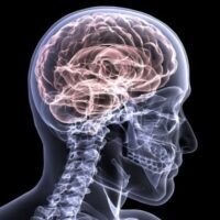
Chronic traumatic encephalopathy (CTE) is a neurodegenerative disease that cannot be diagnosed until death. An update was recently published on defining cases. Rather than a general absence or presence, screeners may note the level of components or “signatures” of neuropathic CTE change.
Study aim
The authors evaluated brain tissue from the Lieber Institute for Brain Development Tissue Bank to examine CTE neuropathological changes in relation to a history of American football youth participation and suicide as a mode of death.
Methods
The authors assessed 186 donated male brains in a tissue bank enriched with samples from people with neuropsychiatric problems and suicide (~66 years old; 58 participated in a contact sport, 67 died by suicide). Two authors screened the brains. Five authors then examined the selected brains for signs of CTE using the 2016 and 2021 consensus definitions. The authors also reviewed clinical documents and interviewed relatives to determine their medical, social, demographic, family and psychiatric history. In addition, they took into account their history of head/brain trauma/injury and their personal history of participation and concussions in sports.
Results
No brain definitively met the criteria for neuropathic CTE change based on the 2016 or 2021 definition. Furthermore, the five authors never unanimously agreed on a case with “hallmarks” of neuropathic changes in CTE. However, three or more authors found 10 cases (5.4%) with “features” of one or both definitions for neuropathic change in CTE.
The authors found no differences between CTE neuropathic change and personal history of playing American football or contact sports (contact sports = 9%, no contact sports = 4%). Furthermore, there was no difference between CTE tissue samples with neuropathic change and samples with a history of brain injury (brain injury = 4%, no brain injury = 6%), mood disorder (mood disorder = 6%, no mood disorder = 6.0%). ), or manner of death (suicide = 6%, non-suicide = 5%).
Viewpoints
Overall, no definitive case of CTE was identified in this sample. Some authors found ‘signatures’ of neuropathic changes in CTE in 1 in 20 brains. Therefore, this condition was very uncommon in men who played amateur football, in people with mood disorders during life and in people with suicide as the cause of death. One of the notable strengths of this study was that the authors used a different brain bank, focusing around the brain tissue donations on psychiatric conditions and not on sports history. The findings at this bank were consistent with low rates at other banks, such as 4% of cases at the Military Brain Tissue Bank. However, these former banks do not represent the general population. It remains unclear how common CTE and “signatures” of neuropathic changes in CTE occur in the general population. This is essential to understand how prevalent CTE is in society and to accurately assess who is at risk for CTE and what the “signatures” of CTE are.
Clinical implications
Medical professionals can reassure people that, despite recent media attention, the prevalence of CTE and its characteristics are relatively small. That said, we need to point out to our stakeholders (e.g. parents, patients) that the level of evidence in this area is weak and there is much for us to learn. Doctors should promote positive brain health by limiting repetitive head impacts by improving rules and regulations in contact sports and limiting contact during exercises.
Questions for discussion
Do you believe that there would be an increased risk of CTE pathology in those who have played contact sports in the past or have a history of repetitive brain injury? How do you inform your patients about CTE?
related posts
- Treatable conditions should be explored in former athletes with CTE-like symptoms
- CTE found in people with no history of contact sports
- When it comes to Chronic Traumatic Encephalopathy (CTE), Mom may not know best
- Can playing contact sports in high school and college increase the risk of CTE?
- Disconnect between concussion education and CTE
- Most military members don’t have to worry about CTE
Written by Jane McDevitt
Reviewed by Jeffrey Driban