Category: Blog

Your blog category

  • Back of knee pain only when fully extended

    Back of knee pain only when fully extended

    Have you ever felt a sharp twinge or dull ache behind your joint while standing or stretching your leg completely? This specific sensation – often overlooked until it becomes persistent – might signal more than temporary strain. Let’s explore why posterior discomfort during full extension demands attention and how it connects to your body’s mechanics.

    Our focus centers on a condition where discomfort arises exclusively when the leg is straightened. Unlike general joint issues, this symptom often points to localized problems in tendons, ligaments, or cartilage. Athletes and active individuals frequently encounter it, but even casual movements can trigger it if underlying factors exist.

    Understanding the knee’s anatomy proves crucial. This complex hinge relies on muscles, tendons, and ligaments working in harmony. When one component faces stress – whether from overuse, injury, or imbalance – targeted symptoms like extension-related discomfort can emerge. We’ll break down common causes and why self-diagnosis often falls short.

    Key Takeaways

    • Posterior knee discomfort during full extension indicates specific mechanical issues
    • Common triggers include tendon strain, ligament stress, and cartilage wear
    • Anatomical knowledge helps identify potential problem areas
    • Persistent symptoms require professional evaluation
    • Early intervention prevents chronic complications
    • Treatment approaches vary based on root causes

    Introduction & Background

    Stiffness or tenderness in the posterior leg area can signal underlying joint issues. Nearly 1 in 4 adults report discomfort in this region during daily activities, according to recent orthopedic studies. Recognizing patterns helps separate temporary strain from chronic conditions.

    posterior knee pain overview

    What Defines Posterior Discomfort?

    This specific discomfort typically appears during leg-straightening motions like standing up or climbing stairs. Common indicators include:

    • Localized swelling behind the joint
    • Reduced flexibility after prolonged sitting
    • Sharp sensations when locking the leg

    Clinical data shows 68% of cases involve multiple symptoms. Early identification prevents minor irritations from becoming mobility-limiting problems.

    Why Knee Health Knowledge Matters

    Understanding joint mechanics transforms how we approach treatment. Misdiagnosed conditions often share similar presentations:

    ConditionKey DifferentiatorCommon Triggers
    Popliteus TendinitisPain during downward stairsOveruse in runners
    Baker’s CystVisible bulge behind jointArthritis complications
    Meniscus TearClicking sound during movementSudden twists/pivots

    We’ll explore these structures in detail next, equipping you with actionable insights for informed health decisions. Proper terminology bridges communication gaps between patients and specialists.

    Anatomy of the Knee: Ligaments, Muscles, and Cartilage

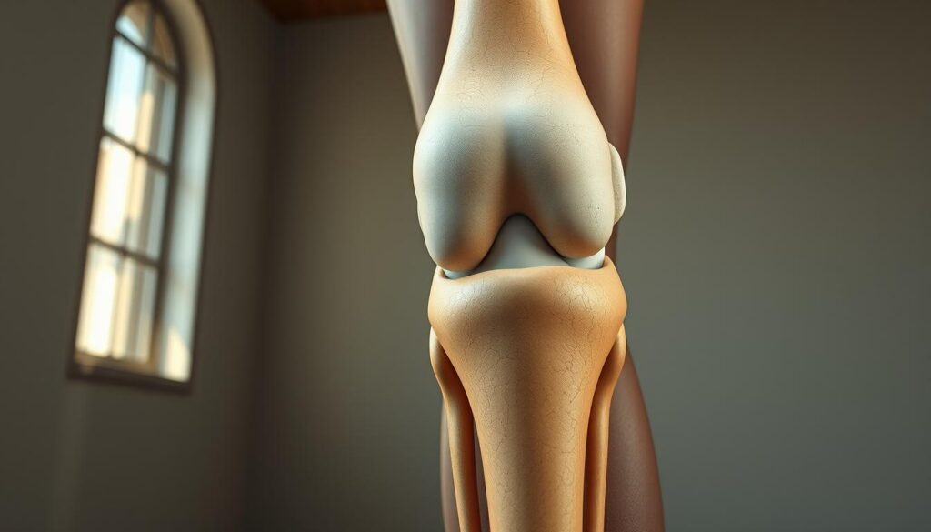
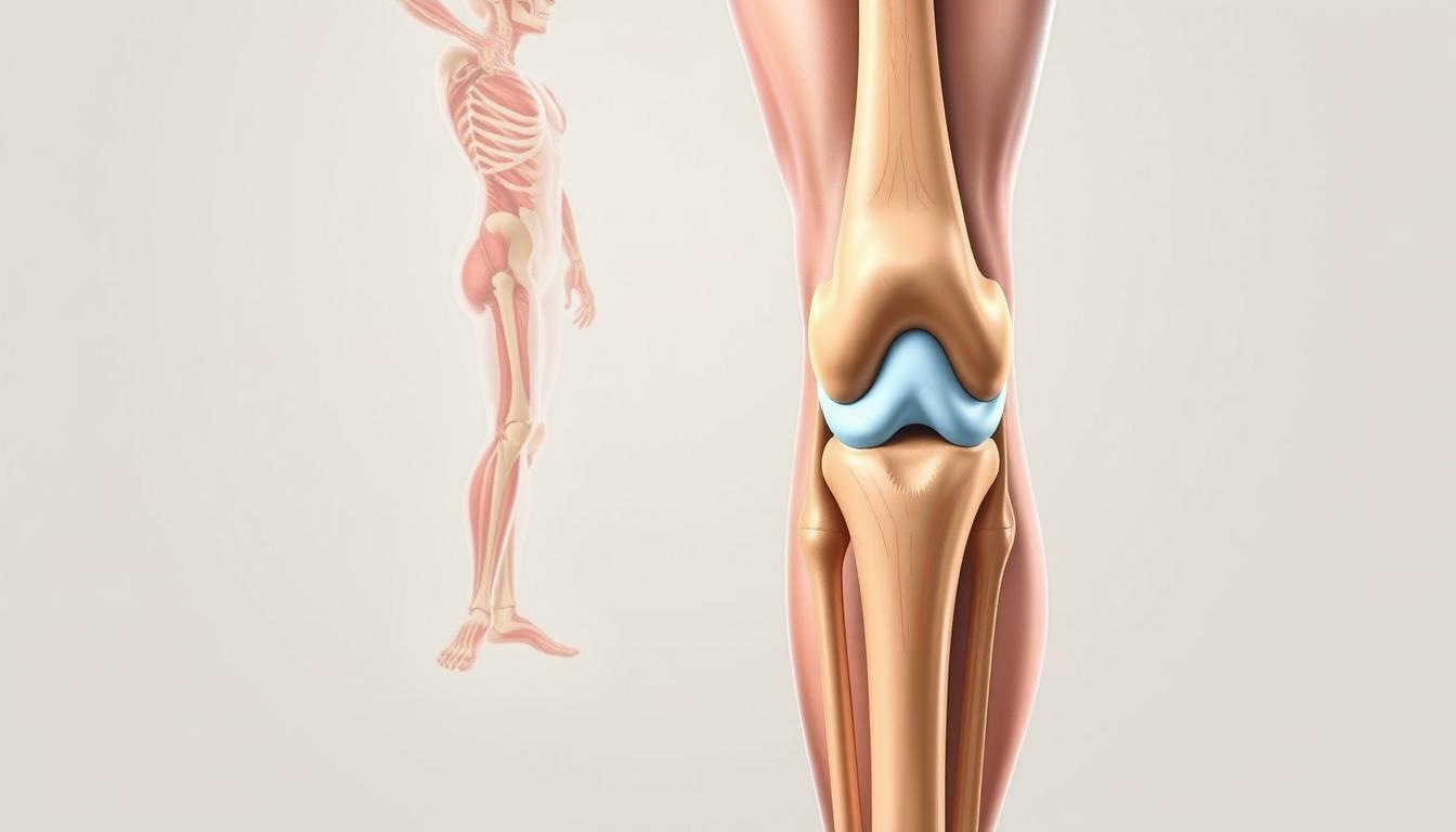
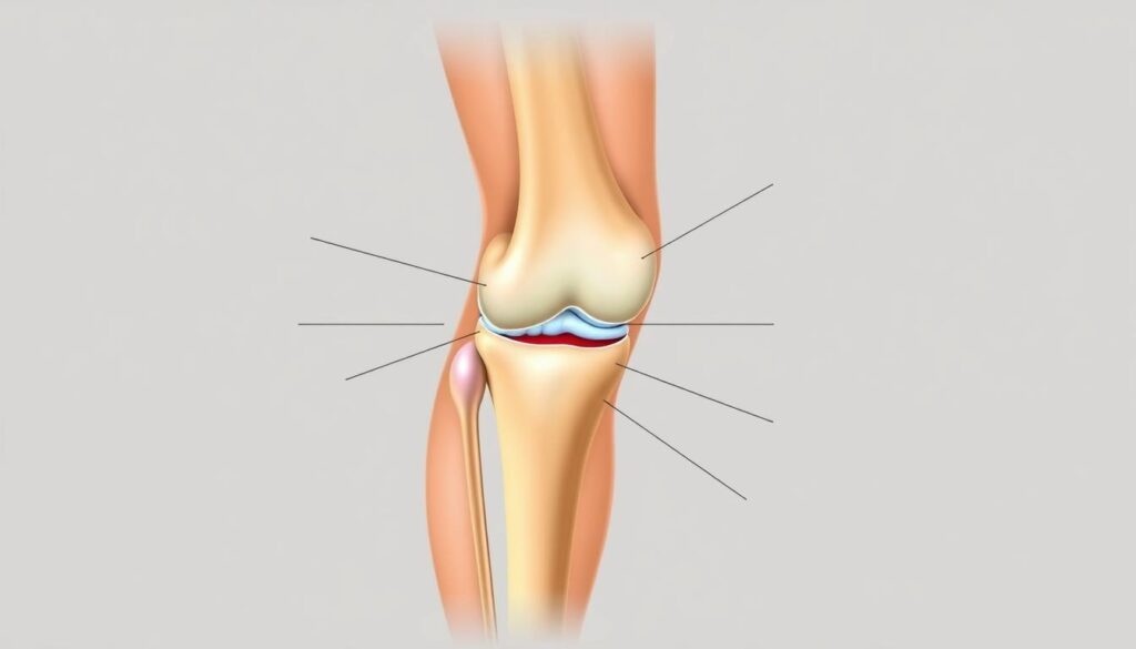
    The human knee operates like a precision-engineered hinge, blending bones with soft tissues for mobility. Three bones form its framework: the femur (thigh bone), tibia (shin bone), and patella (kneecap). These structures rely on ligaments and muscles to maintain alignment during movement.

    Key Structures Involved in Knee Stability

    Four primary ligaments act as biological cables. The collateral ligaments prevent side-to-side shifting, while cruciate ligaments control forward/backward motion. Together, they create a cross-shaped support system inside the joint.

    Muscles like the quadriceps and hamstring groups provide dynamic stability. Tendons anchor these muscles to bones, translating force into movement. Without this coordination, simple actions like walking would strain the joint.

    The Role of the Posterior Cruciate Ligament and Meniscus

    The posterior cruciate ligament (PCL) resists backward tibial movement. It’s thicker than its anterior counterpart, making injuries less common but harder to diagnose. Nearby, crescent-shaped meniscus pads absorb impact and distribute weight evenly.
    StructurePrimary RoleCommon Injuries
    PCLPrevents tibia displacementHyperextension trauma
    MeniscusShock absorptionTwisting motions
    Collateral LigamentsSide stabilityDirect impacts

    Damage to these components often starts subtly. A torn meniscus might only ache during deep squats initially. Similarly, cartilage wear develops gradually, reducing the joint’s natural shock absorption over time.

    Back of knee pain only when fully extended

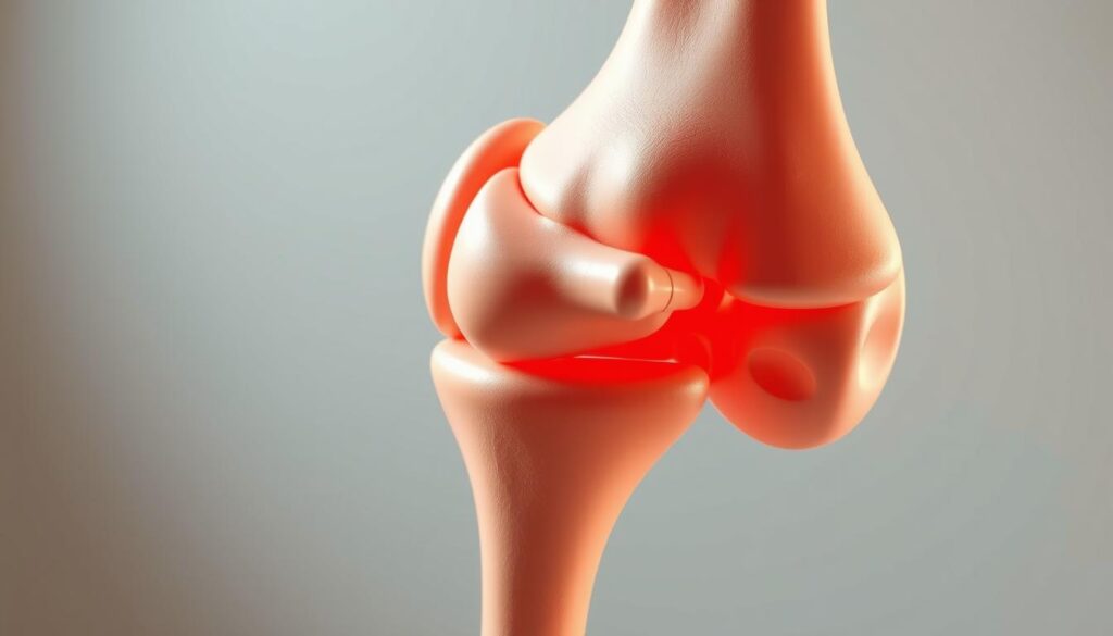
    Many athletes notice a distinct discomfort pattern emerging during movements requiring straight-leg positions. This symptom cluster often serves as the body’s warning system for specific mechanical stress points.

    posterior knee discomfort during extension

    Recognizing Distinctive Symptom Markers

    Full leg extension activates different structures than bent-knee positions. Key indicators include:

    • Sharp resistance when locking the joint
    • Stiffness lasting minutes after standing
    • Swelling concentrated behind the joint capsule

    Unlike bending-related issues, these symptoms typically ease when slightly flexing the leg. This positional variation helps differentiate tendon strain from cartilage damage.

    Condition-Specific Warning Signs

    Specific disorders reveal themselves through extension challenges:

    ConditionExtension SymptomDifferentiating Factor
    Baker’s CystTightness behind jointPalpable fluid-filled lump
    PCL InjuryInstability when standingHistory of hyperextension trauma
    Nerve CompressionElectric-shock sensationsNumbness in lower leg

    Recent studies show 42% of posterior discomfort cases involve multiple coexisting issues. Professional evaluation becomes crucial when symptoms persist beyond 72 hours or limit daily activities.

    Causes and Contributing Factors for Posterior Knee Pain

    Discomfort during leg extension often stems from three primary sources: sudden trauma, repetitive stress, or age-related changes. Athletes and active adults frequently experience these issues, but even routine movements can expose weaknesses in joint structures.

    Muscle Strains, Tendon Issues, and Ligament Tears

    Overexertion during sports or workouts often leads to soft tissue damage. Hamstring tendon strains create localized tenderness, while ligament tears cause instability during weight-bearing activities. These injuries typically worsen without proper rest.

    Common triggers include:

    • Explosive movements like jumping or sprinting
    • Improper warm-up routines
    • Previous untreated injuries

    Injuries, Baker’s Cysts, and Osteoarthritis

    Persistent swelling behind the joint often signals a Baker’s cyst. These fluid-filled sacs frequently develop alongside arthritis or cartilage damage. Unlike acute injuries, cysts may grow slowly, creating pressure that intensifies during extension.

    ConditionPrimary CauseKey Feature
    Hamstring TendinitisOverusePain during acceleration
    PCL TearHyperextensionDifficulty standing upright
    OsteoarthritisCartilage LossMorning stiffness

    Degenerative changes account for 38% of chronic cases according to recent studies. Inflammation from arthritis accelerates tissue breakdown, while prior injuries create weak points prone to reinjury. Early intervention breaks this cycle effectively.

    Diagnostic Methods and the Importance of Medical Evaluation

    Accurate diagnosis forms the cornerstone of effective treatment plans. While discomfort patterns provide clues, modern medicine uses precise tools to pinpoint issues in complex joints. Early detection prevents minor injuries from escalating into chronic conditions.

    Physical Exams and Imaging Tests

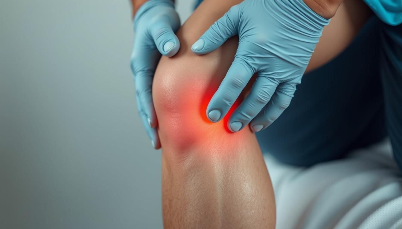
    Clinicians begin with hands-on assessments. They check for swelling, test range of motion, and apply pressure to identify tender areas. Special maneuvers help evaluate cruciate ligament integrity and bone alignment issues.

    Three primary imaging methods reveal hidden problems:

    TestBest ForKey Insights
    X-rayBone fracturesReveals joint spacing and bone spurs
    MRISoft tissue damageShows ACL tears and cartilage wear
    UltrasoundBlood flow analysisDetects cysts and tendon inflammation

    Blood tests occasionally supplement these tools when infection or systemic inflammation is suspected. They help rule out conditions like gout or rheumatoid arthritis that might mimic knee injury symptoms.

    Advanced imaging proves particularly crucial for assessing cruciate ligament damage and meniscus tears. A 2023 Johns Hopkins study found MRI accuracy exceeds 92% for diagnosing ACL injuries compared to physical exams alone.

    Seek immediate evaluation if you notice:

    • Sudden swelling with warm skin
    • Abnormal blood vessel patterns
    • Inability to bear weight

    Treatment Options for Knee Pain

    Effective management starts with understanding your body’s healing potential. Initial approaches prioritize reducing inflammation while restoring mobility. Over 80% of acute cases respond well to non-invasive methods when applied correctly.

    Conservative Treatments and Home Remedies

    The RICE protocol remains foundational for acute care:

    • Rest: Avoid activities stressing the joint
    • Ice: Apply cold packs to reduce swelling
    • Compression: Use elastic bandages for support
    • Elevation: Keep the leg raised above heart level

    Over-the-counter NSAIDs like ibuprofen provide temporary relief. For persistent knee discomfort, physical therapy strengthens surrounding muscles. Targeted exercises improve hamstring flexibility and quadriceps stability, reducing strain on tendons.

    ApproachBest ForDuration
    RICE MethodAcute injuries48-72 hours
    Physical TherapyChronic instability6-8 weeks
    Corticosteroid InjectionsArthritis flare-ups3-6 months relief

    When conservative measures fail, medical providers may also suggest advanced options. Arthroscopic surgery addresses torn cartilage, while joint replacement becomes viable for severe arthritis. Always consult specialists before escalating treatments.

    Recovery and Rehabilitation Strategies

    Rebuilding strength after joint issues requires careful planning. Effective rehabilitation balances tissue healing with progressive challenges to restore full function. Let’s explore methods that help patients regain mobility while minimizing reinjury risks.

    Customized Therapy Protocols

    Physical therapists often design programs targeting specific leg muscle groups. For hamstring-related recoveries, exercises might include:

    • Eccentric curls to rebuild tendon resilience
    • Step-up drills for thigh stabilization
    • Balance boards to improve joint proprioception
    Therapy PhaseFocus AreaDuration
    Initial RecoveryReducing swelling1-2 weeks
    Strength BuildingHamstring activation3-5 weeks
    Functional TrainingSport-specific motions6+ weeks

    Activity Progression Framework

    Returning to normal movements demands gradual exposure. A 2024 sports medicine study showed athletes who followed phased plans had 40% fewer repeat tears. Key progression markers include:

    • Pain-free walking for 48 hours
    • Full range of motion recovery
    • 90% strength in affected leg compared to healthy side

    Monitoring tools like wearable sensors help track thigh muscle engagement during rehab. Therapists adjust programs weekly based on performance data and tissue response. For persistent tears, low-impact alternatives like swimming maintain progress without strain.

    Activity LevelRecommended ExercisesPrecautions
    Early StageStationary bikingAvoid deep squats
    IntermediateLateral lungesMonitor joint clicking
    AdvancedPlyometric jumpsUse shock-absorbing surfaces

    Preventing Future Knee Injuries and Maintaining Joint Health

    Maintaining healthy joints requires more than reactive care—it demands consistent, proactive strategies. Simple daily habits significantly reduce strain on vulnerable areas while improving overall mobility. Let’s explore practical methods to safeguard your body’s most complex hinge system.

    Lifestyle Changes and Injury Prevention Techniques

    Adjusting movement patterns protects delicate tissues during high-impact activities. Athletes should prioritize low-impact cross-training like swimming to balance joint stress. For everyday protection, avoid sudden pivots and wear supportive footwear with proper arch cushioning.

    Strengthening surrounding muscles creates natural armor for the joint. Focus on exercises targeting quadriceps, hamstrings, and glutes. A 2024 sports medicine report found individuals with strong thigh muscles had 65% fewer posterior discomfort episodes.

    Prevention StrategyKey BenefitFrequency
    Dynamic Warm-UpsIncreases blood flowBefore every workout
    Balance TrainingImproves stability3x weekly
    Flexibility RoutinesReduces tendon strainDaily

    Regular check-ups help identify emerging conditions before they escalate. Schedule annual assessments with a knee pain specialist if you engage in repetitive motions. Early detection of cartilage wear or ligament laxity allows for timely interventions.

    Nutrition plays an underrated role in joint preservation. Omega-3 fatty acids from fish and walnuts combat inflammation, while vitamin C supports collagen production. Stay hydrated—synovial fluid depends on adequate water intake to lubricate moving parts effectively.

    Conclusion

    Persistent discomfort during straight-leg movements often signals mechanical stress in critical structures. From tendon inflammation to ligament strain, causes range widely but share a common need for timely care. Our exploration reveals how proper diagnosis separates temporary irritation from chronic conditions requiring specialized treatment.

    Early intervention remains vital. Whether addressing muscle imbalances or cartilage wear, structured rehab plans restore function effectively. Conservative approaches like physical therapy succeed in most cases, while advanced options address severe ACL or cruciate injuries.

    We emphasize consulting specialists when symptoms linger. Diagnostic tools and tailored strategies prevent minor issues from escalating. Remember: joint health thrives on proactive care and informed decisions.

    Our team remains dedicated to delivering clear, research-backed guidance. Trust evidence-based practices – your mobility deserves nothing less.

    FAQ

    Why does the back of my knee hurt only when I straighten my leg fully?

    Discomfort during full extension often stems from tightness or irritation in structures like the posterior cruciate ligament (PCL), meniscus, or tendons. Overuse injuries, arthritis, or cysts may compress tissues when the joint is fully straightened, triggering pain.

    Can a Baker’s cyst cause sharp pain behind the knee during activity?

    Yes. A Baker’s cyst—a fluid-filled sac—often swells with repetitive motion, pressing on nerves or muscles. This can lead to sharp sensations, especially during activities requiring full leg extension, like running or climbing stairs.

    How do I know if my posterior cruciate ligament is injured?

    PCL injuries typically cause instability, swelling, or aching at the back of the joint. Pain worsens when kneeling, squatting, or extending the leg. A physical exam or MRI can confirm damage to this critical stabilizer.

    When should I see a doctor for posterior knee pain?

    Seek evaluation if pain persists beyond 48 hours, limits mobility, or accompanies redness, warmth, or sudden swelling. These could signal tears, blood clots, or infections requiring prompt care.

    What home treatments reduce discomfort from extension-related knee pain?

    Rest, ice packs, and compression help reduce inflammation. Gentle stretches for the hamstrings or calf muscles may relieve tension. Avoid activities that strain the joint until symptoms improve.

    Can physical therapy address chronic pain behind the knee?

    Absolutely. Therapists design programs to strengthen muscles like the quadriceps and improve flexibility, reducing stress on ligaments and cartilage. Techniques may include ultrasound therapy or guided exercises to restore safe movement patterns.

    Are there long-term risks if posterior knee pain is ignored?

    Untreated injuries may lead to chronic instability, cartilage wear, or early-onset osteoarthritis. Conditions like untreated meniscus tears can also worsen, increasing recovery time and complicating future treatment.

    What imaging tests diagnose issues in the posterior knee?

    X-rays detect bone abnormalities, while MRIs provide detailed views of soft tissues like ligaments, tendons, and cysts. Ultrasound may assess fluid-filled structures or guide injections for targeted relief.

  • Pes anserine bursitis symptoms and treatment

    Pes anserine bursitis symptoms and treatment

    Have you ever wondered why inner knee discomfort lingers despite rest or basic care? This guide dives into a common yet overlooked condition affecting athletes, active adults, and anyone experiencing persistent joint issues. We’ll uncover how a small, fluid-filled sac near your knee could hold answers to your mobility struggles.

    Inflammation in this area often develops from repetitive motions or sudden strain. The result? Sharp aches during movement, tenderness when touched, and stiffness that limits daily activities. While these signs might seem vague, recognizing them early can prevent long-term complications.

    Our focus combines insights from leading medical institutions with practical recovery strategies. You’ll learn how simple adjustments to exercise routines or targeted therapies can accelerate healing. We’ve prioritized clear, actionable steps to help you regain comfort without invasive procedures.

    Key Takeaways

    • Inner knee inflammation often stems from repetitive stress or improper movement patterns.
    • Early intervention typically leads to faster recovery through conservative methods.
    • Diagnosis combines physical exams with imaging to rule out similar conditions.
    • Effective management blends rest, targeted exercises, and anti-inflammatory approaches.
    • Trusted medical resources form the foundation of our recommended strategies.

    Let’s explore how understanding this condition’s nuances can transform your approach to joint health. From identifying warning signs to implementing proven relief methods, we’ll walk through each phase of recovery together.

    Introduction to Pes Anserine Bursitis

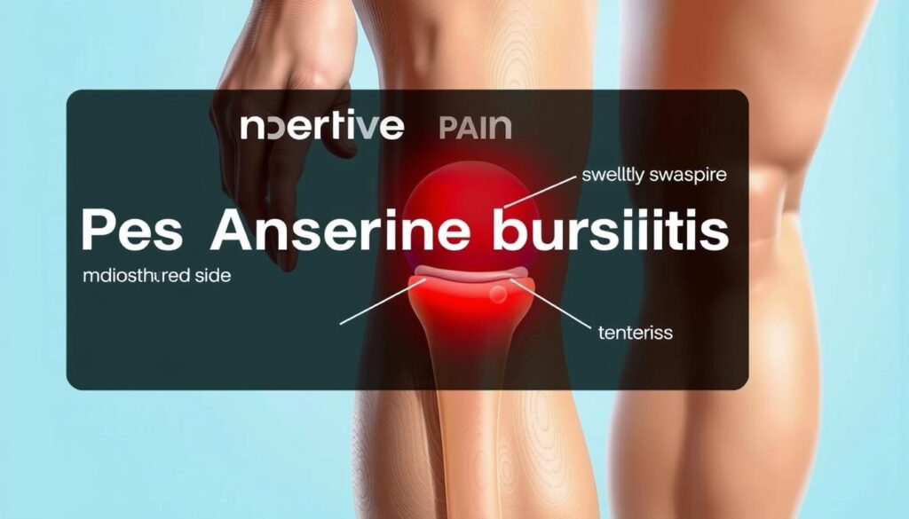
    A tiny sac near the knee can lead to significant mobility issues when inflamed. The pes anserine bursa sits just below the knee joint on the inner leg, cushioning tendons during movement. When irritated, this fluid-filled structure swells, creating friction that disrupts natural motion.

    knee bursa inflammation

    Repetitive strain from activities like running or climbing often triggers this condition. Poor training form and underlying issues such as osteoarthritis amplify risks. Athletes and active adults frequently report tenderness when bending or straightening the leg.

    FeatureHealthy BursaInflamed Bursa
    FunctionReduces frictionCreates painful friction
    Pain LevelNoneSharp during activity
    MobilityUnrestrictedStiffness after rest
    Common TriggersNormal useOveruse or injury

    Proper diagnosis separates this issue from similar knee problems. Healthcare providers assess swelling patterns and pressure points while reviewing activity history. Early identification helps avoid prolonged discomfort and supports targeted recovery plans.

    We’ll explore how strategic care restores function while preventing recurrence. Next sections detail practical steps to address root causes rather than just masking discomfort.

    What is Pes Anserine Bursitis?

    Imagine your knee’s shock absorber failing during routine movements. The pes anserine region houses a critical cushioning structure where three tendons converge near the shinbone. This bursa normally prevents bone-to-tendon friction during walking or climbing.

    knee bursa anatomy

    Anatomy and Function of the Bursa

    Located two inches below the kneecap’s inner edge, this fluid-filled sac separates the tibia from connected hamstring tendons. It acts like biological Teflon® – reducing wear from repetitive motions. When functioning properly, you’ll never notice its presence.

    Common Causes and Risk Factors

    Three primary elements trigger irritation in this sensitive area:

    • Repetitive leg motions (running, squatting)
    • Excessive body weight straining connective tissues
    • Biomechanical issues like bowed legs or flat feet

    Runners often develop issues after sudden mileage increases. Weekend warriors risk inflammation through inconsistent training. Tight thigh muscles compound these problems by pulling excessively on the bursa during activity.

    Understanding these mechanisms helps create smarter recovery plans. Next, we’ll examine how professionals distinguish this condition from similar knee issues.

    Pes anserine bursitis symptoms and treatment

    Recognizing early warning signals of inner knee inflammation helps people seek care before limitations escalate. Many dismiss discomfort as normal soreness until simple tasks like rising from chairs become challenging.

    inner knee pain symptoms

    Recognizing the Symptoms

    Three primary markers distinguish this condition from general joint strain:

    • Persistent ache concentrated 2-3 inches below the kneecap
    • Visible puffiness along the shinbone’s upper edge
    • Sharp flares when bending or straightening the leg

    Movement patterns often reveal hidden triggers. Climbing stairs or hills typically intensifies discomfort due to increased tendon friction. Nighttime stiffness after active days also signals irritated tissues.

    Diagnostic MethodKey IndicatorsPurpose
    Physical ExamLocalized warmth, pressure sensitivityRule out meniscus tears
    Activity AnalysisPain patterns during specific motionsIdentify movement triggers
    ImagingBursa thickness, tendon alignmentConfirm fluid buildup

    Treatment Strategy Foundations

    Initial care focuses on breaking the inflammation cycle. Rest reduces mechanical stress while ice application calms swollen tissues. Over-the-counter NSAIDs provide temporary relief but don’t address root causes.

    Effective plans combine multiple approaches:

    • Activity modifications to protect healing areas
    • Targeted stretches improving tendon mobility
    • Strengthening exercises stabilizing the joint

    Medical professionals often recommend evidence-based non-surgical recovery plans first. Early intervention using these methods typically restores function within weeks while preventing chronic issues.

    Diagnosing Pes Anserine Bursitis

    Modern imaging tools reveal hidden causes of mobility challenges. Healthcare providers start with hands-on evaluations to map discomfort patterns. They press specific areas below the knee while observing reactions to identify tender zones linked to the pes anserinus region.

    Confirming Inflammation Through Testing

    Three-step verification ensures accurate results:

    • Physical assessment: Checking for localized swelling along the upper tibia
    • Movement analysis: Monitoring pain during stair climbing or leg rotations
    • Imaging correlation: Matching symptoms with visual evidence

    X-rays eliminate bone fractures, while ultrasound scans detect fluid buildup in soft tissues. MRI examinations provide detailed views of tendon alignment near the knee joint. These methods help distinguish this condition from meniscus injuries or osteoarthritis.

    Diagnostic ToolKey FunctionAccuracy Rate
    Clinical ExamIdentifies pressure points78%
    UltrasoundVisualizes bursa thickness92%
    MRIAssesses surrounding structures95%

    Definitive diagnosis prevents mismanagement of similar knee issues. Providers combine test results with activity histories to create personalized recovery plans. This precision ensures therapies target the root problem rather than general discomfort.

    Treatment Options and Management Strategies

    Addressing tendon-related discomfort demands methods that target both symptoms and causes. Healthcare teams prioritize approaches that calm irritation while rebuilding strength. We’ll explore proven techniques ranging from basic self-care to advanced clinical interventions.

    Non-Operative Approaches: Rest, Ice, and Medication

    Initial care focuses on reducing strain. Short-term activity changes protect healing tissues – think swapping runs for swimming or cycling. Applying cold packs for 15-minute intervals lowers swelling effectively when done 3-4 times daily.

    Over-the-counter NSAIDs like ibuprofen ease discomfort temporarily. However, prolonged use requires medical supervision. Many find compression sleeves helpful during light activities to support the area without restricting blood flow.

    ApproachKey ActionsAverage Recovery Time
    Rest & Activity ModificationLimit bending/squatting2-4 weeks
    Ice Application15 mins, 3x/dayImmediate relief
    MedicationNSAID regimen3-7 days

    Physical Therapy, Ultrasound, and Injection Therapies

    Structured rehab programs restore mobility safely. Therapists guide patients through gentle stretches that loosen tight hamstrings and improve tendon glide. Ultrasound technology enhances blood flow to accelerate natural repair processes.

    For persistent cases, corticosteroid injections deliver anti-inflammatory agents directly to the affected area. These are often paired with numbing agents for immediate comfort. Clinical studies show 80% of patients report significant improvement within 72 hours post-treatment.

    Every plan adapts to individual needs. Providers monitor progress through follow-up assessments, adjusting techniques as healing advances. This personalized strategy ensures lasting results rather than temporary fixes.

    Practical Exercises and Rehabilitation Guidance

    What if targeted movements could speed up your recovery while protecting vulnerable tissues? Strategic movement plans rebuild strength without overloading healing areas. We focus on methods that restore flexibility while teaching your body safer movement patterns.

    Effective Stretching and Strengthening Exercises

    Hamstring stretches reduce tension pulling on the inner knee. Try seated stretches with legs extended, reaching gently toward your toes. Hold for 20 seconds, repeating 3 times daily. Wall-assisted stretches let you control intensity while standing.

    Strengthen supporting muscles with bridges and side-lying leg lifts. These low-impact exercises build stability without bending the knee excessively. Start with 2 sets of 10 reps, increasing gradually as discomfort decreases.

    Exercise TypeFrequencyBenefits
    Seated Stretch3x dailyImproves tendon glide
    Wall Push Stretch2x dailyReduces muscle tightness
    Bridging4x weeklyStrengthens glutes

    Recovery Tips and Activity Modifications

    Modify daily activities to avoid reinjury. Use handrails on stairs and limit squatting motions during household chores. Swap high-impact workouts for swimming or cycling until symptoms improve.

    Track progress with a simple journal. Note pain levels during specific movements and adjust your program accordingly. Many find compression sleeves helpful during light activity, providing support without restricting circulation.

    Lifestyle Adjustments and Preventive Measures

    Protecting joint health requires smart daily choices that outpace wear and tear. For those recovering from or prone to pes anserine issues, small habit shifts create lasting protection. We’ll explore practical ways to maintain mobility while reducing strain on vulnerable areas.

    Building Sustainable Routines

    Three adjustments significantly lower recurrence risks:

    • Footwear upgrades: Choose shoes with arch support and shock absorption
    • Movement pacing: Alternate high-impact sports with low-stress activities
    • Pre-activity prep: Implement dynamic warm-ups targeting hamstrings

    “Gradual progression in training intensity allows tissues to adapt without overload,” notes sports physical therapist Dr. Elena Martinez.

    Focus AreaAction StepsBenefits
    Footwear SelectionReplace worn shoes every 300-500 milesReduces knee torque by 18%
    Training ModificationsMix running with swimming or cyclingCuts repetitive stress by 40%
    Weight ManagementCombine balanced nutrition with strength trainingLowers joint pressure 5x per pound lost

    Individuals with osteoarthritis management strategies should prioritize consistent strength programs. Focus on quadriceps and hip stabilizers during workouts – these muscles absorb impact before it reaches the knee.

    Weekly activity plans balance challenge and recovery. Sample schedules might include two days of strength training, three days of moderate cardio, and dedicated flexibility sessions. Tracking progress helps identify patterns that trigger discomfort early.

    Conclusion

    Effective management of knee discomfort begins with understanding its origins. Early recognition of pes anserine bursitis allows for swift action, combining rest with targeted therapies to reduce inflammation. Diagnostic tools like ultrasound help confirm fluid buildup while ruling out other joint issues.

    Successful recovery hinges on tailored plans addressing both symptoms and causes. Physical therapy strengthens surrounding muscles, while activity modifications prevent reinjury. Studies show structured exercise programs improve mobility in 89% of cases within six weeks.

    Consult healthcare providers if inner-leg tenderness persists during daily movements. Accurate imaging and professional guidance create roadmaps for lasting relief. Preventive strategies like supportive footwear and gradual training progressions further protect vulnerable areas.

    With proper care, most individuals regain full function without invasive procedures. Small, consistent changes in movement patterns and self-care routines make recovery achievable. Reach out to specialists to design a plan matching your unique needs and lifestyle.

    FAQ

    How does pes anserine bursitis differ from other knee conditions?

    Unlike arthritis or ligament injuries, this condition specifically involves inflammation of the bursa near the hamstring tendons. Pain typically occurs 2–3 inches below the knee joint and worsens with activities like climbing stairs or prolonged sitting.

    Can physical therapy exercises worsen the pain?

    When guided by a licensed therapist, targeted stretches and strengthening routines often reduce discomfort. We recommend avoiding high-impact movements initially and focusing on low-stress exercises like seated leg lifts or gentle hamstring stretches to avoid aggravating the area.

    Are corticosteroid injections safe for long-term use?

    While effective for short-term relief, repeated injections may weaken nearby tissues. We prioritize combining them with rest, ice therapy, and anti-inflammatory medications to minimize risks. Always discuss treatment plans with your healthcare provider.

    What daily habits contribute to flare-ups?

    Repetitive motions like squatting, sudden increases in exercise intensity, or poor footwear choices often trigger inflammation. We suggest modifying workouts, using supportive shoes, and incorporating rest days to manage stress on the knee.

    How long does recovery typically take?

    Most people see improvement within 4–6 weeks with consistent treatment. Chronic cases linked to osteoarthritis or obesity may require longer rehab. Early diagnosis and a structured therapy program improve outcomes significantly.

    Is ultrasound imaging necessary for diagnosis?

    While MRI or ultrasound can confirm inflammation, many providers diagnose based on physical exams and symptom history. Imaging is usually reserved for unclear cases or to rule out tears in the tendons or meniscus.

    Can ice packs replace prescription medications?

    Ice reduces swelling effectively but doesn’t address underlying inflammation. We combine cryotherapy with NSAIDs like ibuprofen for comprehensive management. Always consult a doctor before starting new medications.

    Are there sports to avoid during recovery?

    High-impact activities like basketball or running often strain the knee. We recommend switching to swimming, cycling, or yoga until tenderness subsides. Gradually reintroduce sports under a therapist’s supervision.

  • Knee pain 3 weeks after fall

    Knee pain 3 weeks after fall

    When an unexpected tumble leaves you sore, it’s easy to brush off stiffness as temporary. But what happens when that discomfort lingers for weeks? Hidden damage—like ligament strains or hairline fractures—often reveals itself slowly, masking its severity beneath surface-level soreness.

    Medical studies show that delayed symptoms account for nearly 30% of undiagnosed joint issues. A misstep or awkward landing can twist tissues in ways that aren’t immediately obvious. Without proper care, minor tears may worsen, leading to chronic instability or mobility loss.

    We’ve analyzed cases where patients dismissed early warning signs, only to face complex recoveries later. That’s why understanding your body’s signals matters. Swelling that persists, difficulty bearing weight, or sharp twinges during movement aren’t just inconveniences—they’re clues.

    This guide will help you distinguish between manageable soreness and red flags requiring expert evaluation. From at-home relief strategies to advanced therapies, we’ll equip you with actionable steps to protect your joints and reclaim your active life.

    Key Takeaways

    • Delayed symptoms often indicate underlying joint or tissue damage.
    • Persistent swelling or instability warrants professional assessment.
    • Self-care methods work best when paired with accurate injury identification.
    • Early intervention reduces long-term complications like chronic stiffness.
    • Movement patterns post-accident help clinicians pinpoint hidden issues.

    Overview of Knee pain 3 weeks after fall

    Sudden impacts create complex stress patterns that challenge even resilient joints. While surface wounds heal quickly, deeper structures like cartilage or connective tissues may suffer silent damage that surfaces later.

    Impact of Traumatic Force on Joint Structures

    During a fall, rotational forces and compression can strain ligaments beyond their elastic limit. This creates micro-tears that often evade initial detection. Vulnerable components like the meniscus—a shock-absorbing cartilage—might sustain partial tears that worsen with continued movement.

    Why Symptoms Linger Beyond Initial Injury

    Three factors explain delayed discomfort:

    • Inflammation cycles: Swelling resurfaces as damaged tissues attempt repair
    • Cumulative stress: Daily activities aggravate partially healed injuries
    • Nerve sensitivity: Prolonged irritation heightens pain perception

    Medical literature reveals that 40% of ligament injuries in weight-bearing joints show delayed symptom onset. Persistent swelling often signals ongoing tissue distress rather than routine healing. As one orthopedic specialist notes: “The joint’s layered anatomy allows minor injuries to hide behind temporary stiffness.”

    Recognizing these patterns helps differentiate between normal recovery and emerging complications. Tracking symptom progression—especially changes in mobility or swelling intensity—provides critical clues for timely intervention.

    Common Knee Injuries After a Fall

    The human body’s response to impact reveals hidden vulnerabilities. Collisions with hard surfaces often leave visible marks like scrapes or bruises, but deeper structural harm requires closer inspection. We’ll explore how seemingly minor trauma can mask critical damage needing specialized care.

    common knee injuries

    Abrasions, Lacerations, and Bruises

    Surface wounds account for 65% of immediate post-fall complaints. These include:

    • Road rash: Friction burns from sliding on pavement
    • Contusions: Blood pooling under skin from blunt force
    • Deep cuts: Sharp objects penetrating tissue layers

    While these often heal with basic cleaning and bandaging, persistent redness or pus signals infection. A 2023 Johns Hopkins study found 1 in 5 abrasions develop complications without proper antiseptic care.

    Injury TypeHealing TimeRisk Factors
    Superficial scrape3-7 daysDebris contamination
    Moderate bruise2-4 weeksBlood thinners usage
    Deep laceration4-6 weeksJoint capsule involvement

    Ligament, Meniscus, and Tendon Damage

    Twisting motions during falls strain connective tissues. The ACL and MCL ligaments suffer 78% of sprains in sideways tumbles, while meniscus tears frequently occur during kneeling impacts. As noted in Orthopedic Trauma Journal:

    “Partial tendon ruptures often mimic bruise symptoms initially, delaying diagnosis by 2-3 weeks.”

    Three red flags distinguish severe soft-tissue injuries:

    1. Inability to straighten the joint fully
    2. Audible popping during movement
    3. Instability when shifting weight

    Patellar fractures, though rare, require immediate imaging. They typically occur when kneecaps strike concrete edges or car dashboards at high speed.

    Diagnosing Knee Injuries and When to Seek Help

    Identifying the root cause of ongoing discomfort is essential for effective treatment. While some issues resolve with rest, others demand precise evaluation to prevent long-term damage. Monitoring changes in mobility or sensation helps separate temporary strain from structural concerns.

    Recognizing Warning Signs and Symptoms

    Certain signals demand immediate attention. A loud “pop” during impact often indicates ligament tears. Difficulty straightening the joint fully or bearing weight suggests deeper tissue involvement. Swelling that persists beyond 48 hours—or worsens with activity—points to unresolved inflammation.

    We advise tracking symptom patterns over time. Sharp twinges during rotation, nighttime throbbing, or sudden instability all warrant professional assessment. As one sports physician notes: “Ignoring these clues risks transforming a fixable injury into chronic dysfunction.”

    Medical Imaging and Physical Examinations

    Healthcare providers use hands-on tests to evaluate range of motion and stability. The Lachman test detects ACL tears, while McMurray’s maneuver identifies meniscus damage. Imaging tools like X-rays reveal bone fractures, while MRIs expose soft-tissue injuries invisible to other methods.

    Timely scans matter. A 2023 Mayo Clinic study found early MRI use reduced misdiagnosis rates by 37% in complex cases. Combined with symptom history, these tools create a clear roadmap for recovery.

    Persistent pain swelling or warmth around the joint often signals hidden issues. If self-care fails after 72 hours, consulting a doctor becomes critical. Early intervention curbs complications, letting you regain control faster.

    Managing Knee Pain at Home

    Effective home care can significantly influence recovery timelines following joint trauma. While professional evaluation remains vital for persistent issues, initial management often determines healing efficiency. Let’s explore proven methods to support your body’s repair processes while avoiding common pitfalls.

    Implementing the RICE Protocol

    home treatment for joint injuries

    The RICE method—Rest, Ice, Compression, Elevation—remains the gold standard for acute care. Begin by limiting weight-bearing activities for 24-48 hours. Apply cold packs wrapped in cloth for 15-minute intervals every two hours to reduce swelling. Elastic bandages provide gentle pressure without restricting circulation, while propping the limb above heart level drains excess fluid.

    Balancing Activity and Recovery

    Over-the-counter anti-inflammatories like ibuprofen ease discomfort but shouldn’t mask worsening symptoms. Pair medication with strategic rest periods—use pillows to stabilize the joint during sleep. Gradually reintroduce movement through gentle stretches once tenderness subsides.

    Monitor progress closely. Increased redness, warmth, or throbbing signals potential complications. Combine these steps with natural anti-inflammatory approaches for enhanced results. Most strains improve within 7-10 days with consistent care.

    If stiffness persists beyond 72 hours or weight-bearing becomes impossible, consult a specialist immediately. Early intervention prevents minor setbacks from evolving into chronic limitations.

    Medical Treatment Options for Knee Injuries

    When joint injuries resist home care, targeted medical strategies become essential. Non-surgical approaches often serve as the first line of defense, while advanced cases demand precision interventions. We’ll outline how specialists tailor treatments to injury severity and recovery goals.

    Use of Braces, Medications, and Physical Therapy

    Custom braces stabilize unstable joints, allowing partial tears to heal without strain. Anti-inflammatory medications like naproxen reduce swelling, while corticosteroid injections address persistent inflammation. For mobility restoration, structured physical therapy programs prove vital:

    • Strengthening exercises rebuild muscle support around weakened ligaments
    • Gait retraining corrects compensatory movement patterns
    • Low-impact conditioning maintains joint flexibility during recovery

    As one sports medicine specialist explains: “Therapy isn’t just about healing—it’s about preventing future vulnerability.”

    Surgical Interventions: When It’s Necessary

    Complete anterior cruciate ligament tears or displaced fractures often require surgical repair. Arthroscopic procedures address meniscus damage with minimal scarring, while reconstruction replaces ruptured cruciate ligament tissues using grafts. Recovery timelines vary:

    ProcedureRecovery TimeSuccess Rate
    ACL Reconstruction6-9 months89%
    Meniscus Repair3-4 months78%

    Patients may need surgery if instability persists despite 6 weeks of conservative care. Early intervention prevents cartilage degeneration and chronic instability, preserving long-term joint function.

    Effective Physical Therapy and Rehabilitation

    Structured rehabilitation serves as the cornerstone of recovery for joint-related trauma. Specialized programs bridge the gap between initial healing and full functional restoration, addressing both visible symptoms and underlying weaknesses.

    Guided Recovery Through Expert Intervention

    Licensed therapists design personalized plans based on injury severity and lifestyle goals. They assess movement patterns, identifying compensatory habits that strain the knee joint. As one rehabilitation specialist states: “Our role extends beyond symptom management—we rebuild your body’s natural shock absorption system.”

    Targeted Exercises for Lasting Results

    Progressive routines focus on two key areas:

    • Mobility restoration: Gentle range-of-motion drills combat stiffness
    • Strength rebuilding: Resistance training fortifies muscles supporting the knees

    Common evidence-based activities include:

    ExercisePurposeFrequency
    Straight-leg raisesQuadriceps activation3x daily
    Hamstring curlsPosterior chain balanceAlternate days
    Mini squatsFunctional strength5x weekly

    Athletes may also incorporate sport-specific drills once stability improves. These gradually reintroduce pivoting and jumping motions under controlled conditions. Therapists monitor progress through measurable benchmarks like squat depth or single-leg balance duration.

    Consistent participation in tailored programs yields multiple benefits. Strengthening surrounding muscle groups reduces future injury risks by 42%, according to recent sports medicine research. Patients regain confidence in their body’s capabilities while learning protective movement strategies for daily activities.

    Preventing Future Knee Injuries

    Building joint resilience starts with understanding how daily habits influence tissue strength. Proactive adjustments to movement patterns and conditioning routines can significantly reduce vulnerability to trauma.

    Strategic Conditioning for Joint Protection

    Strengthening muscles around joints creates natural armor against ligament strains. Focus on exercises enhancing quadriceps and hamstring balance:

    ExerciseBenefitFrequency
    Wall sitsBuilds endurance3x weekly
    Step-upsImproves stabilityAlternate days
    SwimmingLow-impact conditioning2x weekly

    Proper technique during physical activities lowers risk factors. Bend hips and knees when lifting heavy objects to avoid excessive joint pressure. Wear supportive footwear during high-impact sports to minimize damage from repeated impacts.

    Daily modifications matter. Replace sudden pivoting motions with controlled turns. Use ergonomic stools for tasks requiring prolonged kneeling. These small changes in movement mechanics protect against cumulative ligament stress.

    Regular check-ups help catch early signs of wear. Preventive care strategies like gait analysis identify imbalance patterns before they lead to fractures or severe damage. Combined with consistent conditioning, these steps build lasting joint health.

    Conclusion

    Recovering from joint trauma demands both patience and awareness. Our analysis shows that delayed symptoms—like those involving the anterior cruciate ligament or cartilage near bones—require precise identification to prevent long-term instability. Early intervention remains critical, especially when discomfort persists beyond initial recovery phases.

    We emphasize three priorities: recognizing subtle warning signs, adhering to structured rehabilitation, and scheduling follow-ups over months for complex cases. Issues affecting the cruciate tissues or surrounding structures often surface gradually, making professional evaluation essential when home care falls short.

    To counter persistent limitations, combine medical guidance with preventive strategies. For detailed guidance on managing joint trauma, visit our resource on knee injury care. Remember—proactive steps today safeguard mobility tomorrow.

    FAQ

    What causes persistent joint discomfort weeks after trauma?

    Lingering issues often stem from unresolved inflammation, undiagnosed ligament tears, or cartilage damage. Conditions like meniscus injuries or anterior cruciate ligament (ACL) sprains may not show immediate symptoms but worsen without proper care.

    How do we differentiate between minor bruises and serious ligament damage?

    Minor injuries typically improve with rest and ice, while severe cases involve instability, audible pops during movement, or inability to bear weight. Persistent swelling or locking sensations warrant imaging tests like MRI or X-rays to assess ligament or bone integrity.

    When should someone consult a specialist for post-fall recovery?

    Seek immediate help if you experience severe swelling, redness, fever, or sudden loss of mobility. Delayed diagnosis of fractures or ACL tears can lead to chronic instability or arthritis if untreated beyond 48–72 hours.

    Can home remedies like compression or elevation speed up healing?

    Yes. The RICE method (Rest, Ice, Compression, Elevation) reduces inflammation and supports early-stage recovery. Pairing this with over-the-counter NSAIDs like Advil or Aleve manages discomfort but doesn’t replace professional evaluation for underlying issues.

    What role does physical therapy play in restoring joint function?

    Therapists design targeted exercises to rebuild strength in quadriceps and hamstrings while improving flexibility. Techniques like manual therapy or resistance training address muscle imbalances, reducing reinjury risks during activities like sports or climbing stairs.

    Are surgical interventions common for chronic instability?

    Surgery becomes necessary for complete ligament tears, displaced fractures, or recurrent dislocations. Procedures like ACL reconstruction or meniscus repair have high success rates, especially when paired with post-op rehab programs from clinics like Mayo Clinic or Johns Hopkins.

    How can lifestyle changes prevent recurring issues?

    Strengthening core muscles, wearing supportive footwear, and avoiding high-impact exercises on hard surfaces protect joints. Brands like ASICS or Brooks offer shoes with cushioning that reduces stress during running or jumping.

  • Ultrasound-Guided Knee Injections

    Ultrasound-Guided Knee Injections

    Are you one of the millions suffering from knee pain and seeking effective relief? The use of advanced technology in medical treatments has revolutionized the way we approach musculoskeletal issues.

    Ultrasound-guided knee injections have emerged as a crucial treatment option, providing targeted pain relief and enabling individuals to progress with exercise therapy. By leveraging the precision of ultrasound technology, healthcare professionals can now administer injections with greater accuracy, ensuring that the medication reaches the exact area of need.

    Key Takeaways

    • Ultrasound-guided injections provide targeted pain relief for knee pain.
    • This treatment enables individuals to engage in exercise therapy more effectively.
    • The precision of ultrasound technology improves the accuracy of injections.
    • Knee pain treatment has evolved with advancements in medical technology.
    • Effective pain management is crucial for overall musculoskeletal health.

    What Are Ultrasound-Guided Knee Injections?

    Ultrasound-guided knee injections represent a significant advancement in the treatment of knee disorders. These injections utilize ultrasound imaging to guide the needle to the precise location, enhancing the effectiveness of the treatment.

    Definition of Ultrasound-Guided Knee Injections

    Ultrasound-guided knee injections involve the use of ultrasound imaging to visualize the knee joint and surrounding structures in real-time, allowing for the precise delivery of therapeutic agents, such as corticosteroids or hyaluronic acid, directly into the affected area.

    Purpose and Benefits of This Procedure

    The primary purpose of ultrasound-guided knee injections is to provide relief from pain and inflammation associated with various knee conditions. The benefits of this procedure include improved accuracy, reduced risk of complications, and enhanced effectiveness of the injected therapeutic agents.

    Evidence suggests that using ultrasound improves the accuracy and effectiveness of injection therapy. Conditions such as knee arthritis benefit significantly from this precision.

    CharacteristicsTraditional Knee InjectionsUltrasound-Guided Knee Injections
    AccuracyLimited by anatomical landmarksEnhanced by real-time ultrasound imaging
    Risk of ComplicationsHigher due to potential misplacementLower due to precise needle placement
    EffectivenessVariable, dependent on injector skillImproved due to accurate delivery

    How Ultrasound-Guided Knee Injections Work

    Ultrasound-guided knee injections represent a significant advancement in the treatment of knee-related conditions, offering a precise and minimally invasive solution. This procedure combines the benefits of ultrasound imaging with the therapeutic effects of injections to provide relief for patients suffering from various knee ailments.

    The Role of Ultrasound in Guiding Injections

    Ultrasound technology plays a crucial role in guiding the injection needle to the exact location within the knee joint. By providing real-time images, ultrasound allows healthcare providers to visualize the needle’s trajectory and placement, ensuring that the therapeutic agent is delivered precisely where it is needed. As noted by medical professionals,

    “The use of ultrasound guidance has significantly improved the accuracy of knee injections, reducing the risk of complications and enhancing patient outcomes.”

    The ultrasound-guided approach enables practitioners to avoid relying on landmark-based or palpation-guided techniques, which can be less accurate. This is particularly important in cases where the knee anatomy is complex or altered due to previous surgeries or conditions.

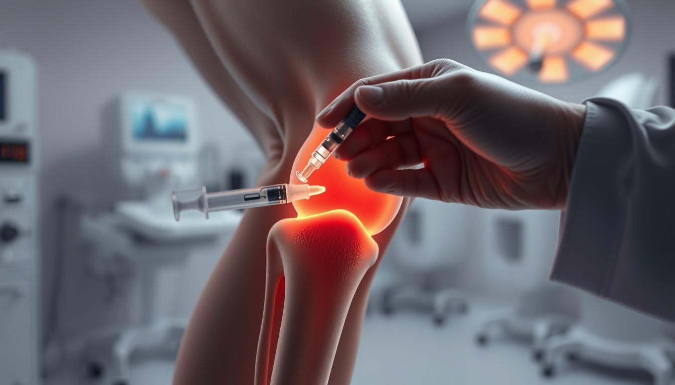
    ultrasound-guided knee injection

    Steps Involved in the Injection Process

    The process of receiving a minimally invasive knee injection typically begins with a thorough preparation of the knee area to minimize the risk of infection. The healthcare provider may clean the area with an antiseptic solution and may use a local anesthetic to numb the skin.

    • The knee is prepared and cleaned to prevent infection.
    • Ultrasound gel is applied to the skin to facilitate imaging.
    • The healthcare provider uses ultrasound to identify the optimal injection site.
    • The injection is administered, and the needle is guided in real-time using ultrasound.
    • The procedure is typically quick, and patients can often resume normal activities shortly after.

    In some cases, the healthcare provider may choose to aspirate excess joint fluid before administering the injection. This not only reduces swelling but also provides a clearer ultrasound image of the joint space, allowing for more accurate placement of the therapeutic agent.

    By understanding the steps involved in knee injection therapy, patients can better appreciate the care and precision that goes into their treatment, ultimately leading to more effective relief from knee pain and associated conditions.

    Conditions Treated with Ultrasound-Guided Knee Injections

    Knee conditions such as osteoarthritis, tendonitis, and bursitis can be effectively managed with ultrasound-guided injections. These conditions often result in significant knee pain and reduced mobility, impacting the quality of life.

    Osteoarthritis and Joint Pain

    Osteoarthritis is a degenerative joint disease that causes the cartilage in the knee to break down, leading to pain, stiffness, and reduced mobility. Ultrasound-guided knee injections, particularly those using hyaluronic acid (viscosupplementation), can help restore joint lubrication, providing relief from osteoarthritis symptoms. For more information on knee pain, visit kneehurt.com.

    The use of ultrasound guidance ensures that the injection is delivered precisely into the affected area, enhancing the effectiveness of the treatment. This approach is particularly beneficial for patients who have not responded well to other treatments.

    Tendonitis and Bursitis

    Tendonitis and bursitis are inflammatory conditions that can cause significant knee pain. Tendonitis involves inflammation of the tendons, while bursitis is the inflammation of the bursae, fluid-filled sacs that cushion the joints. Ultrasound-guided injections can deliver corticosteroids directly to the inflamed area, reducing inflammation and pain.

    By targeting the exact site of inflammation, these injections can provide rapid relief and help restore normal knee function. This targeted approach minimizes the risk of side effects associated with systemic corticosteroid use.

    Other Common Knee Conditions

    In addition to osteoarthritis, tendonitis, and bursitis, other knee conditions such as meniscal tears and ligament sprains can also benefit from ultrasound-guided injections. While these conditions may require additional treatments like physical therapy or surgery, injections can help manage pain and inflammation.

    The versatility of ultrasound-guided knee injections makes them a valuable tool in the management of various knee disorders, offering patients a range of knee pain relief options and improving their overall quality of life.

    Advantages of Ultrasound Guidance

    One of the key advancements in knee pain management strategies is the utilization of ultrasound guidance during injections. This technique has significantly enhanced the effectiveness of knee injections by ensuring that the medication is delivered precisely to the targeted area.

    Improved Precision and Accuracy

    Ultrasound guidance allows healthcare providers to visualize the needle and the target area in real-time, ensuring that the injection is administered accurately. This precision is particularly important in complex knee anatomy, where the proximity of nerves, tendons, and blood vessels can complicate the injection process.

    The accuracy provided by ultrasound guidance means that the medication, often cortisone, is delivered directly to the source of inflammation or pain, such as in cases of osteoarthritis or tendonitis. Cortisone is a potent anti-inflammatory medication that reduces inflammation and swelling in tendons and joints, providing relief to patients.

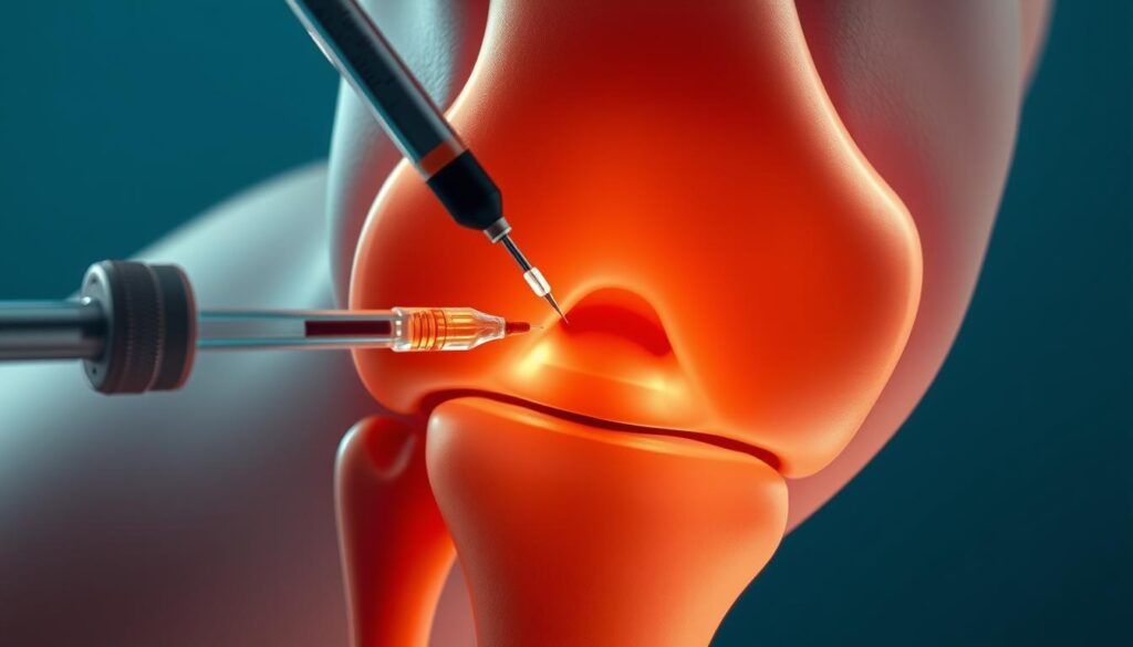
    Ultrasound-guided knee injections

    Reduced Risk of Complications

    By using ultrasound to guide the needle, the risk of complications such as nerve damage or infection is significantly reduced. The real-time imaging allows for adjustments to be made during the procedure, ensuring the needle is placed correctly and avoiding potential complications.

    BenefitsDescriptionOutcome
    Improved PrecisionReal-time ultrasound guidance ensures accurate needle placement.Effective delivery of medication to the target area.
    Reduced RiskMinimized risk of nerve damage and infection.Safer procedure with fewer complications.
    Enhanced EfficacyCortisone is delivered directly to the source of pain or inflammation.Significant relief from pain and inflammation.

    What to Expect During the Procedure

    When undergoing an ultrasound-guided knee injection, several steps are involved to ensure a safe and effective procedure.

    Preparation Steps Before the Injection

    Before the injection, the knee area is cleaned and prepared to minimize the risk of infection. Next, the joint is anesthetized using a local numbing agent like lidocaine to make the procedure more comfortable.

    Preparation is key to a successful ultrasound-guided knee injection. The use of ultrasound imaging allows for real-time visualization, ensuring that the injection is delivered precisely to the targeted area.

    The Injection Experience

    Once the area is numb, the doctor will insert a needle into the knee joint, guided carefully by ultrasound imaging to ensure precise placement. Patients may feel a slight pinch or pressure during the injection, but this is typically minimal due to the local anesthesia.

    The entire procedure is relatively quick, usually taking only a few minutes to complete. The precision of ultrasound guidance enhances the effectiveness of the knee joint injection procedure.

    Aftercare Following Knee Injections

    Post-procedure care is a vital component of knee pain relief options, including ultrasound-guided knee injections. Proper aftercare can significantly influence the effectiveness of the treatment and the patient’s recovery process.

    Immediate Post-Procedure Care

    After receiving knee injection therapy, patients may experience mild soreness, warmth, or swelling at the injection site. These symptoms are typically temporary and resolve within a day or two.

    • Rest the knee and avoid strenuous activities for at least 24 to 48 hours.
    • Apply ice to reduce swelling if necessary.
    • Monitor the injection site for signs of infection, such as increased redness, swelling, or pain.

    knee injection therapy aftercare

    Long-term Recovery Tips

    For optimal recovery and to achieve the best results from knee injection therapy, consider the following long-term tips:

    1. Gradually resume normal activities and exercises as advised by your healthcare provider.
    2. Maintain a healthy weight to reduce stress on the knee joint.
    3. Engage in physical therapy or exercises recommended by your healthcare provider to improve knee strength and flexibility.

    By following these aftercare guidelines, patients can enhance their recovery and improve the overall effectiveness of their knee pain relief options.

    Potential Risks and Side Effects

    While ultrasound-guided knee injections are considered safe, it’s essential to be aware of the potential risks and side effects associated with this procedure.

    These injections, often used as a minimally invasive knee injections treatment for various knee conditions, including knee arthritis treatment, are generally well-tolerated. However, understanding the possible complications can help manage expectations and improve patient outcomes.

    Common Side Effects to Anticipate

    Most patients undergoing ultrasound-guided knee injections experience minimal side effects. Common issues may include:

    • Temporary pain or discomfort at the injection site
    • Mild swelling or redness
    • A temporary flare-up of knee pain

    These side effects are typically mild and resolve on their own within a few days.

    Rare but Serious Complications

    Serious adverse events are rare but can occur. These include:

    • Allergic reactions to the injected medication
    • Infections, which can be serious and require prompt medical attention
    • Joint flare-ups characterized by increased pain, warmth, or swelling
    • In very uncommon cases, a pseudoseptic reaction can happen, marked by significant redness and swelling that typically improves with rest and ice.

    It’s crucial for patients to be aware of these potential complications and to seek medical help if they experience any severe or persistent symptoms.

    By understanding the potential risks and side effects, patients can make informed decisions about their treatment. It’s also important to follow post-procedure care instructions to minimize the risk of complications.

    Candidates for Ultrasound-Guided Knee Injections

    Identifying the right candidates for ultrasound-guided knee injections is crucial for effective knee pain management. This treatment is particularly beneficial for individuals who have not responded well to conservative treatments.

    Who Can Benefit Most from This Treatment?

    Patients with mild to moderate knee osteoarthritis are typically considered good candidates for ultrasound-guided knee injections, particularly those who have not achieved adequate relief from conservative treatments such as physical therapy, oral medications, or other non-surgical interventions. Hyaluronic acid injections, also known as viscosupplementation, are commonly used for these patients.

    Age and Health Considerations

    Age and overall health are significant factors in determining suitability for ultrasound-guided knee injections. While older adults with osteoarthritis may benefit significantly, younger patients with specific knee conditions may also be candidates. Healthcare providers assess each patient’s health status, including the presence of any comorbidities, to determine the appropriateness of this treatment.

    For instance, patients with severe knee damage or certain health conditions may not be ideal candidates. Therefore, a thorough evaluation is necessary to ensure that the benefits of ultrasound-guided knee injections outweigh the potential risks.

    Cost of Ultrasound-Guided Knee Injections

    For individuals considering knee joint injection procedures, understanding the associated costs is essential for making informed decisions. The cost of ultrasound-guided knee injections can vary based on several factors, including the location, healthcare provider, and specific requirements of the procedure.

    Price Range in the United States

    The cost of viscosupplementation, a common treatment involving ultrasound-guided knee injections, can range widely across the United States. On average, the price for a single injection can be between $300 to $1,000. However, the total cost may be higher when considering the series of injections often required for effective treatment.

    Factors influencing the cost include:

    • The expertise and fees of the healthcare provider
    • Geographic location, with urban areas typically having higher costs
    • The specific type of injection or medication used

    Insurance Coverage and Options

    Insurance coverage for ultrasound-guided knee injections varies among providers and policies. Some insurance plans may cover part or all of the procedure if deemed medically necessary, while others may not cover it at all. It’s crucial for patients to check their insurance coverage before undergoing the treatment.

    Options for managing the cost include:

    • Discussing financing options with the healthcare provider
    • Exploring patient assistance programs if available
    • Comparing prices among different providers

    Understanding the financial aspects and exploring available options is crucial for patients considering this treatment. By being informed, patients can better navigate the costs associated with ultrasound-guided knee injections and make decisions that align with their financial situation and healthcare needs.

    Finding a Qualified Provider

    To ensure the best outcomes from ultrasound imaging for knee injections, it’s vital to find a skilled healthcare professional. The expertise of the provider can significantly impact the effectiveness of the treatment.

    Questions to Ask Your Provider

    Before undergoing the knee injection therapy, it’s essential to ask your provider a few critical questions to gauge their expertise and the quality of care you can expect.

    • What experience do you have with ultrasound-guided knee injections?
    • Can you explain the procedure and what I can expect during and after the injection?
    • What are the potential risks and complications associated with this treatment?
    • How will you ensure the accuracy and effectiveness of the injection?

    Asking these questions can help you understand the provider’s level of expertise and their approach to your care. As noted by a medical professional, “Precision is key when it comes to injections; it’s not just about injecting medication, but doing so with accuracy to achieve the desired therapeutic effect.

    Important Credentials to Look For

    When searching for a provider, certain credentials can indicate a higher level of competence in administering ultrasound-guided knee injections. Look for providers who are:

    1. Board-certified in their specialty, such as orthopedics or physical medicine and rehabilitation.
    2. Experienced in performing ultrasound-guided procedures.
    3. Familiar with the latest techniques and technologies in knee injection therapy.

    For more information on ultrasound-guided joint injections and to find a qualified provider, it’s advisable to consult reputable medical resources and professional directories.

    Alternatives to Ultrasound-Guided Knee Injections

    Not everyone may be suited for or prefer ultrasound-guided knee injections, making alternative treatments worth considering. For patients seeking knee pain relief options, there are various other treatments available.

    Other Injection Techniques

    Besides ultrasound-guided injections, other injection techniques can be considered for knee pain management. These include:

    • Fluoroscopically guided injections, which use X-ray guidance to ensure accurate placement.
    • Blind injections, although less accurate, may still offer relief for some patients.
    • Platelet-rich plasma (PRP) therapy, which involves injecting platelet-rich plasma derived from the patient’s own blood to stimulate healing.

    As noted by medical professionals, “The choice of injection technique depends on the specific condition, patient preferences, and the expertise of the healthcare provider.”

    “The key to successful treatment lies in selecting the most appropriate intervention based on a thorough assessment of the patient’s condition.”

    Non-Injection Treatments for Knee Pain

    For those who prefer to avoid injections or are not candidates for them, several minimally invasive knee injections alternatives and non-invasive treatments are available:

    1. Physical therapy to improve knee function and reduce pain.
    2. Medications such as NSAIDs or corticosteroids to manage pain and inflammation.
    3. Alternative therapies like acupuncture or chiropractic care.
    4. Lifestyle modifications, including weight loss and exercise, to reduce stress on the knee.

    It’s essential for patients to discuss these options with their healthcare provider to determine the best course of treatment for their specific needs.

    Conclusion: The Value of Ultrasound Guidance

    Ultrasound-guided knee injections have emerged as a valuable treatment option for knee pain, particularly for individuals suffering from osteoarthritis. By leveraging the precision of ultrasound technology, healthcare providers can deliver injections with greater accuracy, enhancing the effectiveness of knee arthritis treatment.

    Key Benefits and Effectiveness

    The benefits of ultrasound-guided knee injections include improved precision, reduced risk of complications, and a favorable safety profile. These advantages make them an attractive choice for patients seeking knee pain management strategies.

    Discussing Treatment Options with Healthcare Providers

    Patients experiencing knee pain should discuss ultrasound-guided knee injections with their healthcare providers to determine if this treatment is suitable for their specific needs. By exploring this option, individuals can make informed decisions about their knee pain management strategies and potentially improve their quality of life.

    FAQ

    What is an ultrasound-guided knee injection?

    An ultrasound-guided knee injection is a minimally invasive procedure that uses ultrasound imaging to guide the injection of medication into the knee joint, providing precise delivery of treatment for various knee conditions, including osteoarthritis, tendonitis, and bursitis.

    How does ultrasound guidance improve the accuracy of knee injections?

    Ultrasound guidance allows healthcare providers to visualize the needle and the target area in real-time, ensuring accurate placement of the medication and reducing the risk of complications, thereby improving the effectiveness of the treatment.

    What are the benefits of using ultrasound-guided knee injections for osteoarthritis treatment?

    The benefits include improved precision, reduced risk of complications, and enhanced effectiveness in managing osteoarthritis symptoms, such as pain and limited mobility, allowing patients to engage in exercise therapy and other rehabilitation programs.

    What can I expect during an ultrasound-guided knee injection procedure?

    You can expect the procedure to involve preparation steps, such as cleaning and numbing the area, followed by the injection itself, which is typically performed under local anesthesia, and the use of ultrasound imaging to guide the needle to the correct location.

    How should I care for my knee after an ultrasound-guided injection?

    After the procedure, it’s essential to follow immediate post-procedure care instructions, such as applying ice to reduce swelling, and long-term recovery tips, including gradually resuming normal activities and exercises, to ensure a smooth recovery and optimal pain relief.

    What are the potential risks and side effects of ultrasound-guided knee injections?

    Common side effects include temporary pain, swelling, or redness at the injection site, while rare but serious complications, such as infection or nerve damage, can occur; it’s crucial to discuss these risks with your healthcare provider.

    Who is a suitable candidate for ultrasound-guided knee injections?

    Suitable candidates include individuals with knee pain caused by osteoarthritis, tendonitis, or bursitis, who have not responded to conservative treatments or require a more targeted approach; age and overall health will be considered by your healthcare provider.

    How much do ultrasound-guided knee injections cost in the United States?

    The cost can vary depending on factors, such as location, provider, and insurance coverage; it’s essential to check with your insurance provider to determine the extent of coverage and any out-of-pocket expenses.

    How can I find a qualified healthcare provider for ultrasound-guided knee injections?

    To find a qualified provider, look for credentials, such as specialized training in musculoskeletal ultrasound and injection techniques, and ask questions, such as their experience with the procedure and their approach to patient care.

    Are there alternatives to ultrasound-guided knee injections for knee pain management?

    Yes, alternatives include other injection therapies, such as corticosteroid or hyaluronic acid injections, and non-injection treatments, such as physical therapy, bracing, or pain medications; your healthcare provider can help determine the best approach for your specific condition.

    Can ultrasound-guided knee injections be used in conjunction with other treatments?

    Yes, ultrasound-guided knee injections can be used as part of a comprehensive treatment plan, which may include other therapies, such as physical therapy, exercise programs, or pain management strategies, to achieve optimal pain relief and improved joint function.

  • Chronic pain causes

    Chronic pain causes

    Chronic pain affects millions of people around the world. It is a pain that lasts for months or even years. There are many reasons why someone might have chronic pain. In this article, we will look at some of the main causes of chronic pain.

    Key Takeaways

    • Chronic pain can be caused by problems with the nerves, such as nerve damage or central sensitization.
    • Inflammation from autoimmune diseases, infections, or chronic inflammatory conditions can lead to long-term pain.
    • Musculoskeletal issues like arthritis, fibromyalgia, and muscle strain are common causes of chronic pain.

    Neurological Factors

    Nerve Damage

    Nerve damage can lead to chronic pain. When nerves are injured, they might send incorrect signals to the brain, causing pain even without a clear reason. This can happen due to accidents, surgeries, or diseases like diabetes.

    Central Sensitization

    Central sensitization is when the nervous system becomes overly sensitive. This heightened sensitivity makes even normal touch or movement feel painful. It can result from ongoing pain or injury, making the body react more strongly to pain signals.

    Neuropathic Pain

    Neuropathic pain is a type of pain caused by nerve problems. It often feels like burning, tingling, or shooting pain. People with conditions like shingles or multiple sclerosis might experience this. Treating neuropathic pain can be challenging, as it doesn’t always respond to regular painkillers.

    Chronic pain from neurological factors can be hard to manage, but understanding the causes can help in finding the right treatment.

    Inflammatory Causes

    Autoimmune Diseases

    Autoimmune diseases occur when the body’s immune system mistakenly attacks its own tissues. This can lead to chronic pain as the inflammation persists. Conditions like rheumatoid arthritis and lupus are common examples. Managing these diseases often requires long-term treatment.

    Infections

    Infections can also cause chronic pain. When the body fights off an infection, inflammation can linger, causing ongoing discomfort. For instance, some bacterial or viral infections can lead to conditions like post-infectious arthritis. It’s important to treat infections promptly to avoid long-term issues.

    Chronic Inflammatory Conditions

    Chronic inflammatory conditions, such as Crohn’s disease or ulcerative colitis, involve ongoing inflammation in the body. This persistent inflammation can cause significant pain and discomfort. Effective management of these conditions often includes medication, lifestyle changes, and sometimes surgery.

    Chronic inflammation can severely impact quality of life, making daily activities challenging.

    Musculoskeletal Issues

    Arthritis

    Arthritis is a common cause of chronic pain. It involves inflammation of the joints, which can lead to stiffness, swelling, and pain. Osteoarthritis and rheumatoid arthritis are the most common types. Managing arthritis often requires a combination of medication, physical therapy, and lifestyle changes.

    Fibromyalgia

    Fibromyalgia is a condition characterized by widespread musculoskeletal pain. People with fibromyalgia often experience fatigue, sleep disturbances, and memory issues. The exact cause is unknown, but it is believed to involve a combination of genetic and environmental factors. Treatment typically includes medication, exercise, and stress management techniques.

    Muscle Strain

    Muscle strain occurs when muscles are overstretched or torn. This can happen due to overuse, improper use, or injury. Symptoms include pain, swelling, and limited movement. Rest, ice, compression, and elevation (RICE) are common treatments for muscle strain. In severe cases, physical therapy may be needed to regain strength and flexibility.

    Chronic musculoskeletal issues can significantly impact daily life, making it essential to seek appropriate treatment and management strategies.

    Psychological Influences

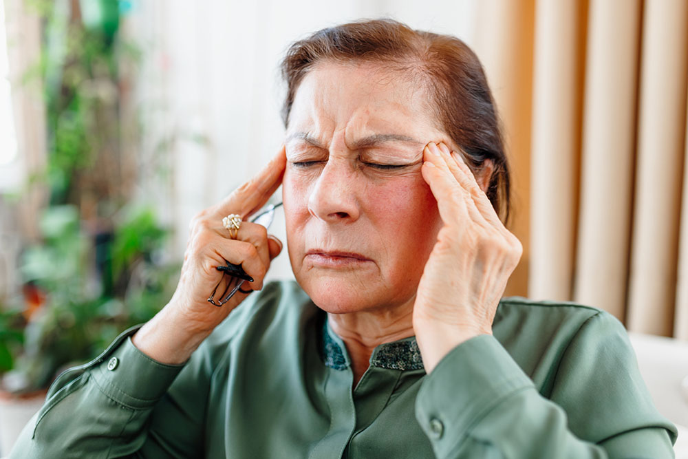
    person in pain

    Stress and Anxiety

    Stress and anxiety can make chronic pain worse. When you’re stressed, your body releases chemicals that can increase pain. Managing stress is important for people with chronic pain.

    Depression

    Depression and chronic pain often go hand in hand. Feeling sad or hopeless can make pain feel worse. It’s important to treat both the pain and the depression to feel better.

    Trauma and PTSD

    People who have been through trauma or have PTSD might feel more pain. The brain and body remember the trauma, which can make pain last longer. Getting help for trauma can also help with pain.

    It’s important to understand that the mind and body are connected. Taking care of your mental health can help reduce chronic pain.

    Conclusion

    Chronic pain can come from many different causes. It might be from an old injury, a disease, or even stress. Understanding what causes chronic pain is important. It helps doctors find the best ways to treat it. If you or someone you know has chronic pain, it’s important to talk to a doctor. They can help find the cause and suggest ways to feel better. Remember, you don’t have to live with pain. There are many treatments that can help.

  • AI promises to be a helping hand for this orthopedic hand surgeon

    AI promises to be a helping hand for this orthopedic hand surgeon

    Like many of my orthopedic colleagues, I’m a bit of a skeptic when it comes to flashy new technology. I didn’t know what to expect when I decided to test a second AI-powered ambient listening solution last fall. I’d tried one before that didn’t quite live up to expectations. Would NextGen® Ambient Assist live up to its promise of helping me save time by reducing the burden of documentation? I quickly found out that this solution was the real deal.

    For about 1/10th the cost of a scribe, Ambient Assist interprets patient-provider conversations in real time to summarize appointments and deliver SOAP notes directly into the EHR. With one tap, Ambient Assist “listens” to the entire patient encounter and then uses AI to generate an accurate medical note in about 30 seconds. I can review and edit it immediately or at the end of the day—no copy-pasting required because it’s fully integrated into my EHR.

    The technology is advanced enough to remove mundane conversations about the weather or a patient’s weekend plans, so visit notes remain focused on medically important details. Audio and transcripts are never stored or retained, providing an additional measure of security.

    I’ve discovered my own set of “best practices” that help me customize the solution to my preferred workflow. For example, before I see a patient, I like to review their x-rays and dictate my treatment plan notes. This allows me to enter each appointment with more context. I’m careful not to use medical jargon when I’m with patients, as I feel it could cause confusion or unnecessary worry. Instead, I dictate the physical exam as I leave the room and quickly review the note on my phone or iPad.

    I have used Ambient Assist for over 500 appointments since I implemented it in the fall of 2023. My patients appreciate the shorter wait times for appointments: I can see at least 5 more patients per day, and my family appreciates the extra time with me. Meanwhile, I feel more energetic and effective at work and at home.

    Can AI really revolutionize modern medicine? For me, it has been a real helping hand.

    Keith Michael Nord, MD

    Meet The Doctors

    Source link

  • You Are Not Alone – Depression, Stress and Anxiety in the Locker Room

    You Are Not Alone – Depression, Stress and Anxiety in the Locker Room

    Trends in student-athlete mental health in the National College Health Assessment, 2011-2019.

    Edwards B, Froehle AW and Fagan SE. J Athl training. 2023,58(4):361-373. doi: 10.4085/1062-6050-0586.21.

    Full text available for free

    Take home message

    College athletes and non-athletes are under a great deal of stress. Reported mental health symptoms and diagnoses are increasing, with 1 in 3 college students reporting a recent diagnosis. Fortunately, more college students are reporting receiving mental health information from institutions and treatment-seeking behaviors over time.

    Background

    Awareness of mental health issues is increasing among the general public, including college athletics. Educational efforts and access to mental health resources have increased, but it is unclear whether these efforts have improved student-athletes’ mental health and treatment seeking.

    Study Objective

    Edwards and colleagues analyzed 8 years of survey data to examine how the mental health of student-athletes, compared to their non-athlete peers, changed between 2011 and 2019.

    Methods

    Researchers obtained data from the National College Health Assessment. The data, which spanned 8 academic years, were reported anonymously by students at participating institutions. Respondents were classified as “athletes” or “non-athletes” based on their responses to specific survey questions. Researchers excluded students who participated only in club or intramural sports. The survey included “yes” or “no” questions grouped into 5 outcome groups: 1) “recent mental health symptoms,” 2) “recent mental health diagnosis,” 3) “mental health treatment seeking behavior,” 4) “mental health information received from institutions,” and 5) “recent impact of mental health on academic performance.”

    Results

    A total of 502,780 students from 607 unique institutions completed the surveys and met the inclusion criteria. This included over 54,400 athletes and 448,300 non-athletes. The main results are described in the table, which show that more students received mental health information from institutions and sought treatment or would consider treatment over time. Meanwhile, more students reported recent mental health symptoms/diagnoses and effects on academic performance over time. Student-athletes were less likely to report mental health symptoms/diagnoses and treatment-seeking behavior. However, more student-athletes reported mental health information from institutions.

    Changes over timeDifferences between groups
    Recent mental health symptomsIncreased over timeAthletes < Non-athletes
    Recent mental health diagnosisIncreased over timeAthletes < Non-athletes
    Behavior Seeking Treatment for Mental HealthImproved over timeAthletes < Non-athletes
    Receiving mental health information from institutionsImproved over timeAthletes > Non-athletes
    The Recent Impact of Mental Health on Academic PerformanceIncreased over timeAthletes < Non-athletes

    Positions

    More than 1 in 5 students reported symptoms of depression. A majority of students reported feeling very sad, very lonely, exhausted, or overwhelmed. Over the 8 years, we went from 1 in 5 students reporting a diagnosis of a mental health condition to 1 in 3 students. It is encouraging that treatment seeking behaviors and access to mental health information have increased over time. This study cannot address whether the increased symptoms/diagnoses of mental health are due to increased prevalence or increased ease of reporting. As access to educational materials increases, it would stand to reason that reporting of mental health issues would also increase. While the trends over time are encouraging, the overall numbers are still discouraging and we need to do more to encourage student-athletes to seek treatment.

    Clinical implications

    Clinicians need to know that athletes and non-athletes are under immense pressure due to the college experience. This requires clinicians to be fully aware of the signs and symptoms of mental health issues and have an appropriate and empathetic plan to help their athletes get the right treatment. We must also continue to reduce stigma and increase awareness among athletes that treatments are available.

    Questions for discussion

    What changes have you observed regarding mental health awareness, recognition, and treatment in student-athletes? What specific strategies do you think have had the most impact on mental health awareness, recognition, or treatment in student-athletes?

    Written by Kyle Harris
    Reviewed by Jeffrey Driban

    related posts

    You are not alone – Depression, stress and anxiety in the sports medicine team
    Specializing in sports in high school may increase risk of depression
    Many NCAA Clinicians Fail to Screen for Mental Health
    Fresh air can improve performance.
    Want to get better? Take a nap!

    Evidence-Based Assessment of Concussion Course - 5 EBP CEUs

    Source link

  • Chronic Pain and the Nervous System

    Chronic Pain and the Nervous System

    Experiencing pain can be a part of life, but living with chronic pain is far from normal. It can be exhausting and affect your ability to live an active life, perform tasks, and get the rest you need. Persistent pain can lead to anxiety, insomnia, and depression. Physical therapy can often reduce or eliminate pain so you can get back to living your life.

    What is chronic pain syndrome?

    There are two primary categories of pain. Acute pain is often a symptom of an underlying health condition. Recovery can take days to weeks and the pain subsides. Chronic pain, or chronic pain syndrome, on the other hand, usually lasts six months or longer. It occurs in addition to the pain caused by the health condition, which may or may not have healed. It is independent of the original injury or illness that began and can persist long after the pain has gone.

    Woman suffers from chronic pain; migraine.

    Chronic pain can present in different forms, such as:

    • Back and neck pain
    • Joint or arthritis pain
    • Cancer pain near a tumor
    • Pain in the testicles
    • Headache, including migraine
    • Pervasive muscle pain (including fibromyalgia)
    • Persistent pain in scar tissue
    • Neurogenic pain due to damage to the nervous system

    More than 20% of Americans have chronic pain that affects your work and lifestyle.

    What causes chronic pain?

    Sometimes chronic pain has a clear cause, such as a long-term illness, cancer, or arthritis. Illness can make your body more susceptible to pain, even after you recover. Injuries, including broken bones, sprains, or infections, can also leave you with chronic pain.

    Pain that does not seem to be related to a physical illness or injury is psychogenic painIt is believed that anxiety, stress and depression lead to low endorphin levels in the blood.

    Regardless of the specific cause, pain is a function of the nervous system. Nerves around an injury site sense the damage and send signals to the spinal cord and brain. The brain registers the pain and the nervous system responds. Muscles tense, emotions run high, and the nervous system becomes hypervigilant.

    This high alert state is beneficial in acute pain. It tells the body that something is wrong, allowing us to protect ourselves from further injury. Once the damage or disease heals, the nervous system functions normally.

    Woman suffers from chronic back pain.

    Symptoms of chronic pain syndrome

    When the nervous system remains in a persistent state of high alert, it often becomes hypersensitive. It can continue the pain even after the original damage has healed. People with chronic pain describe it in different ways:

    • To beat
    • Stiffness
    • To shoot
    • Painful
    • Burning
    • pinch
    • Stinging

    Poor sleep, mood swings, irritability and fatigue often accompany pain, which increases stress levels and nervous system reactivity. As pain increases, a new vicious circle begins.

    PT, Jeff Moyer helps a patient with elbow pain and is a good reason to choose physical therapy first.

    7 Common Treatment Methods for Chronic Pain

    Chronic pain can limit your quality of life and lead to significant long-term health problems. It can make managing daily tasks, maintaining relationships, and maintaining financial stability challenging. Chronic pain is uniquely personal, and finding an effective treatment becomes a priority.

    Your biology and personal history can influence which pain therapies provide relief. However, the treatment combination for one person may not work for another.

    Physiotherapy icon for the topic of chronic pain.

    1. Physiotherapy

    Physical therapy is a highly personalized treatment regimen. Goals may include increased mobility and reduced pain to help you regain full function. Physical therapy includes passive and active treatments. Your program may begin with passive treatments and progress to active methods. Physical therapists play an important role in pain management through various therapies and daily living techniques. Various forms of exercise, including strength, flexibility, balance, endurance, power, and agility, are used to promote and restore normal function, as well as for pain management. While opioids only mask the sensation of pain, physical therapists can also treat pain through hands-on care. Techniques include manipulation, joint and soft tissue mobilization, dry needling, and cupping. This helps to address the pain and target the root of the problem.

    Icon for water; soak foot in water.

    2. Contrast therapy

    Physiotherapists often alternate between cold and hot therapies. Cold therapy constricts blood vessels. Inflammation decreases when muscles contract and pain signals are reduced. Heat therapy increases circulation by dilating blood vessels. Flooding areas with nutrients and oxygen reduces muscle pain and allows the body’s cells to shed waste. Contrast therapy can reduce inflammation and increase circulation, which loosens tight, sore muscles.

    Icon for deep tissue massage.

    3. Deep tissue massage

    Muscle spasms and chronic muscle tension can build up from the stress of a typical daily routine. Cramps can also occur from sprains and strains. The direct pressure and friction applied during massage can release tension in the muscles, ligaments and tendons.

    Icon for ESTIM.

    4. TENS (transcutaneous electrical nerve stimulation)

    A TENS unit uses varying intensities of electrical current to stimulate muscles, which helps to reduce the occurrence and severity of muscle spasms. It can also increase the production of endorphins, the body’s natural painkillers. You can use just the unit that’s in your physical therapist’s office, or you can have a smaller unit made for home use.

    Ultrasound icon for the topic of chronic pain.

    5. Ultrasound

    This deep heating technique is applied to your skin. As the heat waves penetrate your tissues, it is believed to improve blood flow and circulation. An ultrasound helps to reduce cramps, swelling, spasms, pain and stiffness.

    Clipboard for the topic drug therapy.

    6. Drug therapy

    Over-the-counter medications may be enough to relieve mild pain caused by stiffness, inflammation, and muscle soreness. Typical drug therapy includes ibuprofen, aspirin, naproxen, and acetaminophen (Tylenol). Topical pain relievers that come in sprays, creams, and lotions may also provide temporary relief. Stronger medications for debilitating pain range from prescription NSAIDs, muscle relaxants, and anti-anxiety medications to opioids and steroid injections.

    Exercise icon for the topic of chronic pain.

    7. Exercise

    Exercise is an active treatment and an essential part of the PT toolkit for improving mobility and strength. The brain stops sending constant alerts to the nervous system as the pain subsides. You and your physical therapist will work together to develop an exercise regimen to relieve pain. Examples of low-impact exercises include:

    • Swimming
    • Stretching and strengthening exercises
    • Cycling
    • To walk
    • Tai Chi
    • Pilates
    • Yoga

    Exercise makes you stronger and trains your nervous system to trust that exercise is safe, allowing it to go from high alert to normal functioning.

    If you have pain that comes and goes, physical therapy can help you understand the cause and how to treat it. Everyone responds differently to physical therapy. Contact Us Make an appointment today and learn more about the best treatment options.

    Source link

  • When to See a Specialist for Low Back Pain

    When to See a Specialist for Low Back Pain

    A close-up of a woman holding her lower back and struggling with lower back pain

    Lower back pain is a common problem that many people face at some point in their lives.

    Coping with low back pain can be challenging both physically and emotionally. Knowing when to seek specialist care can make a big difference in your recovery and pain relief.

    Discover below some signs that indicate you need to call a professional.

    What is low back pain?

    The lower back, also called the lumbar region, bears a large portion of the body’s weight and is prone to strain and injury. Just causes of lower back pain Involving:

    • Muscle strain or sprain
    • Hernias
    • Degenerative disc disease
    • Spinal stenosis
    • Arthritis
    • Osteoporosis

    When considering consulting a specialist

    Although mild lower back pain can often be treated with rest, ice, and over-the-counter pain relievers, there are certain signs and symptoms that indicate the need for specialized medical attention.

    You should consider seeing a specialist, such as an orthopedic physician, if you experience any of the following:

    Persistent pain

    See a specialist if your lower back pain gets worse over time or if the pain does not go away within a few days with over-the-counter medications.

    Pain radiating to the leg

    Pain radiating from the lower back to one or both legs, often accompanied by numbness or tinglingmay indicate nerve compression and requires immediate investigation.

    Difficulty performing daily activities

    If lower back pain is causing you difficulty performing daily tasks such as walking, standing, or bending over, it is imperative to seek medical attention to identify and address the underlying cause.

    History of trauma

    If your lower back pain is the result of a recent injury, such as a fall or car accident, it is important to see a specialist to rule out serious injuries and prevent long-term complications.

    Progressive weakness or loss of bladder/bowel control

    These symptoms may indicate a serious underlying condition, such as cauda equina syndromerequiring immediate medical intervention.

    Schedule a Consultation for Low Back Pain in Indiana

    If you experience persistent lower back pain and are in a Merrillville, Highland, Hobartor the surrounding areas, please feel free to contact Bone & Joint Specialists.

    Our experienced orthopedic physicians strive to provide you with personalized and compassionate care. We strive to help you relieve your symptoms and improve your quality of life.

    Contact us on 219-795-3360 Unpleasant Schedule your consultation Today.

    Source link

  • Fibromyalgia Quiz | Signs, Symptoms & Treatments – AOTC Jax


    At Arthritis and Osteoporosis Treatment Center, we are committed to the compassionate treatment of all patients. That’s why, this Fibromyalgia Awareness Month, we want to help you learn the signs, symptoms, and available treatment options for this condition.

    Take our fibromyalgia test below and don’t forget to consult our doctors for the best, personalized care.

    What You Need to Know About Fibromyalgia

    What is fibromyalgia?

    Fibromyalgia is a complex and often misunderstood chronic pain condition that affects the muscles and bones. It can cause widespread pain, sleep problems, mood swings, and more. While the exact cause of fibromyalgia remains unknown, it is believed to be a combination of genetic, environmental, and psychological factors.

    Symptoms of Fibromyalgia

    Fibromyalgia is associated with several symptoms that can have a major impact on your daily life, including:

    Pain and stiffness

    One of the most common symptoms of fibromyalgia is pain that spreads throughout the body, often accompanied by stiffness.

    Fatigue and tiredness

    Even after a full night’s sleep, people with fibromyalgia often wake up feeling exhausted and lethargic.

    Trouble sleeping

    Sleep disorders are common in fibromyalgia patients. They often have difficulty falling asleep, staying asleep, and restorative sleep.

    Memory and concentration problems

    Cognitive problems, sometimes called ‘fibro fog’, can cause problems with memory, concentration and mental clarity.

    Headache/Migraine

    Many people with fibromyalgia experience frequent headaches or migraines, which only adds to their discomfort and challenges.

    Is it fibromyalgia or something else?

    Research shows that fibromyalgia is often misdiagnosed. At AOTC, we take the time and care with each patient to ensure the correct diagnosis is made. Once patients begin treatment with our experienced rheumatologists, they finally begin to feel better.

    Click here to learn about other common conditions with symptoms similar to fibromyalgia. Remember to always consult a doctor for a proper diagnosis.

    Best Treatment Options for Fibromyalgia

    While there is no cure for fibromyalgia, several treatment options are available to help manage symptoms and improve quality of life. Treatment plans are often individualized and may include a combination of the following:

    Medicines

    Over-the-counter and prescription pain relievers and certain antidepressants are often prescribed to relieve pain, improve sleep, and control mood. Some medications used to treat seizures, known as anticonvulsants, have also been shown to effectively reduce pain associated with fibromyalgia.

    Lifestyle changes

    Regular physical activity, such as walking, swimming, or yoga, can help improve strength, flexibility, and overall well-being. Stress management techniques such as meditation and deep breathing can also help reduce stress and relieve symptoms.

    Therapy

    Cognitive behavioral therapy (CBT) helps develop coping strategies for pain, sleep, and mood problems. Physical therapy can also improve mobility and reduce pain.

    Visit the Arthritis & Osteoporosis Treatment Center today

    At Arthritis and Osteoporosis Treatment Center, we understand the challenges of living with fibromyalgia. This Fibromyalgia Awareness Month and every month, our compassionate team of healthcare professionals is dedicated to providing comprehensive care tailored to your individual needs. Learn more about our services or contact us today to schedule a consultation and take the first step toward managing your fibromyalgia symptoms.

    Take the fibromyalgia quiz below:

    Fibromyalgia Quiz: Do I Have Fibromyalgia?

    Still not sure if an appointment with a fibromyalgia doctor is right for you? Answer these questions to help determine if it’s time to discuss your symptoms and treatment options with a rheumatologist.


    Source link