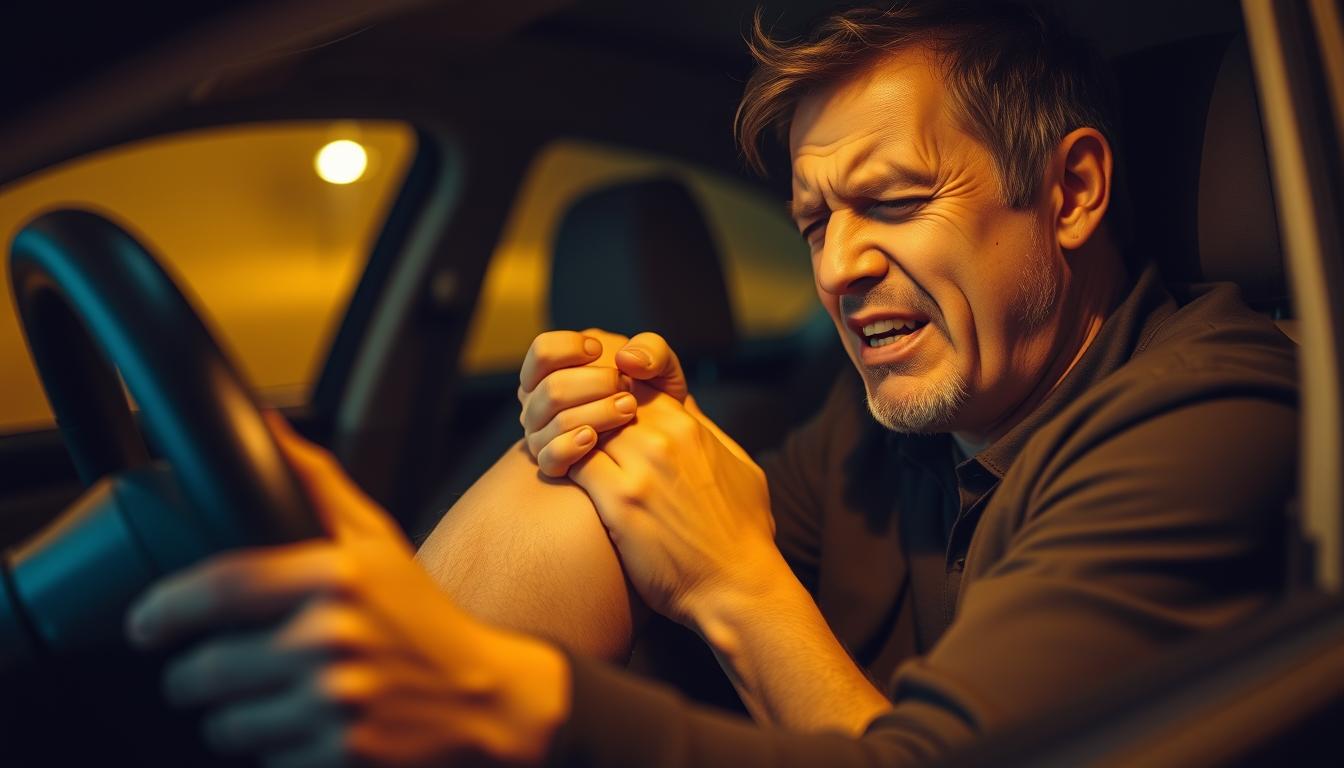
Have you ever hit the slopes with enthusiasm only to be sidelined by knee pain after a day of skiing?
Skiing places unique biomechanical demands on the knee joint, making it particularly vulnerable to both acute injuries and overuse conditions.
Understanding the causes, symptoms, and treatment options for knee pain is essential for both recreational and professional skiers to enjoy the sport without long-term damage.
Key Takeaways
- Understanding the biomechanical stress skiing places on the knee joint.
- Recognizing the difference between normal soreness and serious knee injuries.
- Immediate relief strategies for knee pain.
- Long-term treatment options for skiing-related knee injuries.
- Effective prevention techniques to minimize knee stress while skiing.
Understanding Why Skiing Causes Knee Pain
Knee pain is a common complaint among skiers, and understanding its causes is crucial. Skiing involves various movements that can put stress on the knee joint, leading to pain and discomfort.
The Biomechanics of Skiing and Knee Stress
Skiing involves dynamic movements that can strain the knee. The biomechanics of skiing play a significant role in knee stress. As skiers navigate through turns, their knees are subjected to twisting forces that can affect the ligaments and cartilage. The forward-leaning position in ski boots also loads the muscles, particularly in the quadriceps and calves, contributing to muscle soreness.
Distinguishing Between “Good Pain” and “Bad Pain”
Not all knee pain after skiing indicates a serious problem. It’s essential to distinguish between normal soreness (“good pain”) and potential injury (“bad pain”). Good pain typically presents as general muscle soreness in the patellar tendon, hamstrings, quadriceps, and calves, especially early in the ski season. This type of pain usually improves with time on the slopes and responds well to basic self-care measures.
- Good pain is associated with deconditioning and improves over time with continued skiing and proper care.
- Bad pain, on the other hand, may be sharp, persist longer, or be accompanied by swelling or mechanical symptoms like grinding or clicking of the kneecap.
- Front knee pain with grinding sensations may indicate cartilage damage on the undersurface of the patella.
- Pain on the inside of the knee with mechanical symptoms could suggest meniscus damage or ligament issues.
Recognizing these distinctions early can help prevent minor issues from developing into chronic problems that might require more intensive treatment. By understanding the causes of knee pain and distinguishing between “good” and “bad” pain, skiers can take appropriate measures to manage their discomfort and enjoy their time on the slopes.
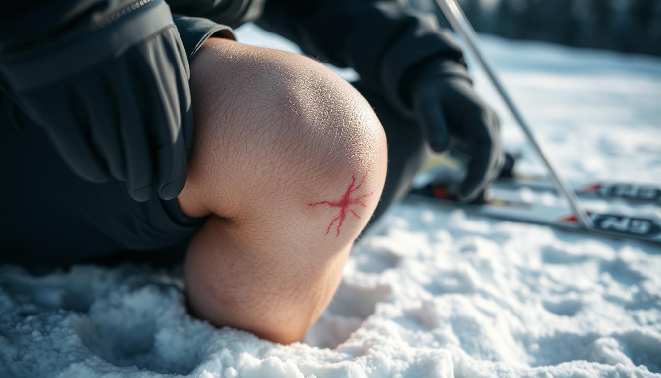
Common Types of Knee Injuries from Skiing
The thrill of skiing comes with a risk: knee injuries that can be painful and debilitating. Skiing puts a significant amount of stress on the knee joint, leading to various types of injuries. Understanding these common injuries is crucial for effective prevention and treatment.
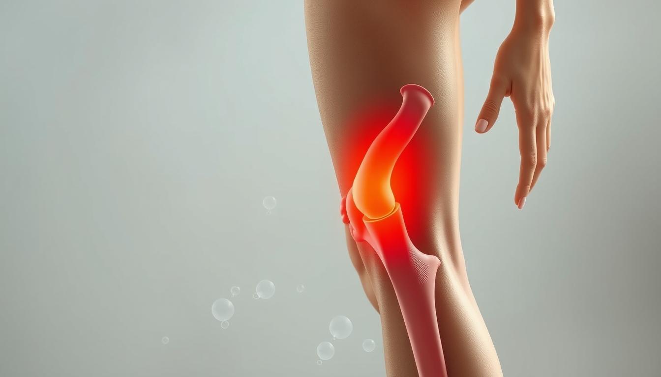
Ligament Injuries: ACL, MCL, and Other Crucial Structures
Ligament injuries are common among skiers, with the Anterior Cruciate Ligament (ACL) and Medial Collateral Ligament (MCL) being particularly susceptible. The ACL is crucial for knee stability, and injuries to this ligament can be severe, often requiring surgical intervention. MCL injuries, on the other hand, can range from mild to severe and typically result from a direct blow to the knee. Prompt diagnosis and appropriate treatment are essential for recovery.

Meniscus Tears and Cartilage Damage
Meniscus tears are another common knee injury associated with skiing. The meniscus is a cartilage structure that cushions the joint, and tears can occur due to twisting or direct impact. Cartilage damage can also occur, leading to pain and reduced mobility. Early diagnosis through imaging techniques like MRI is vital for determining the extent of the damage. Treatment options vary depending on the severity of the tear or damage.
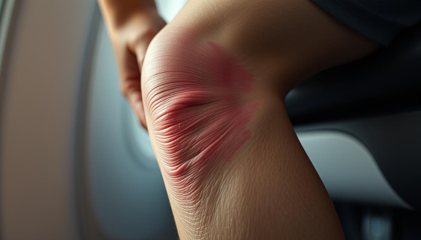
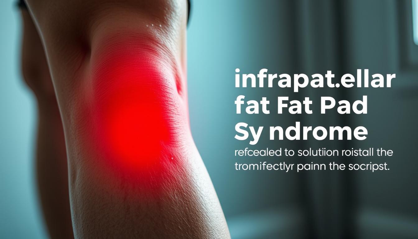
Patellofemoral Pain Syndrome
Patellofemoral Pain Syndrome (PFPS) is a condition characterized by pain behind or around the kneecap, often affecting skiers. This condition was once attributed to improper tracking of the kneecap, but current understanding recognizes it as a complex issue involving multiple factors, including the strength of the hip and thigh muscles. A comprehensive rehabilitation program that includes exercises to strengthen these muscles is essential for recovery. Manual therapies, such as soft tissue mobilization and acupuncture, can also be beneficial when combined with active exercises.
Effective treatment of PFPS involves a graded loading program to strengthen the knee extensors and stabilizers. Interestingly, many of the key muscles involved in knee stability are located in the hip region. Strengthening these muscles through specific exercises can significantly improve knee stability and reduce pain.
Immediate Relief for Knee Pain After Skiing
Knee pain is a common issue for skiers, and understanding how to manage it is crucial for a speedy recovery. When knee pain strikes after a skiing session, it’s essential to take immediate action to alleviate the discomfort and prevent further injury.
The PECH Protocol
The PECH protocol, standing for Pause, Ice, Compression, and Elevation, is a widely recognized method for immediate relief from knee pain after skiing. This protocol is designed to reduce pain, swelling, and further injury.

Pain Management Techniques and Over-the-Counter Options
Beyond the initial PECH protocol, several pain management techniques and over-the-counter options can provide relief for knee pain after skiing. Non-steroidal anti-inflammatory drugs (NSAIDs) like ibuprofen or naproxen can help reduce both pain and inflammation. Acetaminophen is another option for managing pain without anti-inflammatory effects.
Topical analgesics containing ingredients like menthol, camphor, or capsaicin can provide localized pain relief. Continuing cold therapy with ice packs applied for 15-20 minutes several times daily can also manage pain and swelling. Transitioning to heat therapy after the first 48-72 hours can help relax tight muscles and improve blood flow.

Gentle movement within a pain-free range is beneficial for most knee injuries after the initial acute phase. Over-the-counter knee braces or sleeves can provide compression, warmth, and proprioceptive feedback that may help manage pain while the knee heals.
Long-Term Treatment Options for Knee Pain After Skiing
Long-term solutions for knee pain after skiing are multifaceted, offering skiers a range of options to manage pain and resume their favorite sport. The key to effective long-term management lies in accurately diagnosing the cause of knee pain and tailoring treatment accordingly.
Physical Therapy and Rehabilitation Exercises
Physical therapy plays a crucial role in the rehabilitation of knee injuries sustained while skiing. A well-structured physical therapy program can help strengthen the muscles around the knee, improve flexibility, and enhance overall knee function. Physical therapy exercises for knee pain often include squats, lunges, and leg press exercises to strengthen the quadriceps and hamstring muscles.

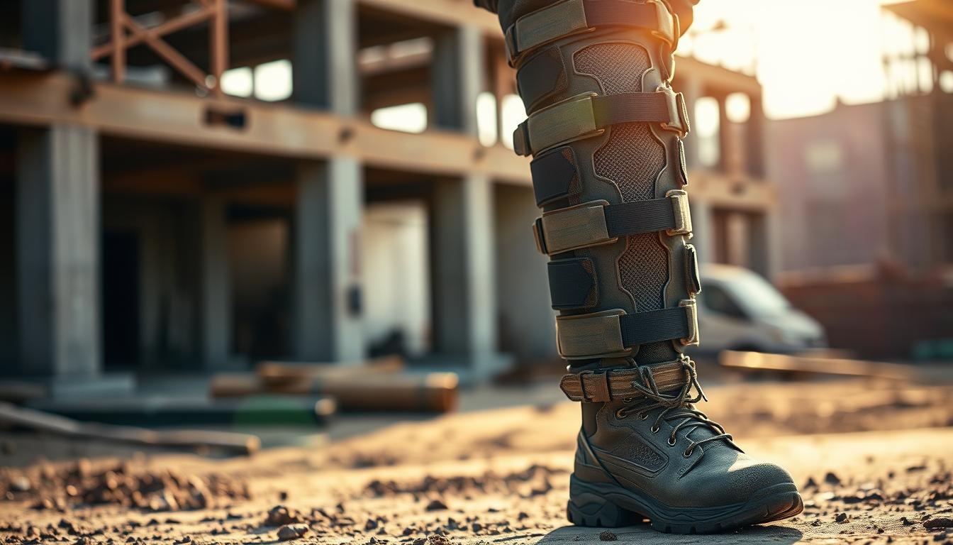
Supportive Devices: Braces, Sleeves, and Orthotics
Supportive devices such as knee braces, sleeves, and orthotics can provide additional stability and protection for the knee, helping to alleviate pain and prevent further injury. When selecting a knee brace for skiing, it’s essential to choose one that offers the right balance of support and flexibility. The best knee braces for skiing are typically those that are designed specifically for high-impact activities and provide medial and lateral support.

Advanced Treatment Options for Persistent Pain
For skiers with persistent knee pain that doesn’t respond to conservative measures, several advanced treatment options are available. These include injectable treatments like hyaluronic acid and platelet-rich plasma (PRP) therapy, which can help improve joint lubrication and promote tissue healing. In some cases, surgical interventions such as arthroscopic procedures or cartilage restoration techniques may be necessary to address underlying damage to the knee joint.
Diagnostic imaging, including MRI and sometimes CT scans, can precisely identify structural damage to bone, cartilage, menisci, or ligaments that may require specialized interventions. Advanced cartilage restoration techniques, including microfracture and autologous chondrocyte implantation (ACI), can address focal areas of cartilage damage. For severe bone-on-bone arthritis, partial or total knee replacement surgery can provide dramatic pain relief.
Prevention Strategies for Future Ski Trips
A proactive approach to knee health while skiing encompasses pre-season conditioning, proper equipment selection, and modifications to skiing techniques to reduce strain on the knees. By addressing these areas, skiers can significantly enhance their overall skiing experience and minimize the risk of knee injuries.
Pre-Season Conditioning and Strength Training
Engaging in pre-season conditioning and strength training is crucial for preparing the muscles around the knee joint for the stresses of skiing. Strengthening the quads and hamstrings through exercises like squats, lunges, and leg curls can help absorb the impact and reduce the strain on the knee. This preparation is vital for withstanding the forces encountered during skiing, especially in challenging terrains.
Proper Equipment Selection and Adjustment
Using the right equipment and ensuring it is properly adjusted is another key factor in preventing knee pain. Ski boots that fit well and are adjusted correctly can help maintain proper alignment and reduce unnecessary stress on the knee joint. Additionally, selecting skis that are appropriate for one’s skill level and the terrain can also contribute to minimizing knee strain.
Technique Modifications to Reduce Knee Strain
Modifying skiing techniques can also play a significant role in reducing knee strain. Maintaining a balanced stance, avoiding the “sitting back” position, and learning to absorb impacts with the entire body are crucial. Working with a qualified ski instructor can help identify and correct technique flaws that may be placing unnecessary stress on the knees. By adopting these strategies, skiers can enjoy their time on the slopes while protecting their knee health.
By combining pre-season conditioning, proper equipment use, and refined skiing techniques, skiers can significantly reduce the risk of knee pain and injuries, ensuring a more enjoyable and sustainable skiing experience.
When to Seek Professional Medical Help
When knee pain persists or worsens after skiing, it’s crucial to determine whether self-care measures are sufficient or if professional medical help is needed. While many cases of knee pain can be managed with rest, ice, compression, and elevation, certain signs and symptoms indicate the need for prompt professional medical evaluation.
If you experience significant swelling, inability to bear weight on the affected leg, visible deformity, or a feeling that the joint is unstable or “giving way,” seek immediate medical attention. A popping or snapping sound at the time of injury, especially when accompanied by immediate pain and swelling, often indicates a significant structural injury that requires professional assessment.
Persistent pain that doesn’t improve with rest, ice, compression, and elevation within 24-48 hours suggests a potentially serious injury that warrants medical evaluation. For more information on when to seek medical help for knee pain, visit https://kneehurt.com/when-to-seek-medical-help-for-knee-pain/. Mechanical symptoms such as catching, locking, or the knee getting “stuck” in certain positions indicate internal derangement that typically requires imaging and possibly intervention.
Even seemingly minor injuries that prevent normal walking or cause pain with basic activities like climbing stairs should be evaluated, as early intervention often leads to better outcomes. For skiers with previous knee injuries or surgeries, any new or changed pain patterns should prompt medical consultation, as these individuals have a higher risk for re-injury or complications.
FAQ
What are the most common knee injuries sustained while skiing?
We often see injuries to the anterior cruciate ligament (ACL), medial collateral ligament (MCL), and meniscus, as well as cartilage damage and patellofemoral pain syndrome. These injuries can be caused by the stress and twisting motions involved in skiing.
How can I manage knee pain immediately after skiing?
We recommend following the PECH protocol: Pause your activity, apply Ice to the affected area, use Compression to reduce swelling, and Elevate your knee above the level of your heart. Over-the-counter pain management options can also be used to help alleviate discomfort.
What are some effective long-term treatment options for knee pain after skiing?
We often recommend physical therapy and rehabilitation exercises to strengthen the muscles around the knee, particularly the quadriceps and hamstrings. Supportive devices like braces and sleeves can also provide stability and relief. In some cases, advanced treatment options like injections or surgery may be necessary.
How can I prevent knee injuries on future ski trips?
We suggest engaging in pre-season conditioning and strength training to build up your strength and endurance. Proper equipment selection and adjustment, as well as technique modifications to reduce knee strain, can also help minimize the risk of injury.
When should I seek professional medical help for my knee pain?
If you experience persistent or severe knee pain, swelling, or instability, we recommend seeking medical attention. A healthcare professional can assess the extent of your injury and provide personalized guidance on treatment and rehabilitation.