I woke up early so I could go to the farmers market. I like looking at the crafts. This time I bought a ring. It is a stainless steel brushed silver ring with very thin rainbow colors along each edge. I had the word ‘resilience’ engraved on it. I like it very much. I also gave my niece a birthday present.
Then I went to the Hawaii Tropical Bioreserve and Garden. It is very beautiful. Jungle and flowers and ocean.
When I got back to my hotel and opened my door, the hotel cat ran into my room. He wouldn’t leave. I tried to ignore him, use a cord to play with him and feed him. Nothing worked. It took more than an hour before he could leave. I used a paper bag! He wanted to climb in. After I got him out, he climbed onto a table outside my door and tried to knock over a vase. I told him to stop and then he lay down in front of my door and scratched it.
New research at ACR Convergence 2023, the annual meeting of the American College of Rheumatology (ACR), found that patients with rheumatoid arthritis (RA) in sustained remission who stopped taking TNF inhibitors (TNFi) had significantly more flares and lower Boolean 2.0 remission rates compared to those who continued treatment. Boolean 2.0 is a revised definition for evaluating disease activity in RA, classifying more patients as achieving remission than Boolean 1.0. It is endorsed by the American College of Rheumatology and the European Alliance for Associations in Rheumatology (EULAR) (Abstract #L07).
As more RA patients achieve durable remission, questions remain about the long-term effectiveness of tapering and stopping TNFi treatment. In the randomized, multicenter, noninferiority ARCTIC REWIND trial, Siri Lillegraven, MD, MPH, PhD at Diakonhjemmet Hospital, Oslo, Norway, and colleagues compared the three-year effect of tapering versus stable treatment in RA patients in sustained remission. It follows a trial last year.
The current study included 92 patients from Norwegian rheumatology centers who were randomized 1:1 to taper off TNF inhibitors until discontinuation or continuation of treatment. During the three-year study period, all received study visits every four months. Patients restarted treatment at the full dose if they experienced a flare, which was defined as loss of remission plus an increase in disease activity score of 0.6 units or more and two or more swollen joints. In lieu of these criteria, a doctor and a patient might agree that a significant flare had occurred. The study also looked at remission status, medication use and serious side effects or complications.
Of the original 92 patients, 80 (87%) completed three-year follow-up. At the end of the study, 75% of patients in the tapering group experienced a flare, compared to 15% in the stable group. Most of those who experienced a flare were in remission by their next office visit (81% in the taper group and 67% in the stable group), although the taper group had significantly lower Boolean 2.0 remission rates throughout the study.
Lillegraven says the researchers were “somewhat surprised by the difference in the proportion of patients in ACR/EULAR Boolean remission in the two groups,” noting that “although most patients in the taper group experienced a flare within the first year and the earlier resume treatment at full dose Boolean 2.0 remission rates were significantly lower in the tapering TNFi group than in the stable group throughout the study period.”
The risk difference for flares observed in this data [-24% over three years] is quite similar to what was observed in the one-year study. That’s a bit surprising, because we might have expected that more patients receiving stable treatment would develop a flare over time, narrowing the difference between the two groups.”
Siri Lillegraven, MD, MPH, PhD at Diakonhjemmet Hospital, Oslo, Norway
Lillegraven notes that the study’s open-label design could influence the evaluation of flares, but says that study staff “were continuously instructed on the importance of recording flares similarly in both groups, a pragmatic approach that will improve clinical care reflects, where patients know which treatment they are receiving. received.”
Lillegraven says her team has many studies planned to better understand how to personalize treatment for RA patients in remission. This includes factors that can help determine which patients should and should not taper off their treatment.
“We have begun planning a 10-year follow-up of the study to better understand the long-term outcomes of different treatment strategies in RA remission. We are [also] consider studies to better understand patient preferences regarding medication tapering.”
Shared decision-making is central to any consideration of tapering, she says.
“The patient should be informed of the risks and benefits of tapering, and the patient’s overall situation should be taken into account before the decision is made. Although the data do not support tapering off TNFi at a group level, factors such as side effects related to the treatment or the patient having a strong preference for tapering will obviously influence such a decision.”
SAINT PAUL, Minn., November 9, 2023–(BUSINESS WIRE)–Spineology Inc. (“Spineology” or the “Company”), the leader in ultra-minimally invasive spine surgery, today announced that John Booth will resign from his position as Chief Executive Officer of Spineology, effective November 10, 2023. The Board of Directors has selected Brian Snider as the next Chief Executive Officer, effective November 13, 2023. Mr. Booth will remain with Spineology through 2024 and serve in an advisory role to enable a smooth transition. Mr. Booth will also resign from the Board of Directors, and the Board of Directors has nominated Mr. Snider as Director.
Snider joins Spineology with nearly two decades of progressive leadership experience in the medical device industry. Most recently, he served as Executive Vice President of Marketing for Alphatec Spine (NASDAQ: ATEC), as a member of the executive leadership team responsible for growing the company’s market capitalization from $20 million to more than $1.2 billion. During his tenure at ATEC, he was responsible for a variety of marketing and product development disciplines. Most recently he was responsible for the Biologics, Cervical and Thoracolumbar business units. Before joining ATEC, Snider spent nine years at NuVasive, Inc., a leader in innovative products and procedures for minimally disruptive spine surgery. During his tenure at NuVasive, Snider held senior-level marketing roles in the Thoracolumbar business segment, including its flagship procedure, XLIF®. Mr. Snider received his BBA in Marketing and Information Systems from George Washington University and his MBA from the Fuqua School of Business at Duke University.
“It has been a great privilege to work with a talented and passionate group of employees within the company, who together have significantly advanced the field of ultra-minimally invasive spine surgery over the company’s history,” said John Booth. “I am confident that the company will continue to grow and deliver disruptive solutions under Brian’s leadership as Spineology’s next CEO.”
Ed Spencer, chairman of the Spineology Board, expressed gratitude for Booth’s leadership. “On behalf of Spineology’s Board of Directors, our employees and shareholders, I express my deep gratitude to John for his success in leading the company, as well as for his service as a member of Spineology’s Board of Directors for more than 20 years .”
“As we prepared for John’s retirement, the Board unanimously agreed that Brian Snider was well suited to lead Spineology,” Spencer continued. “His years of experience growing evolutionary businesses in the minimally invasive spine market will enable him to immediately contribute to Spineology’s continued success.”
“I am excited to join the Spineology team,” said Snider. “Spineology is a company with a unique procedural foundation backed by strong clinical data. I am confident that we will achieve growth and surgical advancements in this next phase. I would like to thank the Board of Directors for the opportunity to serve the Spineology team, shareholders and, most importantly, our patients.”
About Spineology:
Spineology Inc. is at the forefront of ultra-minimally invasive spine surgery, revolutionizing the way spine surgeons treat and heal back pain. Our patented Mesh technology sets us apart from traditional fusion procedures, allowing surgeons to optimize results while minimizing tissue disruption and improving patient recovery. With a strong commitment to patient-centered care and enabling disruptive technologies, Spineology continues to push the boundaries of what is possible in spine surgery today with the tools of tomorrow.
Contacts
Jamison Young Finance Director 651-256-8504 jyoung@spineology.com
A prospective case series study was conducted between 01/01/2018 and 31/06/2020. All patients provided informed consent to participate in the study, which was conducted in accordance with institutional standards.
Patient population
The patients were enrolled consecutively. The inclusion criteria were all adult distance runners diagnosed with iliotibial band syndrome and with a negative response to non-operative treatment after six months. Distance runners were defined as professional or amateur subjects completing medium (1500 m) and long (marathon and ultra-trail runners) distances.
The exclusion criteria were: (i) incomplete clinical reports; (ii) non-distance runners; (iii) additional injuries that interfere with running; (iv) bilateral involvement, (v) negative local anesthetic infiltration test; and (vi) revision surgeries after previous ITB procedures.
The patient must meet all inclusion criteria and none of the exclusion criteria. Before inclusion in the study, all patients completed a preoperative protocol regardless of additional tests performed up to that point.
Preoperative protocol
Complete medical history and physical examination were recorded in all patients. A local anesthetic infiltration test was performed, which consisted of an ultrasound-guided sub-iliotibial bursa infiltration with 2 ml of 2% mepivacaine, immediately followed by a 5 km race. If the patient’s symptoms were temporarily relieved during the race, the test was considered positive.
High-field MRI (≥ 1.5 T) was performed in all cases after the patient had exercised in the 72 hours before the scan, increasing the sensitivity of the imaging technique when edema appeared at the level of the LFC or ITB ( Figure 1 ).
figure 1
Preoperative MRI: coronal (right) and axial (left) images showing edema at the ITB.
Before the surgical indication, a specific rehabilitation program was performed to optimize conservative treatment with techniques not previously used in the patient, including fascia lata stretching exercises, proximal eccentric muscle training, intra-tissue percutaneous electrolysis and at least three focal shock wave exercises. sessions.
Independent variables and outcome variables
Demographic data (age, gender and body mass index -BMI-), comorbidities, athletic discipline, time to surgery and postoperative follow-up time were collected in all patients.
The intraoperative characteristics (time of ischemia, confirmation of ITBS, identification of concomitant lesions and need for drainage) and intraoperative and postoperative complications were also recorded.
The main variables of the study were the rate and time of return to the previous sports level, which were reported by patients during follow-up visits. Return to the previous sport level was considered a dichotomous outcome and was defined as participation after undergoing the PLAR technique in at least one race of the same distance as before the injury, at or above the pre-injury competitive level. The return to sport percentage was calculated from the number of athletes who returned to sport, from the number of athletes who underwent the PLAR technique, and expressed as a percentage.
The secondary variables were the clinical evaluation of the patients based on the Activity Rating Scale (ARS), the International Knee Documentation Committee (IKDC) questionnaire and the level of satisfaction. The results of the ARS and IKDC scales were interpreted as follows: excellent = 95–100 for IKDC and 15–16 for ARS; good = 84–94 for IKDC and 13–14 for ARS; and fair = 65–83 for IKDC and 10–12 for ARS. The level of satisfaction was evaluated in all patients with a poll based on the question: did the operation meet your expectations? The possible answers were: completely satisfied, largely satisfied, somewhat satisfied, dissatisfied.
Surgical procedure
All procedures were performed by the same surgeon. The ITBS diagnosis was confirmed intraoperatively by observing a collapse of the space between the LFC and the ITB due to a combination of bursitis and hard fibrotic adhesions that prevented the passage of the arthroscopy optic (Fig. 2).
Fig. 2
Intraoperative view. Fibrotic adhesions between the LFC and the ITB.
Patients were placed supine on a conventional table with arthroscopic support, during which an ischemia cuff was placed around the thigh and standard aseptic preparation was performed. The LFC, fibular head, Gerdy’s tubercle, and anteromedial (AM) and anterolateral (AL) standard portals were identified and marked.
The procedure began with routine diagnostic arthroscopy through the AL portal. If there was any doubt about additional lesions, an additional AM portal was used to allow tactile examination of the knee structures. Under direct intra-articular view, the superolateral (SL) portal was prepared using a 16G Abbocath spinal needle (Hospira, Lake Forest, IL, USA) as a guide, always passing through the tendon portion of the vastus lateralis muscle or the capsule, taking care not to perforate the quadriceps muscle tissue (Fig. 3). All portals were prepared with a No. 11 scalpel blade.
Fig. 3
Intraoperative view. Superolateral portal (SLP) using a 16G Abbocath spinal needle as a guide
With the knee in 30° flexion, we initially performed debridement and resection of the lateral synovial recess, using a motorized shaver (Fig. 4) and a vaporizer (90 degrees, model 405Q3, Bonss Medical Tech, Taizhou, Jiangsu, China) (Fig. 5). In patients with ITBS, we can observe abnormal anatomy with increased fibrosis in the lateral synovial recess. Therefore, we consider it of utmost importance to perform a wide resection in this area until we obtain a complete view of the iliotibial band externally and the LFC medially, even including the external meniscal wall in the anterior half, and able are to pass the optic from the anterior to the popliteal tendon in the posterior zone, always preserving the meniscal-tibial and meniscal-femoral ligaments. This procedure was performed primarily from the SL portal under visual control from the AL portal, with reversal of the two portals to complete the release.
Fig. 4
Intraoperative view. Loosening the fibrous adhesions in the space between the LFC and ITB using a motorized shaver
Fig. 5
Intraoperative view. Releasing the fibrous adhesions in the space between the LFC and ITB using a vaporizer
The second part of the procedure involved the percutaneous lengthening of the ITB under direct vision by arthroscopy. This was done with controlled knee varus at 30° flexion, seeking a balance between extension and maintenance of muscle function. An 18G 3-mm needle scalpel (Nokor needle; Becton Dickinson and Co., Franklin Lakes, NJ, USA) was used to perform controlled micro-tenotomies as a micro-pie crust technique on the ITB. In all cases they were made longitudinally and parallel to the fibers, and in those cases with greater fibrosis of the ITB, the tenotomies were also made transversely in the posterior third (Fig. 6).
Fig. 6
Intraoperative view. Micro-tenotomies on the ITB with an 18G 3 mm needle scalpel
After completion of the procedure, the skin was closed with Prolene (Ethicon, Inc.) 2/0, and a compressive elastic bandage was placed, with semi-rigid support in the external zone, where a bulge typically forms due to fluid extravasation via the microfibers. -tenotomies. Redon drainage (Fresenius Kabi AG, Bad Homburg, Germany) was used for 12 hours in patients with intraoperative identification of a sub-iliotibial bursa associated with significant vascular infiltration, and in all cases we infiltrated a mixture of corticosteroids and local anesthetic (2 ml Celestone Cronodose + 4 ml 2% mepivacaine).
Postoperative protocol
All patients were discharged with full weight bearing assisted by two crutches depending on tolerance.
Rehabilitation started from the first postoperative day. During the first two weeks, full joint range recovery exercises, isometric exercises, and even post-assisted squats were allowed to minimize muscle atrophy. Between weeks 2 and 4, eccentric muscle training (free, weight-bearing and single-foot squats, as well as frontal and lateral lunge exercises) combined with proprioception exercises using a BOSU ball (both sides up) or an unstable platform was allowed. From weeks 4 to 8, plyometric exercises, elliptical taping, and static cycling exercises were increased, and gentle jumping exercises were allowed depending on tolerance. From the 8th week onwards, and depending on the patient’s muscular and proprioceptive status, we allowed running a distance of 1 km every other day, combining walking and running exercises, and added distance or running exercises every two days. speed increases of 10% if tolerance was found. Good. From the 12th week after the operation, recovery was allowed to continue at the athletics club under the supervision of the coach or physiotherapist.
Follow-up protocol
A minimum follow-up of 12 months was performed. Postoperative data were collected in all patients at 15 days, 1, 3, 6 and 12 months and at the end of follow-up (medical discharge). Complications and clinical course were assessed at all visits, while sports performance and the ARS and IKDC questionnaires were assessed at 3, 6 and 12 months, without access to a copy of the scale during the intervening period, to avoid the patient himself – monitoring the recovery and influencing the final result. The level of satisfaction was recorded at the last follow-up visit.
static analysis
The statistical analysis was performed using the SPSS® version 22.0 package for Mac (IBM, NY, USA). Statistical significance was considered for p ≤ 0.05 and a statistical power of 90%.
Standard descriptive statistics including measures of central tendency (mean/median) and variance (standard deviation). [SD]/interquartile range [IQR]) were calculated, as well as frequencies and ratios.
The preoperative and final follow-up functional scores were compared using the Wilcoxon Signed-Rank test.
A multiple nonparametric analysis comparing the IKDCS and ACS scales preoperatively and at 6 and 12 months was performed using Friedman’s statistical test.
If you’ve been experiencing chronic knee pain and stiffness, you may be wondering if arthritis is to blame. There are various forms of knee arthritis, each with their own causes, symptoms, and treatments. Understanding the differences is key to obtaining an accurate diagnosis and effective relief. This comprehensive guide provides an overview of the most common types of knee arthritis, so you can work with your doctor on the best care plan for your joint health.
What is Knee Arthritis?
Arthritis is inflammation affecting the joints. In a healthy knee, the bones are cushioned by smooth cartilage and lubricated by fluid that allows flexible movement. Arthritis damages this cartilage over time, causing pain, swelling, and stiffness.
Typical blood test findings for different types of knee arthritis:
Type of Knee Arthritis
Common Blood Test Findings
Osteoarthritis
Normal white blood cell count, ESR, CRP. No presence of rheumatoid factor or anti-CCP antibodies.
Let me know if you need any clarification or have additional questions!
The knee is a complex joint containing the femur (thigh bone), tibia (shin bone), fibula, and patella (kneecap). Multiple joints and connective tissues provide stability for standing, walking, running, and other activities. Like other joints, the knees are vulnerable to various forms of arthritis.
General symptoms of knee arthritis include:
Joint stiffness, especially in the morning or after sitting
Osteoarthritis is the most common type of knee arthritis, affecting over 14 million Americans. It occurs when protective cartilage in the joint gradually wears down over time, allowing painful bone-on-bone friction.
OA can be primary (idiopathic) with no known cause, or secondary due to injury, obesity, overuse, or other joint stressors. As cartilage erodes, movement becomes stiff and painful. Fluid-filled cysts and bony growths may also develop around the joint.
Common OA symptoms include:
Aching pain during activity that worsens over time
Morning joint stiffness lasting under 30 minutes
Tenderness, swelling, or inflammation around the knee cap
Hard lumps (bone spurs) around the joint
Gradual loss of flexibility and range of motion
Grating sensation when moving the knee
Risk factors like age, female gender, genetics, and previous joint injury make OA more likely. Treatment focuses on pain relief, anti-inflammatories, physical therapy, weight loss, braces, and if necessary knee replacement surgery. Lifestyle changes are key to preserving joint function.
Rheumatoid Arthritis (RA) in the Knee
Rheumatoid arthritis is an autoimmune disease causing chronic inflammation of the joints and other body tissues. With RA, the immune system attacks the synovial membrane lining the joint. This leads to pain, swelling, and eventual cartilage and bone damage if untreated.
RA typically begins in smaller upper body joints, but knees and other lower extremity joints can be affected as it progresses. Distinct symptoms of knee RA include:
Symmetrical pain in both knees rather than just one
Morning stiffness lasting over 30 minutes
Systemic symptoms like fatigue and fever along with joint pain
More severe pain with movement than at rest
Limping, difficulty walking or standing from kneeling
Joint deformity over time if inflammation isn’t controlled
Medications like DMARDs and biologics aim to stop RA progression and preserve joint health. Low-impact exercise and splints can also help reduce knee symptoms.
Post-Traumatic Arthritis of the Knee
Post-traumatic arthritis develops after an injury damages structures inside the knee joint. Injuries like anterior cruciate ligament (ACL) tears, meniscus tears, or fractures commonly lead to post-traumatic arthritis over time. The initial injury causes instability and extra wear that degrades cartilage and leads to osteoarthritic changes.
Symptoms of post-traumatic knee arthritis may include:
Pain that increases with activity
Recurring swelling and inflammation
Reduced knee extension and flexion
Tenderness along the joint line
Knee buckling or giving way
X-rays, MRIs, and physical examination of the knee help diagnose post-traumatic arthritis. Treatments like icing, immobilization braces, medications, hyaluronic acid injections, and physical therapy can help manage pain in early stages. But if conservative treatment fails, knee replacement surgery may be necessary.
Gout and Pseudogout in the Knee
Gout and pseudogout are inflammatory types of arthritis caused by uric acid crystals and calcium pyrophosphate crystals depositing in joints. This triggers sudden pain, swelling, and stiffness, often in a single joint like the knee.
Gout arises when excess uric acid in the blood crystallizes. Issues like kidney disease, certain cancers, genetics, diet, and some medications can increase uric acid levels. Pseudogout occurs due to abnormal calcium pyrophosphate crystal formation related to aging, joint injury, or metabolic factors.
Flare-ups in the knee joint are excruciatingly painful. Other symptoms include:
Rapid joint swelling, redness, and heat
Extreme tenderness to touch
Decreased range of motion
Fever and chills if infection occurs
Shiny, tense skin over the joint area
Gout and pseudogout require careful diagnosis and management of underlying causes. Typical treatments include NSAIDs, steroids, colchicine, and dietary changes. Draining fluid from the joint may relieve pressure.
Psoriatic Arthritis Affecting the Knee
Up to 30% of people with the autoimmune skin condition psoriasis develop psoriatic arthritis – an inflammatory arthritis distinct from rheumatoid arthritis. The knees are a common location for psoriatic arthritis flare-ups.
Psoriatic arthritis affects joints asymmetrically, often striking just one knee rather than both sides equally. Symptoms include:
Joint pain, swelling, and stiffness
Reduced range of motion, difficulty bending the knee
Pitted, crumbling nails or nail separation from the nail bed
Eye inflammation (uveitis)
Fatigue and loss of appetite when flaring
Sausage-like swelling of fingers or toes
Treatment involves NSAIDs, DMARDs, biologics, and other immunosuppressants to relieve knee inflammation and prevent joint damage. Gentle stretching and exercise is also beneficial once flare-ups subside.
Infectious Arthritis of the Knee
Infectious arthritis, also called septic arthritis, occurs when bacteria, viruses, or fungi enter the joint space and trigger inflammation. Without prompt antibiotic treatment, infectious arthritis can rapidly destroy knee cartilage and surrounding tissue.
Infectious knee arthritis may arise from:
Bacterial spread from infection elsewhere in the body
Penetrating injury introducing pathogens into the joint
Surgery complications
Joint injections with improperly sterilized equipment
Distinct symptoms signaling a possible knee joint infection include:
Prompt medical attention is crucial to avoid permanent joint damage. Treatment involves strong antibiotics, draining the infected fluid, and sometimes surgery to fully clean out the joint space.
Managing Knee Arthritis
Whether you have osteoarthritis, rheumatoid arthritis, or another form, there are many ways to ease knee arthritis symptoms and improve function:
Losing weight to reduce joint stress
Wearing a knee brace for support and stability
Using heat/ice therapy to relieve pain and stiffness
Doing gentle knee stretches and low-impact exercises like swimming or cycling
Physical therapy to improve flexibility and strength
Over-the-counter pain relievers like acetaminophen or NSAIDs
Mind-body practices like yoga, tai chi, and meditation to help cope with chronic pain
Viscosupplementation injections to replenish knee joint fluid
Surgery like arthroscopy, osteotomy, or knee replacement if other therapies fail
Consulting an orthopedist, rheumatologist, or physical therapist can help determine the safest, most effective treatment plan. Don’t resign yourself to living with constant knee pain – explore the many options available to get you moving comfortably again.
Conclusion
Knee arthritis can negatively impact mobility and quality of life. But while there are various types of knee arthritis, there are also a multitude of ways to manage symptoms. Understanding the differences between osteoarthritis, rheumatoid arthritis, and other forms helps you obtain an accurate diagnosis. Work closely with your doctor to find the optimal combination of lifestyle changes, medications, therapies, and possibly surgery to relieve your knee pain and restore function. The more informed you are about your specific type of knee arthritis, the better equipped you’ll be to gain control and get back to healthy, active living.
Rheumatoid arthritis (RA) is one of the first autoimmune diseases to be identified and remains incurable. Despite the discovery of several disease-modifying treatments, the response to each treatment remains unpredictable. This indicates a difference in the pathophysiology of RA between patients.
Study: Deconstruction of the synovium of rheumatoid arthritis defines inflammatory subtypes. Image credits: Oporty786/Shutterstock.com
A new article recently appeared in Nature, reported the examination of synovial tissue from the joints of nearly 80 people with RA, combined with RNA sequencing and surface protein analyses. This allowed the researchers to assemble an atlas of RA synovial changes from more than 314,000 individual cells. This could help develop targeted therapies that recognize the diversity of RA disease processes.
Background
RA affects about 1 in 100 people worldwide. The main feature is the painful swelling of synovial joints that ultimately culminates in joint damage and disability. Recognition of the immunological origins of RA has led to the deployment of therapies that target inflammatory cytokines and pathways, including tumor necrosis factor (TNF), IL-6, stimulation of T and B cells together, and the pro-inflammatory JAK -STAT transcription. regulatory process.
Genetic differences have been identified, as well as diverse clinical features, but these do not fully predict or explain why treatment response varies between patients, nor do they help identify therapeutic targets. The need for a more detailed picture of RA synovial disease activity motivated the current study.
Multiple effector cells participate in RA activity at the synovial level. Previous research suggests that the synovial cellular profile could predict response to treatment. Furthermore, the presence of common cell state compounds could extend the utility of this study to other autoimmune or inflammatory conditions.
What does the research show?
The study was based on 82 synovial tissue samples taken from patients with a spectrum of RA activity from moderate to high. This is measured by the CDAI (clinical disease activity index), which was ten or higher for all participants. The samples came from those who had not yet started treatment, some with a poor response to methotrexate (which stops the proliferation of inflammatory cells), those who responded poorly to anti-TNF agents (to stop pro-inflammatory signaling) and some who had osteoarthritis.
The scientists were able to divide the RA synovium into six groups based on the cell types that were selectively enriched in each group. Each group is accordingly called a cell type abundance phenotype (CTAP) and is defined by specific cell states.
While some samples showed very low levels of lymphocytes, others were abundant in T and B cells, indicating clear synovial differences. Each cell state reflects different disease stages and types, as well as varying cytokine profiles, and the risk genes were differentially expressed between groups.
The researchers created an atlas of RA synovial cell states, consisting of 77 cell states, including 24 T cell clusters, 9 B cell clusters, 14 natural killer (NK) cell clusters, and 15 myeloid clusters. There were also ten stromal cells and five endothelial clusters. This confirmed RA-associated cell states identified in a previous study from more than 5,000 synovial cells.
For example, the CTAP-TB was enriched in TPH and TFH cells, perhaps because these promote the differentiation of B cells into plasmablasts and ABC cells, as opposed to non-TFH/TPH memory CD4+ T cells that only do the latter. Both TFH and TPH cells are enriched in the synovial tissue of all CTAPs, but extra-follicular activation pathways also appear to be present in CTAP-TB.
Conversely, the CTAP-TF mainly involves cytotoxic together with naive CD4 and CD8 T cells, with selective NK cells that can share their transcriptional profile promoted by the tissue microenvironment. Fibroblast subsets were differentially enriched in this CTAP versus CTAP-M. The latter also showed enrichment of myeloid cells, perhaps because inflammatory monocytes were recruited to transform into macrophages as a result of exposure to the specific cell types and soluble factors present in each CTAP.
These cell neighborhoods did not show consistent associations with aggregated RA scores from histology, which are based on the extent and type of inflammatory cell infiltration. This is probably because the former are so diverse. However, the CTAPs each contribute one-fifth of the variance of the histological density and total scores and are associated with inflammation scores.
Interestingly, the CTAPs showed a close relationship with clinical parameters such as the commonly used autoantibodies against cyclic citrullinated peptide (CCP), reflecting increased lymphocyte infiltration into CCP-positive synovial tissue. CTAP-M was associated with CCP-negative synovial tissue. There was no clear association with the strongest genetic risk predictor, HLA–DRB1.
The CTAPs showed distinct cytokine profiles. For example, the T cell neighborhood of CTAP-TB expressed the TFH/TPH highlight genes CXCL13 as expected, while for CTAP-TF the T and NK cell neighborhood was associated with the expression of the genes IFNG And TNF.
As expected, there was little correlation between disease activity and CTAP or treatment response. This supports the theory that inflammatory phenotypes in different types of RA are reflected in the CTAPs and not in clinical disease activity, as reflected by CDAI and other clinical scores.
However, CTAPs change over time, usually to CTAP-F, after anti-inflammatory therapies such as rituximab and the anti-IL-6 agent tocilizumab. CTAP-F is a predictor of poor response to treatment.
What are the implications?
“The CTAP paradigm has the potential to serve as a powerful prototype to classify other types of tissue inflammation.” The subtypes of enriched inflammatory cells in different CTAPs also reveal new research questions about how these interact to produce a range of inflammatory phenotypes in such diseases.
“CTAPs are dynamic and can predict response to treatment, highlighting the clinical utility of classifying synovial phenotypes of rheumatoid arthritis.” It was possible to predict the CTAP using RNA sequencing with different methods. This offers potential therapeutic targets for the future.
Meanwhile, the spectrum of inflammatory changes in RA explains why treatment responses vary so widely among patients treated with anti-TNF agents. This may imply that specific therapies that target the cells and pathways enriched in each CTAP could induce better responses, and advance drug development and precision medicine.
I woke up super early. I had difficulty sleeping. At first it was a bit warm and the fan is very noisy, but the main problems were 1) another guest was snoring and 2) someone was blaring pop music at 3am. It was one song from Judy, but it woke me up. Now back to the snoring. All quests have the windows open because it is hot and there is no air conditioning. A guest snores or snores so loudly that we can all hear it! It kept waking me up. It was loud! It sounded like someone said “ew.” I don’t know, but I hope it’s milder tonight.
Then I drove to the farmers market and met my friend. It started to rain very hard. I never found my umbrella, so all I had was my windbreaker. We walked around and saw all the little stalls. I bought someone a Christmas present. Then we got in our cars and went to another small market. Between the two markets I was given lettuce, a red pepper and a tomato so I can make sandwiches. The only thing I don’t have is vegan Mayo, but I’ll have to suffer through it.
Family drove to my friend’s house. It’s the first time I’ve seen it in real life and it’s beautiful. It’s the kind of house I’d like to live in. He’s doing some renovations and additions, but he gave me a full tour. We sat outside on his lanai for a few hours and talked.
Then I drove back to Hilo where I stopped at a vegan restaurant to get a sandwich. It started raining really hard and I wanted to eat outside, so I took my sandwich back to my hotel room. Since it was almost four in the afternoon, that’s both my lunch and dinner. I’m so tired and I’m not going to get up very late tonight.
I woke up early this morning and headed to Rainbow Falls. I should have seen a rainbow, but I didn’t. I walked up the rock steps to get to the top and see the hot pools. (I think that’s what they’re called). Walking down was a bit slow.
I returned to find that my friend’s cat wouldn’t be seen at the vet until later that day and our outing was postponed. I went shopping for items like t-shirts and a dream catcher.
I came back to my room for lunch. I have a kitchen in my room so I can make my own food.
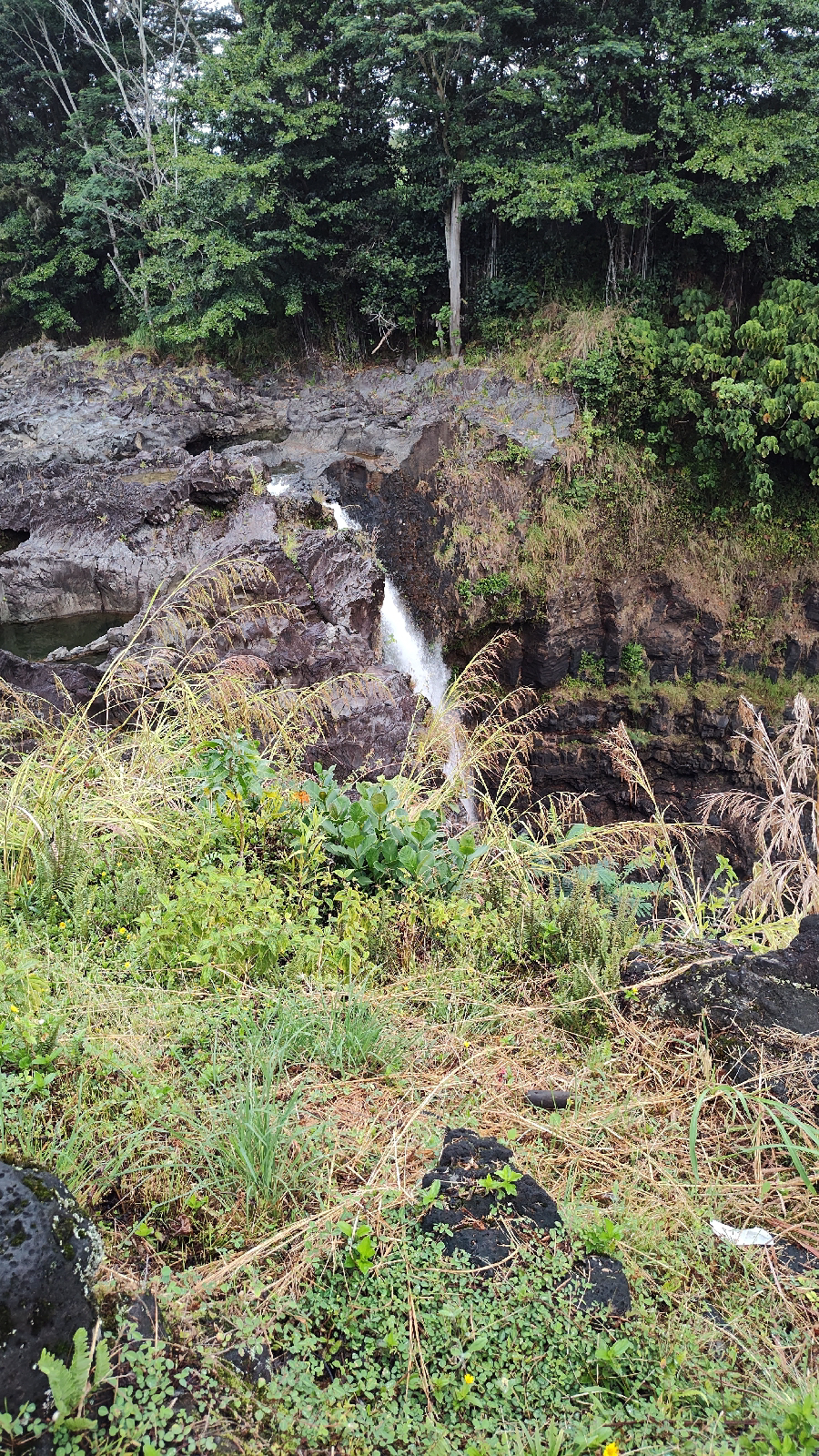
After lunch I wrote out some postcards. I was smart to put them in the car because on my next trip I stopped by a post office and was able to mail them. I drove to another waterfall called ‘Akaka. It was so beautiful.
When I got there, the employee told me to take the shortcut. I hate when people tell me what to do, so I did the whole walk. It was a total of half a mile with a lot of stairs. I took a detour to drive along the scenic route.
I got back to my hotel room just in time to meditate with the other members. When I was done, I drove to a restaurant that had vegan food for takeout.
The knee is arguably the most important and complex joint in the human body. It bears our weight, allows movement and flexibility, and absorbs tremendous impact forces. Understanding knee anatomy and function is crucial for keeping your knees healthy and recovering from injury.
This in-depth knee guide covers everything you need to know about knee anatomy, morphology, function, and common problems. Read on to learn how your knees work so you can keep them in top shape!
Knee Joint Anatomy
The knee joint connects three bones: the femur (thighbone), tibia (shinbone), and patella (kneecap). These bone structures provide the foundation of the knee.
Knee joint components:
Femur
Tibia
Patella
Joint capsule
Cartilage
Synovial membrane
Menisci
Ligaments
Tendons
Muscles
Nerves and blood vessels
The ends of the femur and tibia are covered in articular cartilage, a smooth substance that protects the bones and allows them to glide smoothly against each other.
The whole knee joint is surrounded by a joint capsule lined with synovial membrane. This produces synovial fluid that lubricates the joint and reduces friction.
There are two menisci between the femur and tibia – the medial meniscus and lateral meniscus. These C-shaped discs of cartilage act as cushions or shock absorbers in the knee.
Ligaments connect bones and provide stability to the knee:
Tendons connect muscles to bones. The quadriceps and patellar tendons are key structures that straighten the knee.
Powerful muscles like the quadriceps and hamstrings control knee movement. Smaller muscles provide additional support.
Nerves carry messages between the knee and brain to facilitate movement. The knee joint also has a rich blood supply to provide nutrients.
Knee Morphology
Knee morphology refers to the shape and form of the knee joint structures. Here are some key morphological features:
The femur has two rounded condyles that sit on the flat tibial plateau. This shape allows the knee to flex, extend, and rotate.
The patella is a triangular sesamoid bone embedded within the quadriceps tendon. It protects the knee joint and increases quadriceps leverage.
Menisci are crescent-moon shaped discs between the femur and tibia. This distributes body weight and provides congruency.
Collateral ligaments run vertically on the medial and lateral knee to resist side-to-side motion.
Cruciate ligaments cross each other inside the joint to enable rotation while limiting front-to-back translation.
Muscles like the quadriceps have large attachment sites for strong contraction leverage.
Articular cartilage is smooth and dome-shaped over bony surfaces to facilitate gliding.
The synovial membrane lines the joint capsule and folds into crevices for lubrication access.
Understanding the shape and alignment of knee structures is critical when diagnosing injuries or dysfunction.
Knee Joint Function and Biomechanics
The complex anatomy of the knee allows for specialized motions and weight bearing functions.
Main Knee Functions
Flexion and extension for walking, running, and jumping
Slight internal and external rotation for foot positioning
Weight bearing as the body’s central support joint
Shock absorption to reduce impact loading
Knee Flexion and Extension
The femur and tibia rotate against each other to produce knee flexion and extension. This hinge-like motion ranges from 0° when straight to over 140° during deep flexion.
Key structures involved in knee flexion:
Hamstrings – Flex the knee by pulling the tibia posteriorly
Gastrocnemius – Flexes knee through its connection with the hamstrings
Popliteus – Rotates femur internally during flexion
Key structures for knee extension:
Quadriceps – Straighten the knee by pulling the patella and tibia anteriorly
Soleus – Assists knee extension via synergy with the gastrocnemius
Knee Rotation
The knee joint can rotate slightly inward and outward when flexed to position the foot and lower leg. This is mediated by:
Popliteus – Internally rotates the femur
Iliotibial band – Facilitates external femoral rotation
Rotational stability is provided by the cruciate ligaments and collateral ligaments.
Shock Absorption and Impact Reduction
The knee joint structures work together to reduce damaging impact forces:
Menisci – Act as shock absorbers between the femur and tibia
Cartilage – Compresses to cushion bone articulation
Ligaments – Provide dynamic restraint to overloaded joints
Muscles – Eccentrically contract to dissipate energy
Proper knee biomechanics are essential for absorbing up to 6 times your body weight during activities like jogging!
Common Knee Problems and Injuries
Due to its complexity and heavy use, the knee joint is susceptible to various injuries and pathologies. Some of the most common knee problems include:
Sprains and Strains
Medial collateral ligament (MCL) sprain – Overstretching or tearing of the MCL from impact or twisting. Causes inner knee pain and laxity.
Lateral collateral ligament (LCL) sprain – Injury to the LCL from hyperextension or direct trauma. Results in outer knee pain.
Patellar tendon strain – Overload and inflammation where the tendon attaches to the tibia. Causes anterior knee pain.
Quadriceps/hamstring strain – Muscle overuse leading to partial tears and weakness. Hampers knee stability.
Meniscal Injuries
Meniscus tear – Occurs when abruptly twisting or rotating the knee. Torn menisci cause pain, catching, locking, and swelling.
Discoid meniscus – An abnormal thickened meniscus prone to tearing. More common medially.
Degenerative meniscal tear – Fraying and deterioration seen with aging. Associated with osteoarthritis.
Ligament Tears
ACL tear – Sudden change in direction tears the ACL. Instability, pain, and swelling follow.
PCL tear – Hyperextension injury that strains or tears the PCL. Posterior knee pain and laxity result.
Patellofemoral Problems
Patellar dislocation – The patella dislocates laterally from the femoral trochlear. Causes intense pain.
Patellofemoral pain syndrome – Anterior knee pain from improper patellar tracking. Aggravated by climbing stairs.
Patellar tendinitis (jumper’s knee) – Overuse injury of the patellar tendon. Leads to inflammation and localized pain.
Arthritis
Osteoarthritis – Degenerative joint disease causes knee cartilage to thin and wear down. Results in stiffness, swelling, and activity limitations.
Rheumatoid arthritis – Autoimmune condition that inflames the knee joint linings. Manifests as pain, warmth, and joint destruction over time.
Osgood-Schlatter Disease
Tibial tubercle apophysitis – Inflammation where the patellar tendon inserts into the tibial tubercle. Common in adolescents during growth spurts.
Treatment Options for Knee Injuries
Treating knee injuries and conditions aims to reduce pain, resolve any instability, prevent re-injury, and restore function. Treatment options may include:
RICE – Rest, ice, compression, and elevation for acute knee injuries.
Medications – NSAIDs, analgesics, corticosteroid injections to relieve pain and inflammation.
Bracing – Provides external knee support and stability during ligament injuries.
Physical therapy – Stretches, strengthening, neuromuscular retraining, manual therapy, and modalities to facilitate recovery.
Surgery – Necessary for severe ligament/meniscus tears, joint repairs, or realignment procedures.
Alternative medicine – Acupuncture, massage, cryotherapy, prolotherapy injections are complementary options.
The best treatment approach depends on the specific knee condition as well as patient factors like age, activity level, and expectations.
Knee Injury Prevention Tips
You can help avoid many common knee injuries and keep your knees healthy with these proactive prevention measures:
Maintain appropriate strength, flexibility, and endurance through exercise. Emphasize hamstrings, quads, glutes, calves, hip abductors.
Use proper biomechanics and technique when exercising, running, jumping, or performing manual labor. Avoid locking knees fully straight.
Warm up adequately before activities and sports with dynamic stretches. Cool down and stretch afterwards as well.
Wear supportive footwear designed for your sport or workplace conditions. Replace shoes regularly.
Tape or brace knees as needed for extra support during activity. Especially helpful following prior injury.
Lose excess weight to reduce stress on the knees during weight bearing.
Listen to your body pain signals. Rest and recover from activities that aggravate the knees.
The knee joint is formed by the articulation of the femur, tibia, patella, and associated soft tissue structures.
Normal knee function involves flexion/extension, slight rotation, shock absorption, and weight bearing ability.
Key knee structures include bones, articular cartilage, menisci, ligaments, tendons, muscles, and nerves/blood vessels.
Proper knee morphology like rounded condyles and angled ligaments facilitates biomechanics.
Sprains, strains, arthritis, tendinitis, and ligament tears are common knee problems.
Rehab, medications, bracing, surgery, and preventive measures can help manage knee issues.
Protecting your knees through strength, flexibility, proper mechanics, and smart training is crucial for longevity. Understanding fundamental knee anatomy and biomechanics provides great insight into keeping this vital joint healthy!
Frequently Asked Questions About the Knee Joint
Here are answers to some frequently asked questions about knee anatomy, function, injuries, and care:
What are the four bones of the knee?
The four bones that form the knee joint are the femur, tibia, fibula, and patella. The femur is the thigh bone, the tibia is the shin bone, and the patella is the kneecap. The fibula runs parallel to the tibia but does not directly articulate with the knee.
What does the ACL do?
The anterior cruciate ligament (ACL) runs diagonally through the middle of the knee to prevent the tibia from sliding forward in relation to the femur. ACL tears are unfortunately very common knee injuries, especially in sports that involve pivoting.
Why does my knee make a cracking or popping sound sometimes?
Occasional cracking or popping sounds are fairly normal in the knee and generally nothing to worry about. This can occur from tendons sliding over bony bumps or gases releasing from the fluid-filled joint spaces during movement.
What causes knee buckling?
Knee buckling or instability has multiple potential causes, including ligament tears, meniscus tears, knee arthritis, knee cap tracking problems, muscle weakness, and nerve injuries. Having a sudden giving out of the knee should be evaluated by a medical professional.
What are the symptoms of a meniscus tear?
Common symptoms of a torn meniscus include knee pain, swelling, catching or locking during activity, and a feeling that the knee may give out. Tenderness along the joint line and limited range of motion are also possible.
How long does it take to recover from knee surgery?
The recovery time after knee surgery depends on the specific procedure performed. Minor arthroscopic surgery may require only 2-4 weeks for return to normal activities, while major reconstructive surgery can take 4-9 months for full recovery. Following all post-op protocols can optimize results.
What are the best knee exercises?
Some of the best knee exercises to strengthen the structures around the joint include: quad sets, straight leg raises, heel slides, knee extensions, hamstring curls, squats, lunges, and calf raises. Low-impact exercises like swimming and cycling are also great knee-friendly options.
What causes knee osteoarthritis?
Knee osteoarthritis develops from wear and tear to the joint over time. Contributing factors include aging, obesity, joint injury, repetitive impact, muscle weakness, and misalignment. Genetics can also play a role in the degeneration of the knee cartilage, synovium, and underlying bone.
How can I tell if I sprained my MCL?
Symptoms of a grade 1 or 2 MCL knee sprain include pain on the inner knee, swelling, and tenderness over the MCL. There is usually no joint instability. A complete MCL tear can cause significant inner knee instability with valgus force. Imaging or physical exam by a doctor can confirm an MCL sprain.
I hope this comprehensive guide gives you a great overview of knee anatomy, biomechanics, injuries, treatment, and prevention tips to keep your knees in good working order for life! Let me know if you have any other knee-related questions.
Knee Hurts from Inside: Causes and Treatment Options
Knee pain can be a common issue for people of all ages and can be caused by a variety of factors. One type of knee pain that people may experience is inner knee pain, which can be particularly concerning as it may indicate damage to the knee joint. Inner knee pain is a type of pain that is felt on the inside of the knee joint and can range from mild discomfort to severe pain.
Understanding the causes of inner knee pain can be helpful in determining the best course of treatment. Some common causes of inner knee pain include cartilage deterioration, sports injuries, and trauma to the knee. Other factors that can contribute to inner knee pain include overuse of the knee joint, arthritis, and infections. It is important to seek medical attention if you are experiencing inner knee pain, as early diagnosis and treatment can help to prevent further damage to the knee joint.
Key Takeaways
Inner knee pain can be caused by a variety of factors, including cartilage deterioration, sports injuries, and trauma to the knee.
Overuse of the knee joint, arthritis, and infections can also contribute to inner knee pain.
Seeking medical attention for inner knee pain is important for early diagnosis and treatment to prevent further damage to the knee joint.
Understanding Knee Pain
As we age, our knees can become more susceptible to pain and discomfort. Knee pain can be caused by a variety of factors, including injury, overuse, and underlying medical conditions. Understanding the anatomy of the knee joint can help us better understand the causes of knee pain.
The knee joint is made up of bones, cartilage, ligaments, tendons, and muscles. The bones involved in the knee joint are the femur, tibia, and patella. The patella, or kneecap, sits at the front of the knee and connects the quadriceps muscles to the tibia. The cartilage in the knee joint helps to cushion the bones and prevent them from rubbing against each other. The ligaments and tendons in the knee joint provide stability and support.
Inner knee pain can be caused by a variety of factors, including injury to the medial meniscus, medial collateral ligament (MCL), or pes anserine bursa. Osteoarthritis and rheumatoid arthritis can also cause inner knee pain. In some cases, inner knee pain may be caused by a problem in another part of the body, such as the hip or lower back.
If you are experiencing inner knee pain, it is important to seek medical attention. Your doctor can help diagnose the cause of your knee pain and recommend appropriate treatment options. Treatment options may include rest, ice, compression, elevation, physical therapy, or surgery.
In summary, knee pain can be caused by a variety of factors, including injury, overuse, and underlying medical conditions. Understanding the anatomy of the knee joint can help us better understand the causes of knee pain. If you are experiencing knee pain, it is important to seek medical attention to determine the cause and appropriate treatment options.
Causes of Inner Knee Pain
Inner knee pain can be caused by a variety of factors, including arthritis, injury, and overuse. In this section, we will discuss some of the most common causes of inner knee pain and their associated symptoms, complications, diagnosis, and treatment options.
Arthritis-Related Knee Pain
Arthritis is a common cause of knee pain, particularly in older adults. Osteoarthritis is the most common type of arthritis that affects the knee joint. It occurs when the protective cartilage that cushions the joint wears down over time, leading to pain, swelling, and stiffness. Rheumatoid arthritis is another type of arthritis that can cause knee pain. It is an autoimmune disorder that causes inflammation and swelling in the joint, leading to pain and stiffness.
Injury-Induced Knee Pain
Injuries are another common cause of knee pain, particularly in athletes. Knee injuries can occur due to direct impact or twisting of the joint. Torn meniscus or torn cartilage is a common knee injury that can cause inner knee pain. Other knee injuries that can cause inner knee pain include iliotibial band syndrome, pes anserine bursitis, and plica syndrome.
Other Causes
Other causes of inner knee pain include bursitis, gout, and infection. Bursitis is the inflammation of the bursa, a small fluid-filled sac that cushions the joint. Gout is a type of arthritis that occurs when uric acid crystals build up in the joint, leading to pain and swelling. Infection can also cause knee pain, particularly if the joint becomes swollen, warm, and tender.
Symptoms and Complications
Symptoms of inner knee pain may include swelling, inflammation, popping, warmth, tenderness, and stiffness. Complications of inner knee pain may include mobility issues, instability, and stiffness.
Diagnosis and Treatment
Diagnosis of inner knee pain may involve a physical exam, x-rays, CT scan, ultrasound, or MRI. Treatment options may include arthroscopy, injections, compression, nonsteroidal anti-inflammatory drugs (NSAIDs), arthroscopic surgery, therapy, physical therapy, surgery, knee brace, ice therapy, rest, and ibuprofen.
Prevention and Self-Care
Prevention and self-care tips for inner knee pain may include managing health, preventing overuse, and avoiding stress on the knee joint. Health tips may include eating a healthy diet, getting regular exercise, and managing weight. Preventing overuse may involve avoiding activities that put excessive stress on the knee joint, such as running or jumping. Avoiding stress on the knee joint may involve walking, bending, extending, jumping, and balancing.
Remember to always consult with a doctor if you experience persistent knee pain or swelling. They can help diagnose the underlying cause of your pain and recommend the appropriate treatment options.
Frequently Asked Questions
What are common causes of knee pain on the inside?
Inner knee pain can be caused by a variety of factors, including injuries to the medial collateral ligament (MCL), medial meniscus, or pes anserine bursa. Other potential causes include rheumatoid arthritis, medial plica irritation, and other injuries. It’s important to see a doctor if you experience persistent pain in your inner knee.
What are some exercises to relieve inner knee pain?
Some exercises that may help relieve inner knee pain include stretching and strengthening exercises for the quadriceps, hamstrings, and calf muscles. Low-impact exercises like swimming and cycling can also be helpful. However, it’s important to consult with a doctor or physical therapist before starting any exercise program.
How can I prevent inner knee pain?
To prevent inner knee pain, it’s important to maintain a healthy weight, wear proper footwear, and avoid activities that put excessive strain on the knee joint. Stretching and strengthening exercises can also help prevent knee injuries.
What are the symptoms of bursitis in the inner knee?
Symptoms of bursitis in the inner knee can include pain, swelling, and tenderness in the area around the pes anserine bursa. You may also experience difficulty bending or straightening your knee.
Is inner knee pain a sign of a serious medical condition?
Inner knee pain can be a symptom of a serious medical condition, such as rheumatoid arthritis or a torn meniscus. It’s important to see a doctor if you experience persistent pain in your inner knee.
Can walking worsen inner knee pain?
Walking can worsen inner knee pain if you have an injury or medical condition that is causing the pain. However, low-impact activities like walking can be helpful for strengthening the muscles around the knee joint and improving overall knee health. It’s important to consult with a doctor or physical therapist before starting any exercise program.