Blog

  • Influence of cuff stiffness on hemodynamics and perceived cuff pressure in the upper limbs in men and women: implications for practical training to limit blood flow restriction  BMC Sports sciences, medicine and rehabilitation

    Influence of cuff stiffness on hemodynamics and perceived cuff pressure in the upper limbs in men and women: implications for practical training to limit blood flow restriction BMC Sports sciences, medicine and rehabilitation

    All participants (demographic and anthropometric data are shown in Table 1) successfully completed the experimental sessions without side effects, except for some cases of mild tingling in the fingers at the end of the measurements. Regarding RTD, three men had to be excluded from the data analyzes because the occlusion of arterial blood flow was not possible with the MS and/or LS cuff due to the painful pinching of the skin fold and the stretching of the cuff up to the yield point. In addition, one female was excluded from data analyzes for vsysRPP, SMO2and tHb because arterial blood flow was already occluded at 20% overlap using the HS cuff.

    Table 1 Participant characteristics expressed as means ± standard deviations

    Overlap to occlusion

    There was a main effect of cuff (F2.62 = 175.679, P< 0.001, theP2= 0.850) and post hoc analysis indicated that RTD was lower in the HS compared to the MS (MD = -13.06% (-16.18 to -9.93%), P< 0.001, D= 2.06) and LS cuff (MD = -23.78% (-26.90 to -20.65%), P< 0.001, D= 3.75). In addition, RTD was also lower using the MS compared to the LS cuff (MD = -10.72% (-13.85 to -7.60%), P< 0.001, D= 1.69). Descriptive data are shown in Table 2; Fig. 3.

    Table 2 Hemodynamic, physiological and perceptual responses to progressive practical blood flow restrictions (10%, 20% and 30% overlap relative to the individual’s upper arm circumference) using a high stiffness (HS), medium stiffness cuff ( MS) and low stiffness (LS). Data are expressed as means ± standard deviations
    Fig. 3
    figure 3

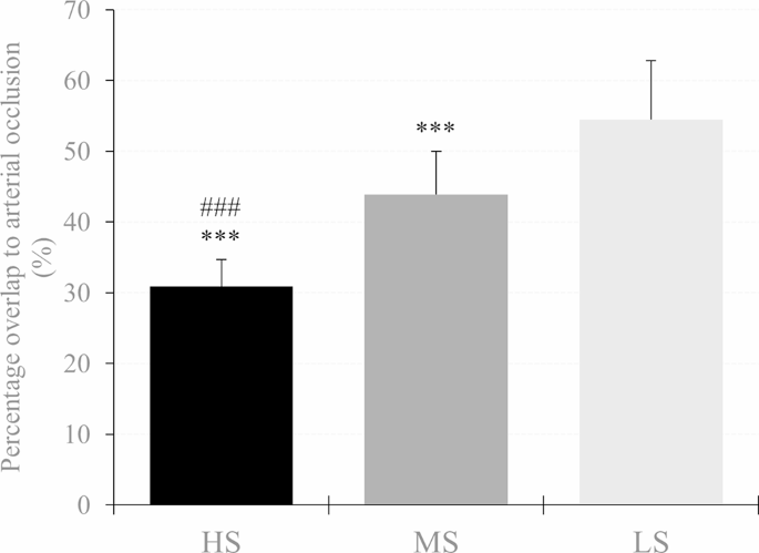
    Percent overlap needed for arterial occlusion in the high stiffness (HS), medium stiffness (MS), and low stiffness (LS) cuff. A significant difference between LS and MS is shown as *p<0.05, **p < 0.01, ***p < 0.001 and #p<0.05, ##p < 0.01, ### p < 0.001 respectively

    Peak systolic velocity of blood flow

    There was an overlap × cuff interaction (F3,642,120,192 = 71.952, P< 0.001, theP2= 0.686) and a main effect of overlap (F1,770,58,422 = 161.427, P< 0.001, theP2= 0.830) and cuff (F2.66 = 50.380, P< 0.001, theP2= 0.604) for vsys. Post hoc analysis showed that vsys was lower at 30% overlap in each cuff compared to baseline (HS: MD = -62.67 cm s− 1(-70.97 to -54.37 cm s− 1 ), P< 0.001, D= 3.95; MS: MD = -21.53 cm s− 1(-29.83 to -13.24 cm s− 1 ), P< 0.001, D= 1.36; LS: MD = -11.02 cm s− 1(-19.31 to -2.72 cm s− 1 ), P< 0.001, D= 0.69). Furthermore, when the HS cuff was applied, vsys was also lower at 20% overlap compared to baseline (MD = -20.51 cm s− 1(-28.80 to -12.21 cm s− 1 ), P< 0.001, D= 1.29). As for cuff differences, vsys was lower at 20% and 30% overlap using the HS cuff compared to the MS (MD = -16.89 cm s− 1(-26.08 to -7.70 cm s− 1 ), P< 0.001, D= 1.07 and MD = -39.91 cm s− 1(-49.11 to -30.71 cm s− 1 ), P< 0.001, D= 2.52, respectively) and the LS cuff (MD = -17.60 cm s− 1(-26.80 to -8.41 cm s− 1 ), P< 0.001, D= 1.11 and MD = -53.99 cm s− 1(-63.18 to -44.79 cm s− 1 ), P< 0.001, D= 3.40 respectively). Furthermore vsys was also lower at 30% overlap using the MS compared to the LS cuff (MD = -14.08 cm s− 1(-23.27 to -4.88 cm s− 1), P< 0.001, D= 0.89). Descriptive data are shown in Table 2; Fig. 4.

    Fig. 4
    figure 4

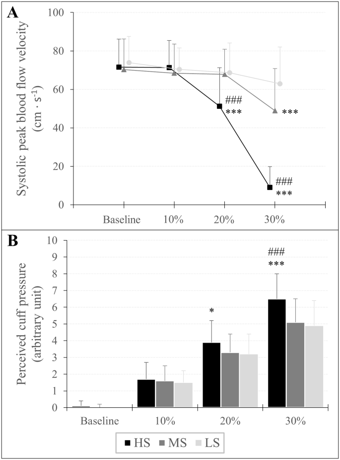
    Peak systolic velocity of blood flow (A) and assessment of perceived cuff pressure (B) in response to progressive practical pressure on blood flow restriction, expressed as percent overlap in relation to the individual’s arm circumference. A significant difference between LS and MS is shown as *p<0.05, **p < 0.01, ***p < 0.001 and #p<0.05, ##p < 0.01, ###p < 0.001 respectively

    Assessment of perceived cuff pressure

    An overlap × cuff interaction (F3,946,130,209 = 13.994, P< 0.001, theP2= 0.298) and a main effect of overlap (F1,668,55,046 = 674.771, P< 0.001, theP2= 0.953) and cuff (F2.66 = 11.067, P< 0.001, theP2= 0.251) was found for RPP. A post hoc analysis showed that RPP increased at each %overlap stage compared to baseline for all three cuffs (HS10%: MD = 1.57 au (0.98 to 2.17 au), P< 0.001, D= 1.51; HS20%: MD = 3.80 au (3.21 to 4.40 au), P< 0.001, D= 3.66; HS30%: MD = 6.41 au (5.82 to 7.00 au), P< 0.001, D= 6.16; MS10%: MD = 1.54 au (0.94 to 2.13 au), P< 0.001, D= 1.48; MS20%: MD = 3.32 au (2.72 to 3.91 au), P< 0.001, D= 3.19; MS30%: MD = 5.03 au (4.44 to 5.63 au), P< 0.001, D= 4.84; LS10%: MD = 1.46 au (0.87 to 2.05 au), P< 0.001, D= 1.40; LS20%: MD = 3.18 au (2.59 to 3.77 au), P< 0.001, D= 3.05; LS30%: MD = 4.95 au (4.36 to 5.55 au), P< 0.001, D= 4.76). Regarding differences between cuffs, RPP was higher using the HS cuff with 20% overlap compared to the LS cuff (MD = 0.71 au (0.06 to 1.37 au), P= 0.016, D= 0.69) and with an overlap of 30% compared to Member States (MD = 1.43 (0.78 to 2.09 au), P< 0.001, D= 1.38) and LS cuff (MD = 1.54 au (0.89 to 2.19 au), P< 0.001, D= 1.48). Descriptive data are shown in Table 2; Fig. 4.

    Oxygenation of the muscles

    SMO2: There was an overlap × cuff interaction (F2,374,78,326 = 3.232, P= 0.037, theP2= 0.089) and a main effect of overlap (F1,297,42,808 = 404,914.= P< 0.001, theP2= 0.925) and gender (F1.33 = 5.096, P= 0.031, theP2= 0.134) for SMO2. Post hoc analysis showed that SMO2 was lower at 20% overlap (HS: MD = -9.94% (-12.43 to -7.45%), P< 0.001, D= 1.15; MS = -8.28% (-10.77 to -5.78%), P< 0.001, D= 0.96; LS: MD = -7.42% (-9.98 to -5.00%), P< 0.001, D= 0.87) and 30% overlap (HS: MD = -17.46% (-19.95 to -14.97%), P< 0.001, D= 2.02; MS: MD = -14.91% (-17.42 to -12.42%), P< 0.001, D= 1.72; LS: MD = -13.79% (-16.28 to -11.30%), P< 0.001, D= 1.59) compared to baseline. Furthermore, the main effect of sex indicated that regardless of overlap and cuff used, SMO2 was lower in men than in women (MD = -5.16% (-9.80 to -0.51%), P= 0.031, D= 0.60).

    tHb: An overlap × cuff (F3,072,101,386 = 6,440, P< 0.001, theP2= 0.163) and overlap × sex interaction (F1,187,39,158 = 14.814, P< 0.001, theP2= 0.310) and a main effect of overlap (F1,187,39,158 = 117,125.= P< 0.001, theP2= 0.780) and gender (F1.33 = 27.981, P< 0.001, theP2= 0.459) was found for tHb. Post hoc tests showed that tHb was higher at 20% overlap (HS: MD = 0.13 au (0.09 to 0.18 au), P< 0.001, D= 0.45; MS: MD = 0.10 au (0.05 to 0.14 au), P< 0.001, D= 0.32; LS: MD = 0.06 au (0.02 to 0.11 au), P< 0.001, D= 0.20) and 30% overlap (HS: MD = 0.22 au (0.18 to 0.27 au), P< 0.001, D= 0.75; MS: MD = 0.17 au (0.12 to 0.21 au), P< 0.001, D= 0.55; LS: MD = 0.15 au (0.10 to 0.19 au), P< 0.001, D= 0.45) compared to baseline. Moreover, tHb was already higher at a 10% overlap using the HS (MD = 0.05 au (0.00 to 0.09 au), P= 0.018, D= 0.16) compared to baseline. Regarding sex differences, post hoc analysis showed that regardless of cuff, tHb was higher by 10% (MD = 0.06 au (0.01 to 0.10 au), P= 0.003, D= 0.20), 20% (MD = 0.15 au (0.11 to 0.20 au), P< 0.001, D= 0.52) and 30% overlap (MD = 0.24 au (0.19 to 0.29 au), P< 0.001, D= 0.80) in men, while in women tHb was only higher during a 30% overlap compared to baseline (MD = 0.12 au (0.07 to 0.16 au), P< 0.001, D= 0.39). In addition, tHb was higher in men compared to women at baseline (MD = 0.51 au (0.31 to 0.70 au), P< 0.001, D= 1.69). Descriptive data are shown in Table 2.

    Source link

  • Can a lappiplasty repair my bunion?

    Can a lappiplasty repair my bunion?

    lapiplastyBunions can be a real pain to deal with on your own. The symptoms of a bunion can be very disabling, making it difficult to walk or get from one place to another. The good news is that some treatment options can help you repair your bunion. With the right approach, your bunion problem can be a thing of the past. A lapiplasty is an approach that many people use to treat their bunion problems. Let’s take a closer look at what exactly a lapiplasty is and how it can help you say goodbye to bunion pain.

    Causes of a bunion

    Symptoms of a bunion?

    • Large bump on the outside of the big toe
    • Redness and swelling around the big toe
    • Corns and calluses on the friction site of the big toe and second toe
    • Limited movement of the big toe

    What is a lapiplasty?

    People often have the misconception that bunions can be fixed by just shaving off the bone, thinking it is an overgrowth. In reality, there are deeper issues that are causing the problems you are experiencing. In the past, bunion surgery approached the problem on a two-dimensional plane, while the problem to be addressed was three-dimensional. This is where Lapiplasty comes into play. With LapiplastyⓇ 3D Bunion Correction, instead of doing what traditional bunion surgery does, which is cutting the bone in half and shifting, Lapiplasty rotates the entire spine of the foot to repair the anatomical deformity that causes the bunions to form. By doing this, the chance of your bunions returning is minimal.

    If you suffer from bunions, Bone and Joint Specialists Orthopedic & Spine Center is the right place for you. With 4 locations in Indiana, we offer extensive access to our physicians who can provide you with world-class service. Working with us means you work with the best. Contact us today at 219.795.3360 or visit us online at www.orthopedicdoctors.com to schedule your consultation.

    Source link

  • Why does my knee feel numb and tingly after ACL surgery?

    Why does my knee feel numb and tingly after ACL surgery?

    It is usually normal for your knee to feel numb and tingly after ACL surgery. This is because some small nerves are cut during the surgical procedure. Don’t worry, we’re not talking about the really important nerves that allow you to bend and straighten your knee, but the nerves that allow you to feel things around your knee. This video below explains all the reasons why your knee may feel numb and tingly after ACL surgery. For more information, read this blog about 5 tips to help you recover after ACL surgery.

    Lauren Youssef, a physiotherapy student at the University of Toronto, explains why your knee may feel numb and tingly after ACL surgery.

    Read Lauren’s blog: Why is my knee numb and tingling after ACL surgery? | Curovat

    There are also 5 great blogs written by Lauren on our site where you can learn more about surgery and injury recovery.

    If you have had an ACL injury or surgery, download Curovate from the links below. Curovate is a physical therapy app that offers daily video-guided exercises, progress tracking, in-app chat with a physical therapist to answer your questions about surgery or injury, and the ability to measure your knee’s range of motion using just your phone.

    If you need more tailored help during your surgery or recovery from your injury, check out our Virtual Physiotherapy page to book your 1-on-1 video session with a physiotherapist.

    acl knee physical therapy 1080x1080 2
    Download it on Google Play

    Other blogs related to ACL injuries:

    References

    1. Horteur, C., Cavalié, G., Gaulin, B., Cohen Bacry, M., Morin, V., Cavaignac, E., & Pailhé, R. (2020). Saphenous nerve damage after anterior cruciate ligament reconstruction: Decreased area of ​​numbness after ligamentoplasty using the quadriceps tendon compared to the hamstring tendon. The Knee, 27(4), 1151–1157.

    2. Inderhaug, E., Strand, T., and Solheim, E. (2015). The impact of sensory deficits after hamstring autograft harvesting for ACL reconstruction. Knee surgery, sports traumatology, arthroscopy: Official Journal of the ESSKA, 23(4), 1060–1064.

    3. Kjaergaard, J., Faunø, L. Z., & Faunø, P. (2008). Loss of sensation after ACL reconstruction with hamstring graft. International Journal of Sports Medicine, 29(6), 507–511.

    4. Nerve Blocks for Surgery – Yale Medicine. (2020, July 02). Retrieved from https://www.yalemedicine.org/conditions/nerve-blocks-for-surgery

    5. Ochiai, S., Hagino, T., Senga, S., Yamashita, T., Oda, K., & Haro, H. (2017). Damage to the infrapatellar branch of the saphenous nerve in anterior cruciate ligament reconstruction using vertical skin incision for hamstring harvesting: risk factors and influence on treatment outcome. Journal of Orthopedic Surgery and Research, 12(1), 101.

    6. Sanders, B., Rolf, R., McClelland, W., and Xerogeanes, J. (2007). Prevalence of saphenous nerve damage after autogenous hamstring harvest: an anatomical and clinical study of tailor branch injury. Arthroscopy: The Journal of Arthroscopic and Related Surgery: Official Publication of the Arthroscopy Association of North America and the International Arthroscopy Association, 23(9), 956–963.

    7. Wang, H. D., Zhang, H., Wang, T. R., Zhang, W. F., Wang, F. S., & Zhang, Y. Z. (2018). Comparison of clinical outcomes after anterior cruciate ligament reconstruction with hamstring tendon autograft versus soft tissue allograft: a meta-analysis of randomized controlled trials. International Journal of Surgery (London, England), 56, 174–183.

    Source link

  • LSU Student Research Fellowship – Adult and Child Tracks

    LSU Ortho Student Research Fellowship

    This opportunity allows highly motivated students interested in orthopedics to be integrated into our academic orthopedics department for a year. You will be in constant conversation with faculty, residents, and department researchers as you learn the basics of research design, methodology, data analysis, and how to navigate the process from IRB application to manuscript submission. In addition to research, there are weekly opportunities for clinical exposure including surgery, clinic and department conferences. There are 3 tracks available: Basic Sciences, Pediatric Orthopedics, centered at the Children’s Hospital of New Orleans, and Adult Orthopedics, centered on the LSU medical school campus (however, all fellows will spend some time at each location).

    • A commitment for 1 academic year (fall to summer).
    • Develop and complete at least 2-3 independent research projects
    • Facilitate ongoing student/resident research projects where necessary.
    • Manage the content of research websites
    • Transporting research material, for example from operating theater to laboratories
      • Personal transportation required
    • Present your progress at monthly department research meetings
    • Attend weekly clinics and/or surgeries with various faculty members
    • Participate in the LSU Ortho student research committee
    • Participate in health disparities research (a department priority), including tissue bank projects, proposals, and grant submissions
    • Attend all department didactic/teaching sessions
    • Assist in developing/maintaining databases for surgical research
    • Effectively communicate the status of each project and progress with research team members
    • Select 1 of the 3 tracks (basic sciences, clinical adults/trauma, clinical pediatrics)
    • Full-time position, approximately $1150/month stipend

    For more information please contact Dr. Vinod Dasa (This email address is being protected from spambots. You need JavaScript enabled to view it.) with regard to the orthopedic direction of adult and basic sciences or Dr. Carter Clement (This email address is being protected from spambots. You need JavaScript enabled to view it.) regarding pediatric orthopedics training.

    https://www.medschool.lsuhsc.edu/ortho/research_fellows.aspx

    Source link

  • You Don’t Look Sick – Living With Rheumatoid Arthritis: DAY 3 IN YOSEMITE

    This morning started early after a long day yesterday. The burning sensation disappeared. I’m pretty sure it was something in the bath oil that caused me to have a bad reaction.

    I drove to Yosemite (it’s an hour away) via the one-way bridge that cars cross in both directions. I got into the Yosemite gate pretty quickly. I drove another 20 minutes to the meeting spot. I was early so I stopped to take some photos at some take-out points.

    AVvXsEj8fQFzX37CqWuTC2jscUzBzo I312LPh1ehpBTflwEXhv8EQw8ghDh4l0lGd6L7hi3LibIaKRMDGybeyNloXfPggwjTVRYa49EGOLV3wOTtOgcntGmT3g9JYih2wwNq 9hShgJZG73Y7fd 4skaxWXZjW0IDtE9 eMZD3KQLCpQXW4aN1Wg8r8AKWzh14

    I met up with Katherine and we got in her car to go to the next spot. It was Tuolumne Grove to see the gigantic majestic redwood trees. We walk through the forest.

    AVvXsEi4yLA 7L1du3 K46ylU7j5SvD yAZuB PcPY3leZ5wvYjyLCgMgzLTKxxNApCOCmLLxKoC4WfJ0Cvx8vOVGkyKLLWDMajXIcZ1neG33sTLuycIYTPOI3BMA e1A9CVfREjQ g6lEIjV sCsq80Zy6xQMhcuRym GMRySi5shIGkV TjujrLfFx7 7T3k

    Then we got in the car and drove to Olmsted Point where we walked a bit, took pictures and meditated for a while. There’s a fire in the park, so today the air was smoky.

    AVvXsEgli5LrIkMuER1svV8Afg5S2DwmfmPD8EDb2k hevvJd7rvJ5mdvAu4YofgKd5eTPeYyzqNld6n7 Bh9PYPRZloBfRKmqiDMv1Xn0fHbOreTQxZ0kpxE4F5VHAB7wroXm94phIgnHiv0vraqZr6P0OPHJebfIwSve5b8m6Q4RfayBh0ncreR2E cdc JzM

    Then we went to Taneya Lake where we ate a packed lunch she brought. It was nice to sit in her camp chairs and have lunch by a beautiful lake. One interesting fact: the boulders and trees all had rings around them. It was a trail left by the lake this year after the historic snowfall. The water rose so high!

    AVvXsEizIOYmk5AWD5gXMKAzsQPtpSg4BZMq5b2iEWbwKLCC8YLqvEwgCVc6M4sOd0qkbBj 39ssfPqRitZs295 zH3ZdJVvZrhj fPU VGu0yXPR0Tv2BVWPUEtBmejSsT6AzcZ

    AVvXsEheUXX pzP96brRcZT5f li9hN3vFO3FenrbrrCQhqcyJOXfaNsgdGo6A6RiJ3m75jHxg fRJbBpnGVmKV pHhLA031onSIpFQBj2AEoOeGqsJJJW1mz3dcjbS QN1GhP3M1E YrYhJCahM tIrJP tcgYl8Nl

    Then we went to Tuolumne Meadow to Soda Springs. It’s a nice walk to see water that is actually naturally carbonated. Scientists can’t figure out why. I also used an outhouse for the first time in a long time.

    AVvXsEgLrqKSsDFTG0SRqWl65m3NylngC9bpIYz3YDzG6dhKVQJDsvXvuGZ EQIdFK4m1vjhnjdDziLLD72P2YbDPfGAil2aPaVrfMjm4a3SWC3Ey4W cbKiXHDcjYRcEubfRqut00DbaFcq8WrXwgpOXLZLweW3iJ5fFmS2l7vEqBvJQjHvLj7cCRWMyRGQuaI

    AVvXsEjGh2TrFwFIhCeOjW3UE7wWj r 04fD5SAPcaf71xAF kIEO3bt1TZxdLwddPHBW

    We then headed back to Olmsted Point because the smoke cleared and I was able to get a better photo.

    AVvXsEhMPTTAwnqOxw37KDMnNfwdUynBY6N05oGBhITFyJPKn5E nAEmo5 o5zr8 iNsJZCXEG2eE 2MiSEO4oLxixb3dgeeJl8J23GedmGH9OZU98kXesVsojpFIbnb0vV5mH0WAb5JPBIux3EsllMMe7 kDaSNY4ft0z0LT yq8Q1ZDEHJksH2jHXBo4M8q2s

    That was the end of my tour/walk for the day. I drove back to the room to shower and get ready for dinner. Tonight I decided to get take out and eat on my porch.

    AVvXsEjjJNyKfZ7BaJuiiVniaRil5J9oayHFJ7W5rLRj3OuoFThoH5g2u3H QA66v8V47s49rsdV8K1RIYaIm9ozldos176TSRRQnojaiObZxEiipaPlbl3UaERkjbEN tYyD1Lyp0zS59VdR8opLX7VVvayIodsIPwEBwr2KRoG088gMTz34zWOXF22a3fZU w

    Now I have to go to bed, because tomorrow is coming soon. It was going to rain, so this walk will be interesting!

    By the way, if you’re interested in the dog, she was caught watching football with her boyfriend.

    AVvXsEgoL2D1V0m6Nm X7m0BkPpkfM5v2V96SidcOAtZEFsuJBpd p3ReujvzosbB87uzyWuA9kFGfb2Un 19osec3fDLg4 b4REONLUbo11JcCLswGrVCud5RiOwI3VyILSED

    Of course she comes from Chihuahua Mexico….

    See you tomorrow…

    Source link

  • Menopause and sleep disorders – better bones

    Menopause and sleep disorders – better bones

    Most of us know from experience that tossing and turning at night means you’ll feel bad the next day. And many of us understand that there are long-term health consequences when we get too little sleep.

    But recently I discovered something new from a fascinating study into sleep and its effect on how quickly women’s genes age after menopause – what’s known as their ‘epigenetic age’. (Carroll et al. 2017)

    In the study, postmenopausal women with five insomnia symptoms were biologically almost two years older than comparable women without insomnia symptoms. The more symptoms of insomnia – Waking up during the night, not being able to fall asleep, disturbed sleep and so on – the greater the effect. However, if you only sleep six hours a night, it appears that your epigenetic age does not increase, as long as it is a good night’s sleep. And that, of course, is what many of us have problems with.

    So what can we do about this? I would like to point out that the research shows that our genes are not completely out of our control, and that we can influence how they respond by taking better care of ourselves. For those of us who have gone through menopause and may be having trouble sleeping, this means looking at ways we can improve our rest.

    dr-browsn-favorite-sleep-remedies4 sleep tips to try

    • Keep a regular schedule. Your brain likes consistency, so go to bed at 11 p.m. and at the same regular time every night and in a dark room. When you get up, immediately soak up the morning light by going outside or opening a window. Ensuring good light health is key to your circadian health.
    • Turn off the electronics! Computers, TVs, phones: they’re all the enemy of sleep. Even a short duration of light shining into your eyes tricks your brain into thinking it’s time to wake up. Every night at least an hour before bed, turn off the phone and put it on the charger in another room so you aren’t tempted to look at it when you can’t sleep.
    • Eat early and in moderation. Heavy meals just before bed disrupt sleep, while avoiding caffeine and stimuli (such as dramatic/stressful TV shows) can also help with sleep.
    • Once you’re in bed, stay relaxed. Once you’re in bed, consider practicing relaxing belly breathing or guided visualizations like that of Yoga Nidra.

    In the checklist above you will see some of my favorite sleep remedies, including of course L-Tryptophan, which can be combined with magnesium for an even more relaxing effect.

    Reference:
    Carroll, JE et al. 2017. Epigenetic aging and immune aging in women with insomnia symptoms: findings from the Women’s Health Initiative Study, Biological Psychiatry 81(2):136-144.

    Dr.  Susan BrownI am Dr. Susan E. Brown. I am a clinical nutritionist, medical anthropologist, writer and motivational coach speaker. Learn my proven 6-step natural approach to bone health in my online courses.


    Source link

  • Women heal less quickly from ACL injuries than men

    Women heal less quickly from ACL injuries than men

    Injuries to the anterior cruciate ligament (ACL), located in the knee, are commonly thought to be caused by acute traumatic events, such as sudden twists. Led by researchers at Penn State, new work analyzing an animal model of ACLs suggests that such injuries may also occur as a result of chronic overuse, specifically due to a reduced ability to repair microtraumas associated with overuse. Importantly, according to the team, women are less able to heal from these microtraumas than men, which could explain why women are two to eight times more likely to tear their ACL ligaments than men.

    “ACL tears are one of the most common injuries, affecting more than 200,000 people in the US each year, and women are known to be particularly susceptible,” said lead researcher Spencer Szczesny, associate professor of biomedical engineering and orthopedics and rehabilitation at Penn . Stands. “Although recent research suggests that chronic overuse may lead to ACL injuries, no one has yet examined the different biological responses of female and male ACLs to applied force.”

    From the research, published in the Journal of Orthopedic Researchresearchers placed ACLs from deceased male and female rabbits in a custom-built bioreactor that simulated the conditions of a living animal but allowed for direct observation and measurement of the tissue. They then applied repetitive forces to the ACLs that mimicked the forces that would naturally occur during activities such as standing, walking and trotting, and measured the expression of genes related to healing.

    In male samples, the team found that low and moderate applied forces, such as those that would occur while standing or walking, resulted in increased expression of anabolic genes, which are related to building molecules needed for healing. In contrast, greater applied forces, such as would occur with repetitive trotting, reduced the expression of these anabolic genes. However, in female samples, the amount of force applied did not affect the level of anabolic gene expression.

    “It didn’t matter whether there was low, medium or high activity in women,” said Lauren Paschall, a biomedical engineering graduate student at Penn State and first author of the paper. “Female ACLs exposed to chronic use simply did not heal as well as male ACLs, which may explain why women are prone to injuries. This supports the hypothesis that non-contact ACL injuries are attributed to microtraumas associated with chronic overuse that predisposes the ACL make for injuries.” .”

    According to the researchers, one explanation for the gender differences the team observed could be due to the higher amounts of estrogen in women.

    “Some studies have shown that the overall effect of estrogen on ACL injury is negative,” Paschall said. “Specifically, studies have shown that human women are more likely to tear their ACLs during the preovulatory phase, when estrogen levels are high, than during the postovulatory phase, when estrogen levels are low.”

    She said the team plans to further investigate the role of estrogen in ACL injuries.

    Szczesny noted that although the team’s research did not take place in humans, the findings may indicate that providing women with additional recovery time after injuries could be beneficial.

    “Ultimately, this work could also help identify targets for therapies to prevent ACL injury in women,” he said.

    Other Penn State authors on the paper include Sabrina Carrozzi, graduate student; Erdem Tabdanov, assistant professor of pharmacology; and Aman Dhawan, professor of orthopedics and rehabilitation.

    The Orthopedic Research and Education Foundation and the Congressional Directed Medical Research Program supported this research.

    Source link

  • Lemon Garlic Chicken |  GF, DF |  BoneCoach™ Recipes – BoneCoach™

    Lemon Garlic Chicken | GF, DF | BoneCoach™ Recipes – BoneCoach™

    Looking for a weeknight-friendly dinner recipe?

    Try this!

    With just six ingredients, our Lemon Garlic Chicken recipe is a one-pot meal that is delicious and easy to prepare.

    Plus, it’s packed with bone-strengthening proteins and essential vitamins and minerals like selenium and phosphorus.

    You’ll find yourself returning to our Lemon Garlic Chicken recipe again and again…

    Try it tonight!

    2B RECIPE Lemon Garlic Chicken 1

    SERVES: 4

    TOTAL TIME: 40 minutes

    Ingredients

    1 whole flat chicken (or 8 chicken thighs with skin and bone)

    2 tablespoons (30 ml) freshly pressed extra virgin olive oil

    3 cloves garlic, minced

    2 tablespoons (30 ml) freshly squeezed lemon juice

    1/2 teaspoon (2 ml) dried oregano

    1/2 teaspoon (2 ml) sea salt

    Directions

    1) Place the oil, garlic, lemon juice, oregano and salt in a large oven dish and mix together. Dip the chicken in the lemon-garlic mixture.

    2) Bake in the oven at 375F for 40 minutes. Let the cooked chicken rest for 10 minutes before enjoying it.

    Recipe created by BoneCoach™ Team Dietitian Amanda Natividad-Li, RD & Chef.

    Medical disclaimer

    The information shared above is for informational purposes only and is not intended as medical or nutritional therapy advice; it does not diagnose, treat or cure any disease or condition; it should not be used as a substitute or substitute for medical advice from physicians and trained medical professionals. If you are under the care of a healthcare professional or are currently taking prescription medications, you should discuss any changes in your diet and lifestyle or possible use of nutritional supplements with your doctor. You should not stop prescribed medications without first consulting your doctor.

    Source link

  • Clearing up a confusing diagnosis – ACL Strong

    Clearing up a confusing diagnosis – ACL Strong

    Osteoarthritis is the most common joint disease in the United States and can cause pain, stiffness, and limitation in daily activities. Unfortunately, many people feel forced to give up their favorite sport or lifestyle as a result of being labeled “osteoarthritis.”

    A diagnosis of ‘osteoarthritis’ can be confusing.

    What does it mean? Does this mean that you cannot cycle, walk, hike or exercise without pain? How should you treat it and what can you do to prevent it from getting worse?

    What is osteoarthritis?

    Osteoarthritis, also known as degenerative joint disease, occurs when the cartilage that covers the ends of the bones in your joints gradually deteriorates. The degenerative process, which typically affects weight-bearing joints such as the knees, hips and hands, causes thinning of the cartilage, narrowing of joint spaces and reduced cushioning between the bones. This breakdown process puts more pressure on the sensitive surface of the bones in the joint, causing inflammation, pain, bone spurs and further wear and tear of the joint.

    knee arthrosis

    Being diagnosed with osteoarthritis does NOT mean that you cannot or should not have an active lifestyle. On the contrary, activity can actually improve the symptoms of osteoarthritis and slow or stop its progression. Many people with osteoarthritis lead an active, healthy lifestyle, without restrictions.

    How is osteoarthritis diagnosed?

    Osteoarthritis affects more than 32.5 million Americans and is usually diagnosed by physical examination and an X-ray. Based on the X-ray images, doctors will look for a smaller distance between the bones in the joint and for signs of inflammation. They will look at how much cartilage there is in the joint to protect the underlying bone from further wear and tear. They will also look for bone fragments or ‘spurs’ that are sticking into the joint and could cause more pain.

    unnamed

    X-ray findings should be compared with a physical examination to assess actual loss of range of motion, stiffness, weakness, and activity limitations before determining a treatment plan.

    Did you know that there are people with osteoarthritis who are NOT symptomatic? They have no pain, stiffness, swelling, or disability associated with the condition. In fact, they may not even know they have it!

    Evidence of degenerative joint changes can be seen in young adults to older adults, but the risk of developing osteoarthritis increases with age.

    Some of the risk factors that contribute to the development of osteoarthritis include:

    • Age (over 60 years)
    • Genetics
    • Being a woman
    • Being overweight
    • Have a pre-existing knee injury
    • Overuse of the knee or hip joint
    • Muscle weakness around the joint

    Does osteoarthritis always get worse?

    Osteoarthritis is a progressive condition that keeps getting worse if nothing is done to reduce pressure and wear on the joint.

    Joint wear and tear that causes inflammation, swelling and stiffness leads to more weakness, further breakdown, more stiffness and more disability. The cycle continues if no change is made to break the cycle.

    Although there is no cure for osteoarthritis, it is possible to slow or stop the progression of osteoarthritis through appropriate interventions…and most people don’t realize this!

    Someone with osteoarthritis may experience periods of ‘flare-ups’, where the joint suddenly becomes swollen, stiff and/or painful. There may also be periods when symptoms disappear, range of motion is restored, and the joint feels relatively normal.

    Controlling the “controllable factors” is an important tactic in managing osteoarthritis so that flare-ups don’t become worse than they need to be. Knowing what to do to calm a flare-up is an important step in long-term management and will help you continue doing the activities and sports you love.

    Normal range of motion (ROM) helps spread the forces on the joint; Good STRENGTH helps support the joint; and good MOVEMENT PATTERNS help reduce abnormal forces on the joint. These concepts form the basis of what will SLOW or STOP the worsening of degenerative joint disease.

    Losing weight if you are overweight, strengthening the muscles around the hip and knee, maintaining mobility and flexibility, and controlling inflammation and pain are important strategies to prevent osteoarthritis from worsening.

    Common treatments for osteoarthritis

    Activity

    While it may seem like activity may initially cause more pain and swelling, physical activity has actually been proven to reduce symptoms and improve the quality of life for people with degenerative joint disease. Exercise promotes circulation and can lead to more pain-free days. Of course, you need to make sure you choose the right activity, progress the right way, and listen to your body.

    A great place to start is by improving mobility and flexibility with simple stretching exercises.

    Screen Shot 2018 07 22 at 9.49.16 AM
    Heel slides

    Physiotherapy

    Physical therapy has been proven to relieve symptoms, increase activity tolerance and improve the quality of life for people with osteoarthritis using a structured approach to restore range of motion, functional strength and balance.
    If you want to live an active lifestyle, exercise and enjoy the outdoors, ACL STRONG provides an expert training progression to strengthen your legs and core, improve balance and body control, and learn how to benefit your body and lifestyle in the long term. can protect. This is an online program and an excellent option if you want the convenience and freedom of exercising independently at home, while still receiving expert guidance and support.

    Monster Walk zig zag side2
    Skaters 1

    Injectable products

    Your doctor may be willing to try hyaluronic acid which can be injected into the affected arthritic joint. This fluid is similar in makeup to your body’s synovial fluid, which is like oil in your car. An injectable can lubricate affected bone and joint tissue to reduce the symptoms of osteoarthritis, making you feel more comfortable and delaying the need for surgery.

    Arthroscopy

    Arthroscopic surgery is an option when an orthopedic surgeon is confident that symptoms can be reduced by clearing out the surface tissue in the affected joint. Arthroscopic surgery is performed by placing small incisions in the joint to remove damaged tissue and create a smoother surface. The surgery will facilitate increased blood flow to the joint, which can help ‘restart’ the healing process and reduce the symptoms of osteoarthritis.

    Arthroscopy

    Joint replacement surgery

    In cases where more conservative treatments have failed to relieve symptoms and improve quality of life, joint replacement surgery may become the best option. Replacing the degenerated joint surface with mechanical components can give you the recovery you need to be as active as you want.

    Recovery from joint replacement surgery will involve several months of activity progression and physical therapy. Although the timeline varies for everyone, most patients return to normal activities and exercise, such as walking, biking, golfing, tennis, weight lifting, housework, and yard work. With hard work, dedication and consistency, many can return to their previous sports and activities at a higher level.

    Osteoarthritis should NOT be daunting or intimidating. Receiving a diagnosis of osteoarthritis does not have to be confusing.

    👉 How you deal with your condition is up to you. You CAN lead an active, healthy and sporty lifestyle, with the presence of degenerative joint diseases. Manage it with exercise, nutrition, making good choices and expert support, and you can continue living the lifestyle you choose. 💪

    Source link

  • Real-world treatment patterns of rheumatoid arthritis in Brazil: analysis of DATASUS national administrative claims data for pharmacoepidemiological studies (2010-2020)

    Study design and database

    This was a descriptive, retrospective claims database study using the DATASUS database. The study identified patients with RA who sought care within SUS between January 1, 2010 and December 31, 2020.

    The administrative claims data in DATASUS is presented as procedure codes from billing data and includes demographic information, all procedures (inpatient and outpatient), costs, and additional information23. Hospital admission (SIH [Sistema de Informações Hospitalares])24 and outpatient (SIA [Sistema de Informações Ambulatoriais])11 data exists separately and is linked at the patient level through multiple steps with different combinations of individual level information (date of birth, gender, city and zip code) for a probabilistic linkage approach. This Brazilian Healthcare Record Linkage (BRHC-RLK) methodology has been used in previous studies to enable a more comprehensive recording of each patient’s health record and thus allow a more complete evaluation of their journey through the SUS system.25. The method is based on multiple steps with different combinations of patient information from both databases, making it possible to identify or link patient data in both systems, while maintaining the anonymized nature of the database.

    Patient-level data within DATASUS is anonymized and encrypted before being made available to researchers. DATASUS is publicly available and does not require further approval from ethics committees, according to Brazilian Ethics Resolution No. 510/2016.

    Study population

    The study population included patients with at least one claim of RA (according to the International Classification of Disease, 10th edition). [ICD-10] codes: M05.0, M05.3, M05.8, M06.0, M06.8, or M08.0) and ≥ 2 claims for disease-modifying antirheumatic drugs (DMARDs) ≥ 1 month apart in the 2010 survey– 2020 period. This study examined a treated population where the index date was the first DMARD claim and followed until the end of the study period (December 31, 2020) or the last available information. Detailed DMARD definitions can be found in Supplementary Table S1.

    To capture initial treatment and address the potential for misclassification common in claims data, patients with a DMARD claim without an RA ICD-10 code 12 months prior to the index date were excluded. The index date was defined as the date of the first RA ICD-10 and DMARD prescription in the public health system during the study period. Patients with RA with less than six months of follow-up were excluded, in an effort to reduce the number of individuals with a false diagnosis or lack of follow-up in the database.

    Because SUS is a healthcare system with universal coverage, patients with additional private health insurance can also receive medications (such as expensive drugs) covered by SUS at no out-of-pocket cost. This is often observed in other therapeutic areas26. For this reason, we stratified the results across the following cohorts: Cohort 1 is the entire study population, Cohort 2 is SUS-exclusive (i.e., dependent on SUS for all healthcare-related encounters, procedures, and treatments), and Cohort 3 represents SUS + private patients ( i.e. depending only on SUS for prescription drug coverage)26.

    Measurements of DMARD treatment

    DMARD treatments measured using procedure codes (see Supplementary Table S1) were grouped into the following categories: csDMARD for conventional synthetics and/or immunosuppressants (ciclosporin, cyclophosphamide, chloroquine, hydroxychloroquine, leflunomide, methotrexate, azathioprine and sulfasalazine), bDMARD for biological drugs (adalimumab, abatacept, etanercept, infliximab, rituximab, tocilizumab, golimumab, certolizumab) and tsDMARD for a synthetic, oral target therapy [Janus kinase (JAK) inhibitor, tofacitinib].

    Treatment patterns were evaluated by specific drug (independently of monotherapy or in combination) as provided for RA treatment and the order of available treatments in SUS, by line of therapy (LOT), time point of each drug, previous and subsequent DMARD treatments in SUS. The first treatment was the first therapy from the admission according to RA ICD-10 code. LOT was defined as at least three claims (dispensation) of the same drug (b/tsDMARD) in a row. A new series of at least three claims (dispensation) of the same drug in the correct order was considered a new line of treatment. Thus, the switch to a treatment was identified as at least three claims for drugs other than the previous one, which are not included in the definition of drugs used in combination. Gaps were allowed regardless of time and did not constitute a new LOT. First-line (LOT1) refers to initial treatment, first b/tsDMARD claim of RA during the study period. Second-line (LOT2) refers to the second b/tsDMARD used for RA treatment, when the first b/tsDMARD was stopped. Third line (LOT3) refers to the third b/tsDMARD, when the previous b/tsDMARD was terminated. csDMARDs used before b/tsDMARD were also assessed. Switching treatment was defined as at least three claims for drugs different from the previous one (new LOT), and not part of drugs used in combination.

    static analysis

    Derived variables included age and distance to the clinic. Age was defined as the age at the first claim of an ICD-10 code for RA in the database. Distance was calculated as the Euclidean distance (km) between two zip codes: the patient’s place of residence and the health care facility or tomography or antiangiogenic treatment facility, as applicable. Treatment switch, discontinuation and end of follow-up were the main outcomes of censoring events of interest, also relevant in defining LOT and creating Sankey diagrams.

    Continuous variables (e.g., age) are summarized by central tendency (means, medians) and dispersion (variance, range); and for categorical variables (e.g. gender) based on absolute number and percentage. Stratifications and/or sensitivity analyzes were performed to evaluate differences in gender, age groups, patient region of residence, drug use, treatment line, and others.

    Stratified analyzes for mainstream and new users were prespecified, and for SUS-exclusive and SUS+ private cohorts. Frequent users were patients with RA who were currently receiving bDMARD treatment, and new users were patients with RA who were starting a new bDMARD treatment (i.e., their first prescription). To describe the use and sequential patterns of RA bDMARD treatments, patients were stratified by treatment type, LOT-specific drug, and SUS-exclusive status.

    In multivariable logistic regression analyses, age, SUS-exclusive status, distance to clinic (160+ km), and pre-index cs/imsDMARD and other independent predictors were included to evaluate initiated therapy (LOT1) with b/tsDMARD (JAKi). Multivariable analyzes were performed using Cox regression models evaluating predictors by time to switch to tsDMARD (JAKi) compared to bDMARD (LOT2+), applying the same independent predictors from multivariable logistic regression analyzes (age, SUS-exclusive status , distance to clinic, pre-index cs/imsDMARD, other), plus the number of previously used bDMARDs. Sankey diagrams were used for visualizations of treatment patterns. Sankey diagrams quantitatively illustrate the sequence of treatment (and/or duration of treatment) and allow stratification by subpopulations of interest with censoring based on different treatment, discontinuation, or end of follow-up. Kaplan Meier survival analyzes and plots were generated for time to switch from LOT1 to LOT2, among those treated with b/tsDMARD, analyzed by drug type and by SUS-exclusive status.

    The visual representation of the time-to-event of the switch from LOT1 to LOT2 in patients receiving b/tsDMARD therapies was presented in Kaplan-Meier curves. The last available patient information or end of study period was considered censored for patients who did not switch from LOT1 to LOT2.

    All analyzes were performed using Python version 3.6.9 and statistical significance was set at p < 0.05.

    Source link