Every month, millions of women experience a range of physical and emotional changes during their menstrual cycle. While many are aware of the common symptoms like cramps and mood swings, some women also experience joint pain, including knee discomfort.
The fluctuation of estrogen and progesterone levels throughout the month can lead to changes in the body’s inflammatory responses, affecting joints and potentially causing pain.
Understanding the connection between the menstrual cycle and knee pain is crucial for proper management and treatment. We will explore how hormonal shifts can impact joint function and stability, and what can be done to alleviate the discomfort.
Key Takeaways
- Understanding the link between menstrual cycle and knee pain
- The role of hormonal fluctuations in joint pain
- How estrogen and progesterone affect the body
- Strategies for managing knee pain during menstruation
- When to seek professional help for persistent pain
Understanding the Connection Between Menstruation and Joint Pain
Women often report experiencing joint pain during their period, highlighting the need to understand the underlying causes. This connection between menstruation and joint pain is more common than many might think, affecting various aspects of women’s lives.
How Common is Joint Pain During Periods?
According to a 2023 study, many girls and women experience pain of differing severity at certain times during their menstrual cycle. For some, the pain is debilitating enough to hinder their regular activities, such as attending work or school for one to three days each month.
The prevalence of joint pain during menstruation varies, but it is recognized as a significant symptom among women of reproductive age. Understanding the extent of this issue is crucial for addressing the associated health concerns.
The Impact on Daily Activities
Joint pain during menstruation can significantly impact daily activities, from exercise routines to work performance and social engagements. The severity of the pain and its effect on activities can vary greatly among individuals.
Recognizing the connection between menstrual cycle and joint pain is essential for developing effective management strategies and improving overall health and quality of life.
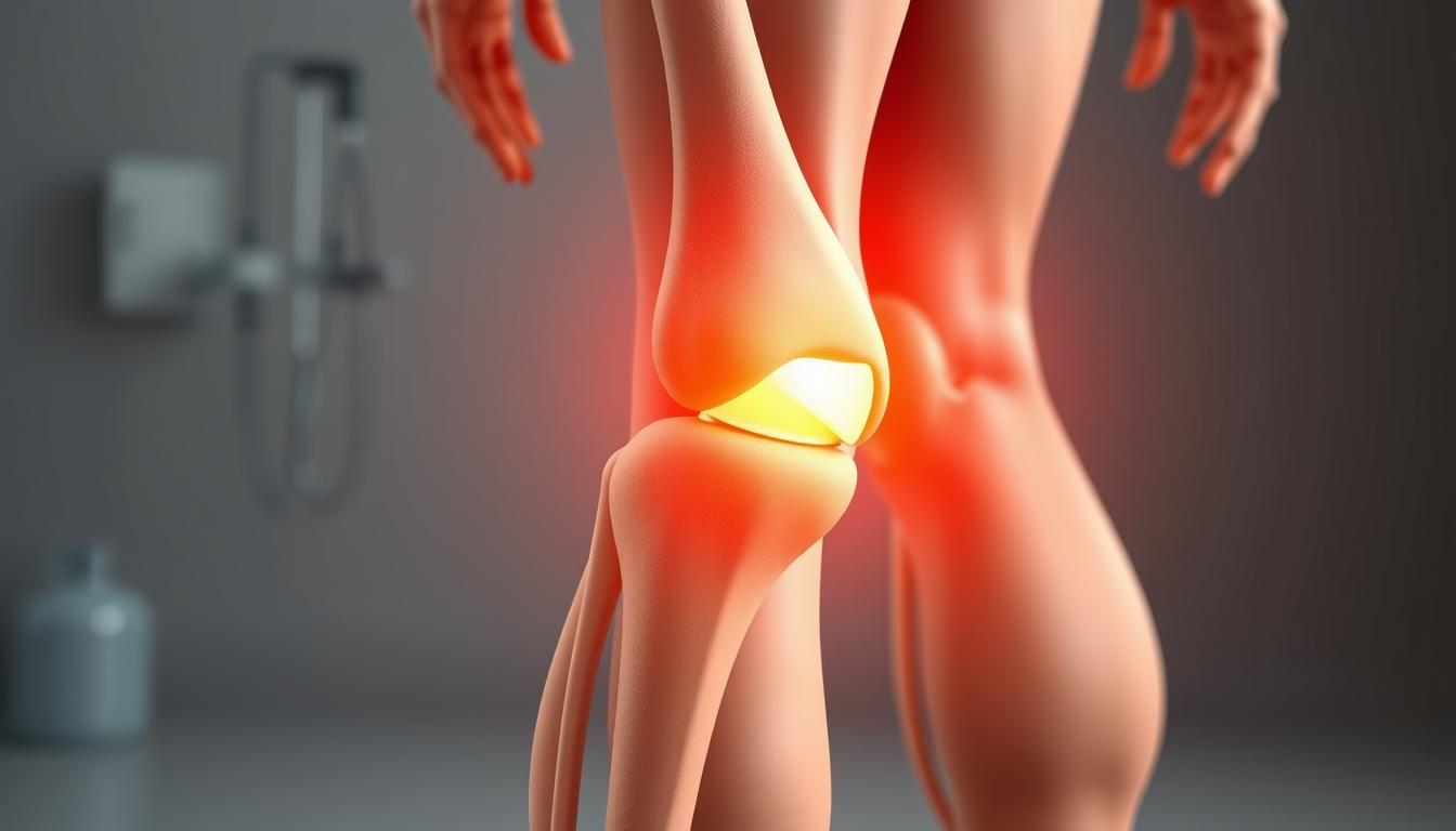
The Role of Hormones in Knee Pain During Menstruation
Understanding the role of hormones in knee pain during menstruation requires a deep dive into the menstrual cycle and its effects on the body. The menstrual cycle is characterized by fluctuations in hormone levels, particularly estrogen and progesterone, which can influence various physiological processes.
Estrogen and Progesterone Fluctuations
Throughout the menstrual cycle, estrogen and progesterone levels surge and drop at different times, affecting the body in multiple ways. Estrogen levels, for instance, have been shown to impact collagen synthesis and joint laxity, potentially leading to increased vulnerability in knee joints. As study author Matthew Tenan notes, “We know that progesterone and estrogen affect how the nervous system functions, so we theorized that the menstrual cycle might be affecting how women use their muscles.”
A study found that the firing rates of muscle fibers were significantly higher later in women’s cycles, about a week before their next period, compared to earlier in the menstrual cycle. This change is attributed to the decrease in progesterone and the maintained levels of estrogen.
| Hormone | Effect on the Body | Impact on Knee Joints |
|---|---|---|
| Estrogen | Influences collagen synthesis | May increase joint laxity |
| Progesterone | Affects inflammation responses | Can contribute to joint discomfort |
How Hormonal Changes Affect Muscle Function
Hormonal fluctuations during the menstrual cycle also affect muscle function around the knee. The changes in estrogen levels and progesterone influence muscle fiber recruitment patterns and neuromuscular control. As Tenan explains, “The way the brain activates the neurons that cause the muscle to move are altered specifically at the latter part of the cycle right before the start of the next period.”
This alteration in muscle function can lead to periods of increased joint vulnerability, potentially resulting in knee pain during menstruation. Understanding these changes is crucial for developing effective management strategies for menstrual knee pain.
“The menstrual cycle might be affecting how women use their muscles,” says Matthew Tenan, highlighting the complex interplay between hormonal changes and musculoskeletal function.
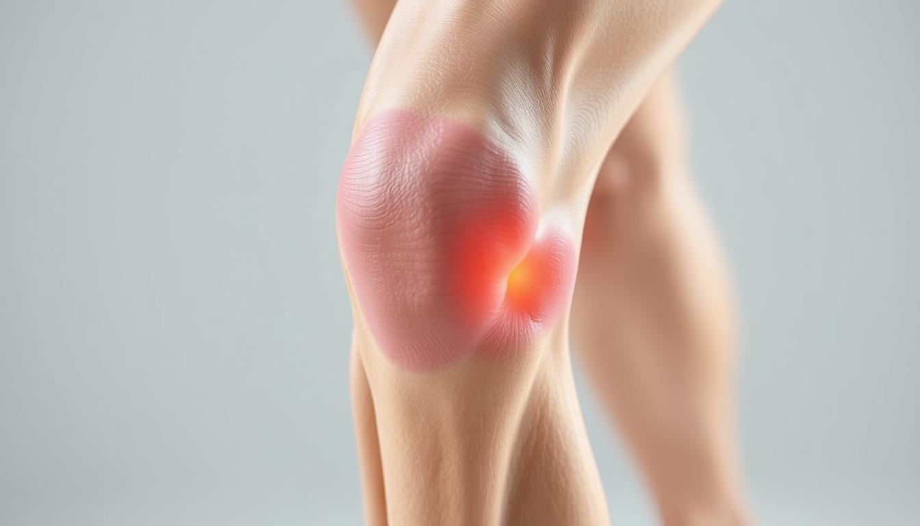
Causes of Knee Pain During Menstruation
Knee pain during menstruation is a common complaint among many women, and understanding its causes is crucial for effective management. We will explore the various factors that contribute to this condition, helping women identify the underlying reasons for their discomfort.
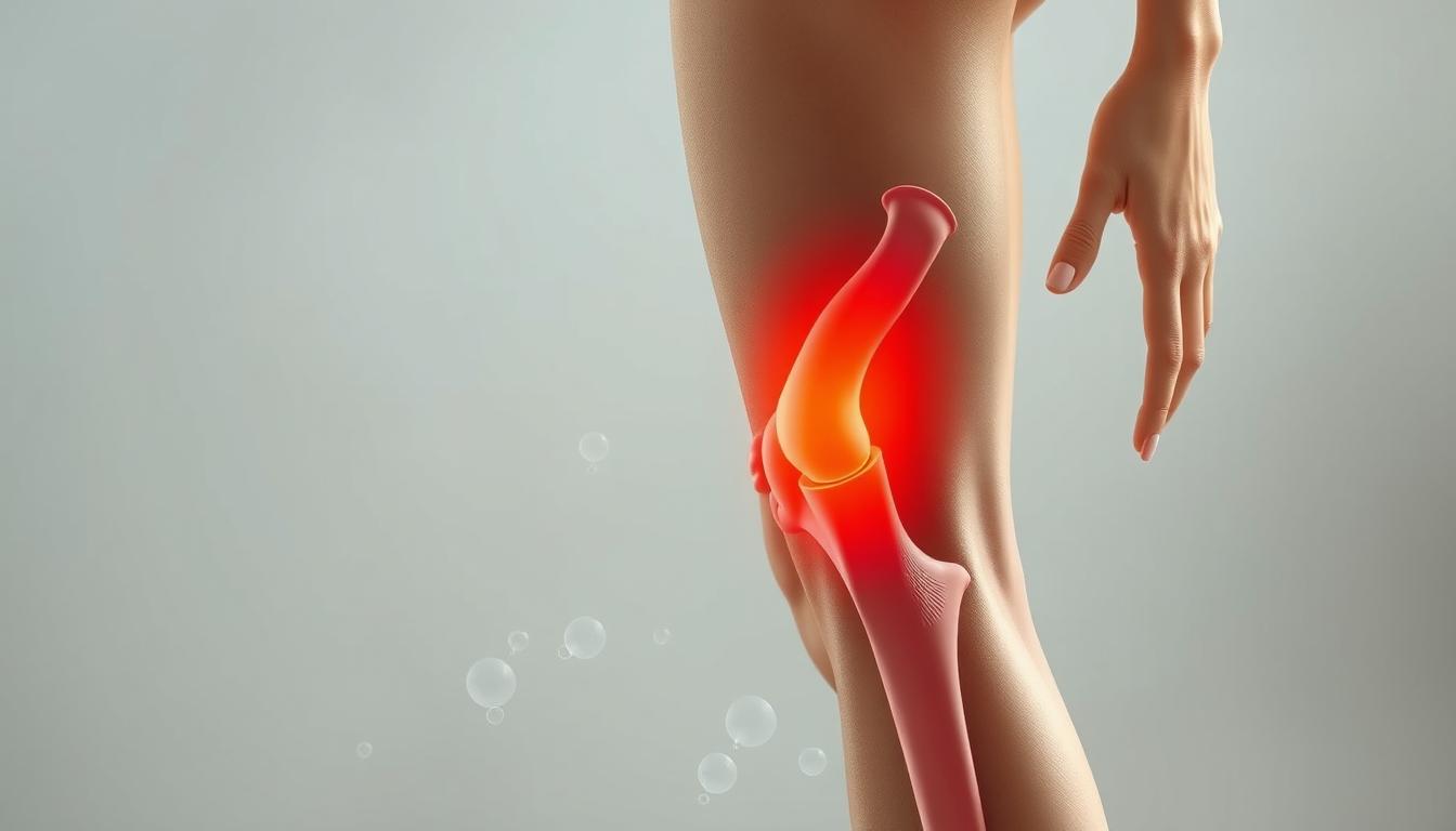
Prostaglandins and Inflammation
Prostaglandins are hormone-like substances that play a significant role in the menstrual process. They are responsible for uterine contractions during menstruation and can also triggerinflammationthroughout the body, including in the knee joints. As prostaglandin levels increase, they may heighten sensitivity and pain in the joints.
The body’s inflammatory response changes during different phases of the menstrual cycle, directly impacting joint comfort and function. During the late luteal phase, the drop in estrogen levels can lead to increased inflammation, potentially affecting joints and causing pain.
Fluid Retention and Joint Pressure
Some women experience fluid retention during their periods, which can put additional pressure on joints and exacerbate discomfort. Hormonal fluctuations affect the body’s water balance, leading to joint swelling and increased pain. This fluid retention can worsen existing knee pain or create new discomfort.
Underlying Conditions That May Worsen Symptoms
Women with conditions like endometriosis or Polycystic Ovary Syndrome (PCOS) may be more prone to joint pain during periods. A family history of joint issues or autoimmune conditions can also contribute to menstrual-related joint pain. Understanding these underlying conditions is crucial for managing symptoms effectively.
| Condition | Effect on Knee Pain |
|---|---|
| Endometriosis | Increased inflammation and pain |
| PCOS | Hormonal fluctuations exacerbating pain |
| Autoimmune Disorders | Heightened sensitivity and pain |
By understanding the causes of knee pain during menstruation, women can take the first step towards effective management and relief.
How Long Does Menstrual Knee Pain Last?
Understanding how long menstrual knee pain persists is crucial for managing symptoms effectively. The duration of knee pain during menstruation can vary significantly among women.
Typical Duration Patterns
Typically, knee pain associated with menstruation follows a specific pattern. It often begins just before or at the start of the menstrual period and can last for a few days. The pain usually peaks during the first day or two of bleeding and then gradually subsides as prostaglandin levels decrease.
We will outline the typical timeline of knee pain during menstruation, explaining when it usually begins in relation to the start of bleeding and how long it typically persists. The normal progression of joint pain symptoms includes peaking and then gradually subsiding as hormone levels stabilize.
Variations Among Different Women
There are significant variations in the duration of menstrual knee pain among different women. While some may experience brief discomfort, others might have pain throughout their menstrual cycle. Factors such as age, overall health status, fitness level, and genetic predisposition can influence the duration of knee pain.
- Some women may experience knee pain only during the first few days of their period.
- Others may have discomfort throughout their entire menstrual cycle.
- The intensity and duration of pain can also vary from one cycle to another.
It’s essential to distinguish between normal, temporary menstrual knee pain and pain that might indicate a more serious underlying condition. If the pain is persistent and severe, individuals should consult a doctor for proper evaluation and guidance.
Potential Complications of Untreated Menstrual Joint Pain
Failing to address menstrual joint pain can lead to complications that interfere with daily activities and overall well-being. If left untreated, the condition may result in both physical and psychological issues that can significantly impact one’s quality of life.
Physical Complications
Untreated menstrual joint pain can lead to decreased mobility and physical function over time. Prolonged inflammation in joints may cause long-term damage to joint structures, potentially resulting in chronic conditions. Persistent joint pain can also lead to compensatory movement patterns, creating additional musculoskeletal problems. For instance, altered gait patterns or avoiding certain activities can strain other parts of the body, leading to further discomfort.
Psychological Impact
The psychological impact of recurring menstrual joint pain should not be underestimated. Chronic pain can contribute to increased anxiety and depression, particularly if the pain is severe or persistent. The anticipation of monthly pain can also affect mental health, leading to mood disturbances. Furthermore, chronic pain can disrupt sleep quality, energy levels, and overall quality of life, making it essential to seek proper diagnosis and treatment to mitigate these effects.
Ignoring menstrual joint pain might delay the diagnosis of underlying conditions such as arthritis or endometriosis, which require specific treatment. Thus, proper management of menstrual joint pain is crucial to prevent both physical and psychological complications, ensuring overall health and well-being.
Effective Management Strategies for Knee Pain During Periods
Managing knee pain during menstruation requires a multi-faceted approach that incorporates lifestyle changes and targeted treatments. We will explore various strategies to help alleviate this discomfort.
Exercise and Movement Recommendations
Engaging in low-impact exercises such as yoga, swimming, or walking can significantly improve joint flexibility and reduce pain. These activities maintain joint mobility without exacerbating the discomfort. Regular exercise can also help reduce inflammation and pain by promoting blood flow and strengthening the muscles around the knee.
- Gentle stretching exercises to maintain flexibility
- Low-impact aerobics like swimming or cycling
- Yoga poses that target the knee and surrounding muscles
Heat Therapy and Self-Care Techniques
Applying heat to the affected area is a simple yet effective way to soothe achy joints and muscles. Using a heating pad or taking warm baths can provide significant relief. Additionally, practicing relaxation techniques such as meditation, deep breathing, or massage can help reduce stress-related joint pain.
Dietary Considerations
A well-balanced diet rich in calcium, magnesium, omega-3 fatty acids, and antioxidants can support joint health and potentially reduce knee pain during menstruation. Foods that are high in anti-inflammatory compounds can help mitigate inflammation and fluid retention.
- Increasing consumption of fatty fish for omega-3
- Eating leafy greens for calcium and magnesium
- Avoiding processed foods that may worsen inflammation
Medications and Pain Relief Options
For many women, NSAIDs (non-steroidal anti-inflammatory drugs) such as ibuprofen are effective in managing menstrual knee pain by reducing both pain and swelling. Alternatively, acetaminophen can be used for pain relief. It’s essential to follow the recommended dosing and be aware of potential side effects.
We recommend consulting a healthcare provider before starting any new medication regimen, especially if considering hormonal therapies for severe pain or hormonal imbalances.
When to Consult a Healthcare Provider About Menstrual Knee Pain
Seeking medical help for menstrual knee pain is a crucial step in managing symptoms and improving quality of life. Although menstrual-related joint pain is often temporary and manageable, individuals should consult a doctor if the pain is persistent and severe. If your period or PMS symptoms interfere with your ability to partake in your usual activities, such as going toschool or work, it’s time to talk to a healthcare provider.
Some people hesitate to discuss period problems with healthcare providers out of shame or fear. However, early diagnosis helps expand the number of remedies and reduce the risk of invasive treatments. A healthcare provider can help overcome these barriers by being empathetic and listening to your concerns.
Specific warning signs that indicate when menstrual knee pain requires professional medical attention include severe pain that doesn’t respond to over-the-counter NSAIDs (non-steroidal anti-inflammatory). If you notice significant changes in your usual period symptoms, such as recurrent vaginal yeast infections, you should also seek medical help.
By understanding the causes of menstrual knee pain and seeking appropriate treatment, individuals can alleviate their discomfort and prevent potential long-term complications. Effective communication with healthcare providers is key to receiving the right diagnosis and treatment plan.
FAQ
What causes knee discomfort during my menstrual cycle?
We experience knee discomfort during our menstrual cycle due to hormonal fluctuations, particularly the changes in estrogen and progesterone levels. These changes can lead to increased prostaglandin levels, causing inflammation and pain in our joints.
How do prostaglandins contribute to knee pain?
Prostaglandins are hormone-like substances that our body produces. They can cause our uterus to contract, leading to cramps, and also trigger inflammation in our joints, resulting in knee pain.
Can exercise help alleviate menstrual knee pain?
Yes, engaging in gentle exercises like yoga can help us manage knee pain during our periods. Exercise can improve joint mobility and reduce stiffness, making it easier to perform daily activities.
Are there any medications that can help with menstrual knee pain?
We can consider taking non-steroidal anti-inflammatory drugs (NSAIDs) to help alleviate knee pain and reduce inflammation. However, it’s essential to consult our healthcare provider before taking any medication.
How long does menstrual knee pain typically last?
The duration of knee pain during our menstrual cycle can vary from woman to woman. Generally, it subsides once our period ends, but some of us may experience it for a longer duration.
Can underlying conditions worsen menstrual knee pain?
Yes, pre-existing conditions like arthritis or previous injuries can exacerbate knee pain during our menstrual cycle. It’s crucial to consult our healthcare provider if we’re experiencing persistent or severe knee pain.