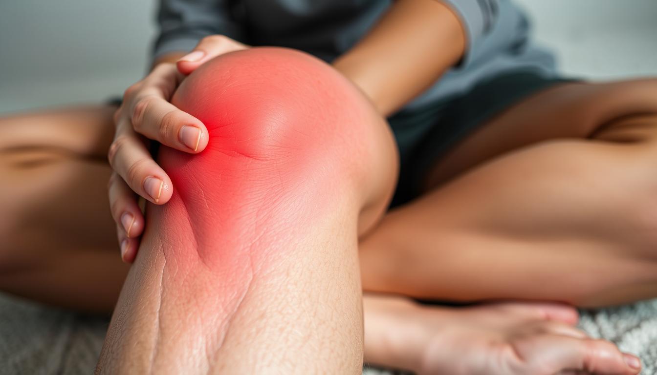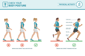

Every runner dreads that telltale pain front knee that signals something is wrong. The ache around kneecap intensifies with each step. Stairs become challenging. Yet stopping your training feels impossible.
Runner’s knee affects millions of active individuals each year. The good news? You can treat this condition without sidelining your passion.
This comprehensive guide reveals proven strategies to heal patellofemoral pain syndrome while maintaining your running routine. You will discover practical modifications, targeted exercises, and expert techniques that address the root causes of knee pain.
Understanding Runner’s Knee and Patellofemoral Pain Syndrome
Runner knee represents one of the most common overuse injuries in sports medicine. Medical professionals call this condition patellofemoral pain syndrome. The name describes pain occurring where your kneecap meets your femur.
The patella normally glides smoothly within a groove on your thigh bone. When this tracking system fails, friction develops. This mechanical problem creates the characteristic pain front knee that defines the condition.
What Causes Patellofemoral Pain
Multiple factors contribute to developing patellofemoral pain syndrome. Biomechanical issues top the list of causes. Your body mechanics during activities create stress patterns that affect the joint.
Muscle imbalances frequently lead knee pain development. Weak hip muscles fail to stabilize your leg properly. Tight leg structures pull the kneecap off its intended path. Poor foot mechanics transfer abnormal forces upward through your lower body.
Training errors accelerate the condition. Increasing mileage too quickly overwhelms your tissues. Running on cambered surfaces creates uneven loading. Worn footwear provides inadequate support for your unique biomechanics.
Biomechanical Risk Factors
- Weak quadriceps muscles affecting kneecap stability
- Tight iliotibial band pulling patella laterally
- Overpronation altering lower leg alignment
- Hip muscle weakness reducing pelvic control
- Poor core strength compromising running form
Training-Related Causes
- Rapid increase in running volume or intensity
- Excessive downhill running creating braking forces
- Inadequate recovery between hard sessions
- Sudden changes in running surface or terrain
- Inappropriate footwear for your gait pattern
Recognizing the Symptoms of Runner’s Knee
Pain around kneecap serves as the hallmark symptom. This discomfort typically feels dull and achy rather than sharp. The sensation concentrates behind or around your kneecap.
Certain activities provoke symptoms more than others. Running downhill commonly aggravates the condition. Navigating stairs challenges the joint. Sitting with bent knees for extended time creates stiffness.
Some individuals notice grinding sounds when bending their knee. These sounds indicate cartilage irregularities. Swelling may develop around the front knee area. The joint might feel unstable during certain movements.
Key Symptom Pattern: Pain front knee that worsens with activity, improves with rest, and returns when you resume running characterizes patellofemoral pain syndrome. Early recognition allows faster treatment response.

Smart Strategies for Running Through Recovery
Stopping running completely often proves unnecessary for treating runner’s knee. Strategic modifications allow continued activity while promoting healing. The key involves reducing strain on the patellofemoral joint without eliminating movement entirely.
Your tissues need controlled loading to recover properly. Complete rest can lead to muscle weakness and delayed healing. Intelligent training adjustments provide optimal stimulus for tissue adaptation without pain.
Reducing Running Volume and Intensity
Cutting your weekly mileage represents the first modification. Reduce your total distance by thirty to fifty percent initially. This reduction decreases cumulative stress on the joint while maintaining cardiovascular fitness.
Intensity matters as much as volume. Replace speed work with easy-paced runs. Your effort should allow comfortable conversation throughout. Hard intervals and tempo runs create excessive joint loading during recovery.
Frequency adjustments help manage symptoms. Instead of running six days weekly, reduce to three or four sessions. Insert rest days between runs. This spacing allows inflammation to settle between training bouts.
Volume Reduction Guidelines: If your typical week includes forty miles, temporarily decrease to twenty or twenty-five miles. Monitor your pain levels during and after each run. Gradually increase distance only when you complete sessions without pain.
Modifying Your Running Surface and Terrain
Surface selection significantly impacts joint stress. Hard pavement creates higher impact forces than softer alternatives. Seek out grass fields, dirt trails, or rubberized tracks for your runs.
Downhill running particularly aggravates patellofemoral pain. The braking mechanics place enormous strain on the front knee structures. Choose flat routes or slight inclines during your recovery phase.
Cambered roads force asymmetrical loading patterns. Running consistently on road shoulders creates uneven stress. Select level surfaces or alternate your running direction on crowned roads.
Preferred Running Surfaces
- Soft dirt trails with minimal technical features
- Well-maintained grass fields or parks
- Rubberized outdoor tracks
- Treadmills with cushioned decks
- Flat asphalt roads without camber
Surfaces to Avoid Temporarily
- Concrete sidewalks and hard pavement
- Steep downhill sections or descents
- Technical rocky trails requiring jarring movements
- Heavily cambered road shoulders
- Uneven surfaces with holes or obstacles

Incorporating Cross-Training Activities
Cross-training maintains fitness while reducing knee stress. Swimming provides excellent cardiovascular work without impact. The water supports your body weight completely. Focus on freestyle or backstroke for best results.
Cycling offers another low-impact alternative. Proper bike fit becomes crucial for knee health. Your saddle height should allow slight knee bend at the bottom of each pedal stroke. Avoid high resistance that strains the joint.
Deep water running mimics running mechanics without ground impact. Special flotation belts keep you upright in the pool. This activity preserves running-specific muscle patterns during recovery.
Optimize Your Recovery with Professional Guidance
Struggling to balance training and healing? A sports medicine specialist can create a personalized plan that keeps you running while addressing your specific biomechanical issues.
Running Form Adjustments to Reduce Knee Strain
Biomechanical modifications during running significantly decrease patellofemoral stress. Small changes in your movement patterns redistribute forces away from vulnerable structures. These adjustments feel awkward initially but become natural with practice.

Increasing Your Cadence
Step frequency directly affects joint loading. Most runners naturally select cadences between one hundred sixty and one hundred seventy steps per minute. Increasing this rate to one hundred eighty or above reduces impact forces.
Higher cadence means shorter stride length. Your foot lands closer to your center of mass. This positioning decreases braking forces and knee flexion angles. Both changes reduce strain on the patellofemoral joint.
Implement cadence changes gradually. Increase your step rate by just five percent initially. Use a metronome app or music with appropriate beats per minute. Your body needs time to adapt to new movement patterns.
Adjusting Your Foot Strike Pattern
Landing mechanics influence knee stress significantly. Heel striking with your foot far ahead creates excessive braking. This pattern increases force transmission through the front knee structures.
Transitioning toward a midfoot strike reduces these forces. Your foot contacts the ground more beneath your body. The change decreases the lever arm affecting your kneecap. Knee flexion at initial contact also increases slightly.
Make strike pattern changes cautiously. Abrupt transitions cause calf and Achilles tendon issues. Practice new mechanics for short intervals during easy runs. Gradually extend the duration as your muscles adapt.
Transition Warning: Changing your foot strike pattern too quickly can cause different injuries. Limit modified running to ten minutes during your first week. Add five minutes weekly as your lower leg muscles strengthen.
Maintaining Proper Body Alignment
Posture affects force distribution throughout your kinetic chain. Forward lean from your ankles rather than your waist. This alignment engages your core muscles and maintains efficient mechanics.
Hip positioning influences knee tracking. Avoid excessive pelvic drop on your stance leg. Strong hip abductors prevent this compensatory motion. Practice single-leg balance exercises to develop this stability.
Arm swing contributes to overall efficiency. Keep your elbows bent at ninety degrees. Swing from your shoulders rather than across your body. Proper arm mechanics reduce rotational forces at the knee.

Essential Strengthening Exercises for Runner’s Knee Recovery
Targeted muscle strengthening addresses the root causes of patellofemoral pain. Weak muscles around your hip and knee allow poor movement patterns. Building strength in specific areas improves joint mechanics and reduces pain.
Consistency matters more than intensity with these exercises. Perform them three to four times weekly. Quality movement trumps high repetitions. Focus on controlled motions throughout each exercise.

Quadriceps Strengthening Protocol
The quadriceps muscles control kneecap movement directly. The vastus medialis oblique muscle particularly influences patella tracking. Weakness in this area allows lateral kneecap drift.
Terminal knee extensions target this critical muscle. Sit with your leg extended and a rolled towel under your knee. Straighten your leg completely while squeezing your thigh muscles. Hold for five seconds and repeat fifteen times.
Wall sits build isometric quadriceps strength. Stand with your back against a wall. Slide down until your knees bend to ninety degrees. Hold this position for thirty to sixty seconds. Complete three sets with rest between.
Step-downs develop eccentric quadriceps control. Stand on a small step or platform. Slowly lower your opposite foot toward the floor. Control the descent for three seconds. Return to start and repeat ten times per leg.
Hip Strengthening Exercises
Hip abductor weakness allows excessive knee valgus during running. This inward collapse increases lateral forces on the kneecap. Strengthening these muscles improves lower extremity alignment.
Clamshell exercises isolate the hip abductors effectively. Lie on your side with knees bent. Keep your feet together while raising your top knee. Perform fifteen repetitions on each side. Add resistance bands as you progress.
Side-lying leg raises target the same muscle group. Lie on your side with your bottom leg bent. Raise your straight top leg toward the ceiling. Control the motion in both directions. Complete twelve repetitions per side.
Single-leg bridges strengthen your glutes and improve hip stability. Lie on your back with one knee bent. Extend your other leg straight. Push through your bent leg to lift your hips. Hold for two seconds at the top. Perform ten repetitions per side.
Beginner Exercise Sequence
- Terminal knee extensions – 2 sets of 15 reps
- Wall sits – 3 sets of 30 seconds
- Clamshells – 2 sets of 15 reps per side
- Side-lying leg raises – 2 sets of 12 reps per side
- Single-leg bridges – 2 sets of 10 reps per side
Advanced Exercise Progression
- Single-leg squats – 3 sets of 10 reps per side
- Bulgarian split squats – 3 sets of 12 reps per leg
- Step-downs with control – 3 sets of 12 reps per leg
- Lateral band walks – 3 sets of 20 steps each direction
- Weighted single-leg bridges – 3 sets of 12 reps per side
Core Stability Work
Core strength influences your entire kinetic chain. Weak abdominal and back muscles allow excessive trunk motion during running. This instability transfers to poor lower extremity mechanics.
Planks build foundational core endurance. Hold a front plank position for thirty to sixty seconds. Maintain a straight line from shoulders to ankles. Progress to side planks for oblique engagement.
Dead bugs improve core stability while moving your limbs. Lie on your back with arms extended upward. Slowly lower opposite arm and leg toward the floor. Return to start and alternate sides. Complete ten repetitions per side.

Flexibility and Mobility Work for Knee Pain Relief
Tight muscles alter joint mechanics and increase patellofemoral stress. Regular stretching improves tissue flexibility and reduces strain on vulnerable structures. Dedicate time daily to mobility work for best results.
Perform stretches after running when your muscles are warm. Hold each position for thirty seconds minimum. Breathe deeply and avoid bouncing movements. Consistency produces lasting flexibility improvements.

Quadriceps and Hip Flexor Stretches
Tight quadriceps pull on the kneecap and increase joint compression. Standing quad stretches effectively address this tightness. Stand on one leg and pull your opposite foot toward your buttocks. Keep your knees together and push your hips forward.
Hip flexor tightness affects pelvic positioning during running. Perform kneeling hip flexor stretches regularly. Place one knee on the ground and the other foot forward. Push your hips forward while maintaining upright posture. You should feel stretching in the front of your hip.
IT Band and TFL Mobility
The iliotibial band connects to your kneecap through lateral structures. Tightness in this tissue pulls the patella outward. Foam rolling helps release tension in the IT band and tensor fasciae latae.
Lie on your side with a foam roller under your thigh. Roll from your hip to just above your knee. Spend extra time on tender spots. Perform this mobility work for one to two minutes per leg daily.
Cross-leg IT band stretches complement foam rolling. Stand and cross one leg behind the other. Lean toward the side of your front leg. Hold this position while feeling stretch along your outer thigh.
Hamstring and Calf Flexibility
Tight hamstrings affect knee mechanics during running. Perform standing hamstring stretches by placing your heel on a low step. Keep your leg straight and lean forward from your hips. Maintain a neutral spine throughout the movement.
Calf tightness limits ankle mobility and alters running mechanics. Wall calf stretches address both gastrocnemius and soleus muscles. Place your hands on a wall and step one foot back. Keep your back heel down and lean forward. Bend your back knee slightly to target the soleus muscle.
Essential Daily Stretches
- Standing quadriceps stretch – 30 seconds per leg
- Kneeling hip flexor stretch – 30 seconds per side
- IT band foam rolling – 2 minutes per leg
- Standing hamstring stretch – 30 seconds per leg
- Wall calf stretch – 30 seconds per leg (straight and bent knee)
Stretching Best Practices
- Stretch after workouts when muscles are warm
- Hold each position for minimum 30 seconds
- Breathe deeply and relax into stretches
- Avoid bouncing or forcing movements
- Perform daily for cumulative flexibility gains
Footwear and Equipment Considerations for Knee Health
Running shoes significantly influence lower extremity mechanics. Worn or inappropriate footwear contributes to poor alignment and increased joint stress. Proper shoe selection and replacement schedules support healthy movement patterns.

Selecting Appropriate Running Shoes
Your foot type determines optimal shoe characteristics. Visit a specialty running store for gait analysis. Experts will observe your running mechanics and recommend suitable options.
Overpronators benefit from stability shoes that limit excessive inward rolling. These shoes feature firmer midsole material on the inner edge. The support prevents the collapse that stresses knee structures.
Neutral runners typically perform well in cushioned shoes without excessive support features. These designs allow natural foot motion while providing impact absorption. High-arched feet particularly need adequate cushioning.
Replace running shoes every three hundred to five hundred miles. Track your mileage using a training log or GPS watch. Worn shoes lose cushioning and support properties. This degradation increases injury risk significantly.
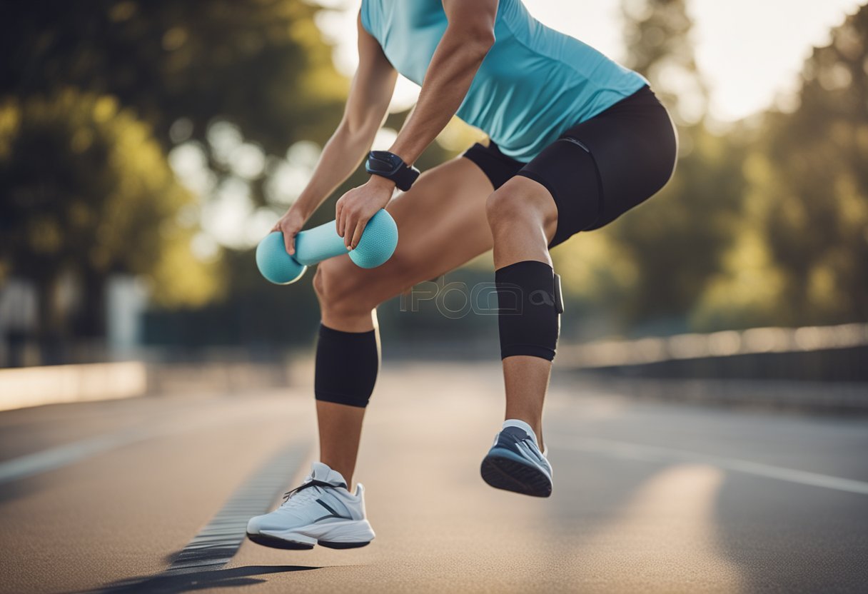
Knee Braces and Supportive Gear
Patellar tracking braces may provide temporary symptom relief. These devices feature a small pad that guides kneecap alignment. Use them during runs if they reduce your pain without creating dependency.
Compression sleeves offer support and proprioceptive feedback. The gentle pressure increases awareness of knee position. Some runners find this helpful during the recovery phase.
Kinesiology tape represents another supportive option. Proper taping techniques can improve patellar tracking. Consult a physical therapist for instruction on effective application methods.
Equipment Note: Supportive devices should complement your strengthening program, not replace it. Use braces or tape during the acute pain phase while building muscle strength. Gradually reduce reliance as your muscles improve.
Training Surface Equipment
Treadmills with good cushioning reduce impact forces compared to roads. Modern machines offer shock absorption systems that protect your joints. Adjust the incline to one percent to simulate outdoor running conditions.
Quality insoles improve shoe fit and foot support. Custom orthotics address specific biomechanical issues. Over-the-counter arch supports help many runners with mild overpronation or flat feet.

Get Expert Gait Analysis and Footwear Recommendations
Professional running gait analysis identifies your specific biomechanical patterns. Our specialists provide personalized footwear recommendations and custom orthotic solutions that address your unique needs.
Additional Treatment Modalities for Pain Management
Self-treatment techniques complement your exercise program. These modalities reduce inflammation and promote tissue healing. Incorporate them into your daily routine for optimal recovery.
Ice and Heat Therapy
Ice application reduces inflammation and numbs acute pain. Apply ice packs for fifteen to twenty minutes after running. Place a thin cloth between ice and skin to prevent tissue damage.
Heat therapy relaxes tight muscles and improves blood flow. Use heating pads before stretching sessions. Avoid heat during acute inflammation periods. Wait until initial swelling subsides before applying warmth.
Contrast therapy alternates ice and heat exposure. This technique may enhance circulation and recovery. Use three minutes of heat followed by one minute of ice. Repeat this cycle three to four times.
Massage and Self-Myofascial Release
Regular massage addresses muscle tightness and trigger points. Focus on your quadriceps, IT band, and calf muscles. Professional sports massage accelerates recovery when budget allows.
Self-massage using foam rollers or massage balls provides daily relief. Spend extra time on tender areas without creating excessive pain. Roll slowly and breathe deeply during the process.
Percussion massage devices offer targeted muscle release. These tools deliver rapid pulses that reduce muscle tension. Use them on your thighs and calves for two to three minutes per area.

Anti-Inflammatory Strategies
Over-the-counter anti-inflammatory medications reduce pain and swelling. Ibuprofen or naproxen taken as directed can help during acute phases. Consult your physician before extended medication use.
Natural anti-inflammatory approaches include dietary modifications. Foods rich in omega-3 fatty acids reduce systemic inflammation. Turmeric and ginger possess anti-inflammatory properties.
Adequate sleep supports tissue recovery and reduces inflammation. Aim for seven to nine hours nightly. Poor sleep impairs healing processes and increases injury risk.
Long-Term Prevention Strategies for Runners
Preventing recurrence requires ongoing attention to training principles. Smart progression and consistent strength work keep patellofemoral pain syndrome at bay. Implement these strategies permanently rather than just during recovery.

Progressive Training Load Management
The ten percent rule guides safe mileage increases. Add no more than ten percent to your weekly volume. This gradual progression allows tissues to adapt without overload.
Hard-easy training patterns prevent cumulative fatigue. Follow intense workouts with recovery runs or rest days. Your body needs time to repair and strengthen between challenging sessions.
Periodization organizes your training into distinct phases. Build base mileage before adding speed work. Include recovery weeks with reduced volume every third or fourth week. This structure optimizes adaptation while preventing overuse.
Maintaining Strength and Flexibility
Continue strengthening exercises even after pain resolves. Perform your exercise routine two to three times weekly indefinitely. This maintenance program preserves the improvements you achieved.
Regular stretching prevents flexibility losses over time. Dedicate ten to fifteen minutes daily to mobility work. Consistent practice maintains the range of motion needed for healthy mechanics.
Reassess your form periodically as fatigue develops. Video yourself running when tired during long runs. Compare this footage to your fresh running form. Address any mechanical breakdowns with targeted drills.
Regular Biomechanical Assessments
Annual gait analysis detects developing issues early. Changes in strength, flexibility, or footwear affect your mechanics. Professional evaluation identifies problems before they cause pain.
Monitor for asymmetries in your training response. One-sided tightness or weakness indicates compensatory patterns. Address these imbalances promptly through targeted exercises.
Replace running shoes before they fully break down. Track mileage carefully and retire shoes around four hundred miles. Maintaining proper footwear prevents biomechanical regression.
Weekly Prevention Checklist
- Perform strengthening exercises 2-3 times
- Complete daily stretching and mobility work
- Track weekly mileage and intensity
- Follow hard-easy training pattern
- Monitor for early warning signs of pain
- Maintain adequate sleep and recovery
Monthly Prevention Tasks
- Review training progression and adjust as needed
- Check running shoe wear and mileage
- Assess any developing aches or tightness
- Schedule recovery week if training intensively
- Perform self-assessment of running form
- Update training log with patterns and trends
When to Seek Professional Medical Help
Self-treatment works for many cases of runner’s knee. However, certain symptoms require professional evaluation. Recognizing these warning signs prevents minor issues from becoming serious injuries.

Red Flag Symptoms Requiring Immediate Attention
Severe pain that prevents normal walking demands immediate evaluation. This intensity suggests structural damage beyond typical patellofemoral pain syndrome. Significant swelling appearing rapidly also warrants urgent assessment.
Locking or catching sensations indicate possible cartilage problems. These symptoms suggest loose bodies or meniscus tears. Knee instability or giving way raises concerns about ligament injury.
Numbness or tingling in your leg represents nerve involvement. These neurological symptoms require prompt medical investigation. Fever accompanying knee pain might indicate infection.
Seek Immediate Care If You Experience: Inability to bear weight on your leg, knee deformity, severe swelling within hours of injury, audible pop followed by instability, or symptoms of infection including fever and warmth.
Experiencing Severe or Persistent Symptoms?
Don’t let runner’s knee sideline your training permanently. Our sports medicine specialists provide comprehensive evaluation and treatment plans designed specifically for active runners.
When Conservative Treatment Fails
Persistent pain despite six to eight weeks of proper self-treatment requires professional assessment. Your condition may need imaging studies or specialized interventions. Continuing to train through unresponsive pain risks worsening tissue damage.
Progressive worsening despite activity modification indicates the need for expert help. Your pain syndrome might involve factors beyond typical biomechanical issues. Professional evaluation identifies these complicating elements.
Inability to perform daily activities without pain suggests significant dysfunction. Climbing stairs or sitting should not cause severe discomfort. This level of limitation warrants medical intervention.
Professional Treatment Options
Physical therapists provide hands-on treatment and exercise prescription. They identify specific muscle imbalances and movement dysfunctions. Guided rehabilitation accelerates recovery beyond self-directed programs.
Sports medicine physicians offer comprehensive evaluation including imaging when necessary. X-rays rule out structural abnormalities. MRI scans visualize soft tissue damage if symptoms warrant advanced imaging.
Injection therapies may help in resistant cases. Corticosteroid injections reduce severe inflammation temporarily. Platelet-rich plasma treatments might promote tissue healing in chronic situations.
Surgical intervention remains rare for patellofemoral pain. Operations become consideration only after exhausting conservative treatments. Procedures address structural problems like severe malalignment or cartilage damage.
Real Recovery Success Stories from Runners
Many runners successfully overcome patellofemoral pain while maintaining their training. These experiences demonstrate that runner’s knee does not mean the end of your running career. Learning from others’ journeys provides motivation and practical insights.

Marathon Training Through Recovery
Sarah developed knee pain eight weeks before her goal marathon. Instead of abandoning her training, she implemented strategic modifications. She reduced her mileage by forty percent and moved all runs to soft trails.
Daily strengthening exercises became non-negotiable in her routine. Hip strengthening particularly helped her mechanics. She replaced one weekly run with pool running to maintain fitness.
Her pain diminished within three weeks of starting this approach. She gradually rebuilt mileage while maintaining her exercise program. Sarah completed her marathon successfully and remains pain front knee.
Returning to Competitive Running
Michael faced patellofemoral pain that threatened his collegiate running career. Medical evaluation revealed significant quad and hip weakness. A structured twelve-week strength program transformed his condition.
He performed exercises daily without exception. His running volume stayed reduced during the initial six weeks. Professional gait analysis identified form issues that he systematically corrected.
Michael returned to full training after three months. His race times actually improved due to better mechanics and strength. He attributes his success to patience and consistent effort with strengthening exercises.
“Runner’s knee forced me to address weaknesses I had ignored for years. The strengthening work made me a better, more resilient runner. My pain completely resolved, and I’m running faster than ever.”
Your Path Forward: Running Smart While Healing
Fixing runner’s knee without stopping running requires patience and strategic planning. The condition responds well to biomechanical corrections and targeted strengthening. Most runners successfully return to pain front knee activities within two to three months.
Your recovery depends on addressing root causes rather than just managing symptoms. Weak muscles, tight tissues, and training errors all contribute to patellofemoral pain syndrome. Comprehensive treatment targeting these factors produces lasting results.
Start your recovery program today with volume reduction and surface modifications. Begin strengthening exercises immediately even if you feel unmotivated. Consistency with these interventions determines your success more than any single factor.

Remember that professional guidance accelerates recovery when needed. Sports medicine specialists and physical therapists offer expertise beyond self-treatment. Seeking help early prevents minor issues from becoming chronic problems.
Your running future remains bright despite current knee pain. Thousands of runners overcome this condition annually. Implement the strategies outlined here and trust the recovery process. You will return to the activities you love stronger than before.
Start Your Personalized Recovery Plan Today
Take the guesswork out of healing runner’s knee. Our comprehensive assessment identifies your specific biomechanical issues and creates a customized treatment plan that keeps you running while you recover.


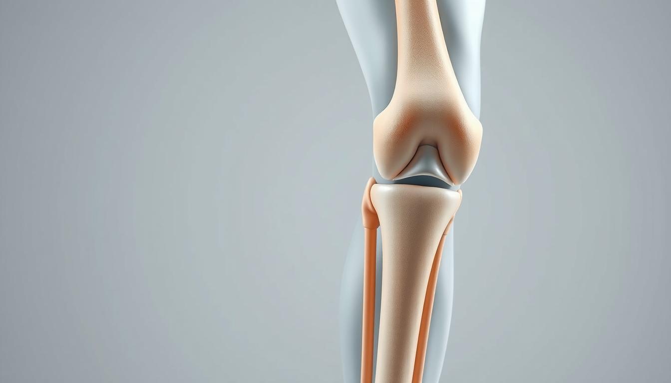
 in the human knee joint, rendered in highly realistic medical illustration style. The MCL is prominently featured in the center foreground, shown with its characteristic fan-like structure and attachments to the femur and tibia. The surrounding musculature, tendons, and bony landmarks are clearly visible, allowing for a comprehensive understanding of the MCL's anatomical context. The lighting is soft and directional, creating subtle shadows that enhance the three-dimensional form. The background is minimalist, with a plain, neutral color palette to avoid distractions and focus the viewer's attention on the MCL structure. The overall tone is educational and informative, suitable for use in a medical article on knee instability.")