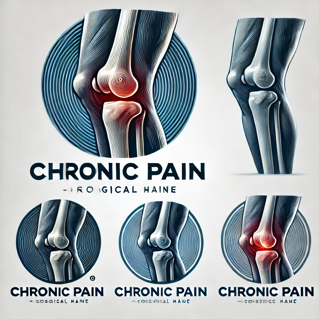
Iliotibial Syndrome: Causes and Treatment
Iliotibial syndrome, also known as IT band syndrome, is a common overuse injury that affects the iliotibial band, a thick band of fascia running along the outside of the thigh. This article explores the anatomy and function of the iliotibial band, the causes of iliotibial syndrome, diagnosis methods, and treatment options.
Key Takeaways
- Iliotibial syndrome is often caused by biomechanical factors and training errors.
- Clinical evaluation is an important diagnostic tool for identifying iliotibial syndrome.
- Conservative management, including rest, stretching, and physical therapy, is the primary treatment approach for iliotibial syndrome.
- Surgical interventions may be considered for severe or persistent cases of iliotibial syndrome.
- Prevention strategies, such as proper warm-up, cross-training, and gradual progression of activity, can help reduce the risk of developing iliotibial syndrome.
Anatomy and Function of the Iliotibial Band

Structure of the Iliotibial Band
The iliotibial band (ITB) is a thick, fibrous band of connective tissue that extends from the iliac crest in the pelvis down to the tibia in the lower leg. Its primary composition is fascial tissue, which is dense and fibrous, providing both flexibility and tensile strength. The ITB plays a crucial role in the stabilization of the knee, especially during activities that involve running or cycling.
Anatomically, the ITB is not a discrete structure but rather a thickening of the tensor fasciae latae muscle’s fascia. This integration with muscle tissue is what allows the ITB to assist in hip abduction, flexion, and rotation. The distal portion of the ITB also contributes to lateral knee stabilization by attaching to the Gerdy’s tubercle on the tibia.
- The ITB originates from the iliac crest.
- It is comprised of fascial tissue.
- It attaches to the Gerdy’s tubercle on the tibia.
Tip: A well-balanced strength and flexibility training program can help maintain the proper function of the ITB and prevent overuse injuries.
Function of the Iliotibial Band
We recognize the iliotibial band (ITB) as a critical structure in lower limb biomechanics, particularly for those engaged in repetitive activities such as running and cycling. Its primary function is to stabilize the knee during gait, working in concert with other muscles around the hip and thigh.
Stabilization is not the ITB’s only role; it also assists in the abduction of the hip, which is essential for maintaining pelvic balance during single-leg support phases of walking or running. This secondary function underscores the ITB’s importance in coordinated movement and injury prevention.
- The ITB contributes to lateral knee stabilization.
- It aids in hip abduction.
- The band plays a role in shock absorption, reducing the impact on the knee joint.
Tip: A well-conditioned ITB is less susceptible to overuse injuries, emphasizing the need for targeted strength and flexibility exercises in preventative care.
Causes of Iliotibial Syndrome

Biomechanical Factors
We recognize that the development of Iliotibial Syndrome is often closely linked to various biomechanical factors. These factors can include abnormalities in leg length, muscle imbalances, and foot structure, which may contribute to the stress on the iliotibial band during activity. For instance, a discrepancy in leg length can lead to an uneven gait, placing more strain on one side of the body.
- Overpronation of the foot, where the foot rolls inward excessively when walking or running, can also increase tension in the iliotibial band. Similarly, weak hip abductor muscles may fail to stabilize the pelvis, resulting in increased movement and stress on the IT band. It’s essential to identify and address these biomechanical issues to prevent or alleviate symptoms.
Here is a list of common biomechanical factors that may lead to Iliotibial Syndrome:
- Leg length discrepancy
- Muscle imbalances
- Overpronation of the foot
- Weak hip abductor muscles
Tip: Regular strength training and stretching exercises targeting the hip abductors can help in correcting muscle imbalances and reducing the risk of Iliotibial Syndrome.
Training Errors
Training errors, such as overtraining or sudden increases in training intensity, can contribute to the development of iliotibial syndrome. It is important to gradually increase training volume and intensity to prevent overuse injuries. Additionally, improper footwear and running on uneven surfaces can also exacerbate the condition. Here’s a list of common training errors that may lead to iliotibial syndrome:
- Overtraining
- Sudden increases in training intensity
- Improper footwear
- Running on uneven surfaces
It is crucial to address these training errors to reduce the risk of developing iliotibial syndrome. As a reminder, proper training techniques and gradual progression are key to preventing this condition.
Diagnosis of Iliotibial Syndrome

Clinical Evaluation
Following the initial consultation, we conduct a thorough clinical evaluation to diagnose Iliotibial Syndrome. This involves a detailed history taking to understand the onset and progression of symptoms. We pay close attention to the patient’s activity level and any recent changes in training routines or equipment.
Physical examination is pivotal. We assess for tenderness along the iliotibial band, especially at its insertion on the lateral epicondyle of the knee. The Ober test, which evaluates the tightness of the iliotibial band, is a key component of our assessment. Additionally, we may perform the Noble compression test to provoke symptoms and confirm the diagnosis.
It’s essential to rule out other conditions that can mimic Iliotibial Syndrome. Therefore, we consider the differential diagnosis carefully. Here’s a simplified list of conditions we consider:
- Lateral meniscus tear
- Lateral collateral ligament injury
- Popliteal tendonitis
- Bursitis
Tip: A comprehensive clinical evaluation is crucial for an accurate diagnosis. It’s important not to rush this process and to consider all potential contributing factors.
Imaging Studies
Following a thorough clinical evaluation, we often turn to imaging studies to confirm the diagnosis of iliotibial syndrome. While MRI scans are not routinely used, they can be particularly helpful in visualizing the soft tissue structures and any associated inflammation of the iliotibial band. Ultrasound imaging is another tool at our disposal, offering the advantage of dynamic assessment while the joint is in motion.
Typically, imaging is reserved for cases where the diagnosis is uncertain or if there is a lack of response to conservative treatment. In such scenarios, imaging can provide valuable insights into the extent of the injury and rule out other potential causes of lateral knee pain.
Remember, imaging should complement, not replace, a comprehensive clinical evaluation.
When interpreting imaging results, it’s crucial to correlate findings with the patient’s symptoms and physical exam results. This ensures a holistic approach to diagnosis and subsequent treatment planning.
Treatment Options for Iliotibial Syndrome

Conservative Management
After exploring various conservative management techniques, we have found that rest and stretching are crucial components of recovery. Additionally, we recommend incorporating strength training exercises to address muscle imbalances. Below is a table summarizing the recommended exercises and their frequency:
| Exercise | Frequency |
|---|---|
| Stretching | Daily |
| Strength Training | 3 times a week |
We advise patients to gradually increase activity levels and to avoid overtraining, as this can exacerbate symptoms. It is important to monitor progress and adjust the treatment plan accordingly. We emphasize the significance of a comprehensive approach that addresses both the symptoms and underlying causes of the syndrome.
Surgical Interventions
After considering the various conservative management approaches, surgical interventions may be recommended for individuals with severe and persistent symptoms. These interventions typically involve the release or lengthening of the iliotibial band to alleviate tension and reduce friction. In some cases, a table summarizing the success rates and potential risks of surgical procedures may aid in decision-making. It’s important to note that surgical interventions should be considered as a last resort and only after thorough evaluation and discussion with a healthcare professional. Post-surgery, a comprehensive rehabilitation program is essential for optimal recovery and long-term success.
Conclusion
In conclusion, Iliotibial Syndrome is a complex condition that can be caused by a variety of factors, including overuse and biomechanical issues. Treatment typically involves a combination of physical therapy, rest, and targeted exercises to address muscle imbalances. It is important for individuals suffering from this syndrome to seek professional medical advice and adhere to a comprehensive treatment plan in order to achieve long-term relief and prevent recurrence.
Frequently Asked Questions
What are the common symptoms of Iliotibial Syndrome?
Common symptoms include pain on the outside of the knee, swelling, and a clicking sensation during movement.
Can Iliotibial Syndrome be prevented?
Yes, it can be prevented by avoiding sudden increases in physical activity, maintaining proper running form, and using proper footwear.
Is stretching helpful for Iliotibial Syndrome?
Yes, stretching the iliotibial band and surrounding muscles can help reduce tension and alleviate symptoms.
What are the risk factors for developing Iliotibial Syndrome?
Risk factors include overuse, excessive running or cycling, improper footwear, and muscle imbalances.
Can I continue physical activity if I have Iliotibial Syndrome?
It is recommended to modify physical activity to avoid exacerbating symptoms, and to consult with a healthcare professional for personalized recommendations.
How long does it take to recover from Iliotibial Syndrome?
Recovery time varies depending on the severity of the condition and individual factors, but with proper treatment, most individuals can recover within a few weeks to a few months.



One Comment