Author: Mokhtar

  • Meniscus Tear Rehab & Exercises (Stretching | Strength | Plyometrics)

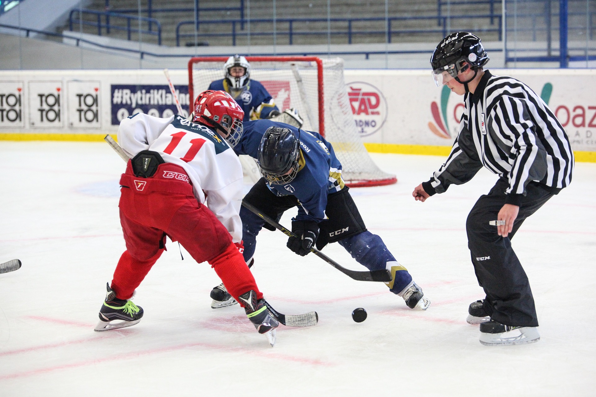
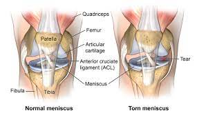
    We will be discussing meniscus tears, their function, and why surgery might not be the best option. The meniscus is a fibrocartilaginous structure located between the femur and tibia in the knee joint. There are two menisci in each knee, the medial meniscus, and the lateral meniscus. They function to provide shock absorption, force transmission, and stability to the knee joint.

    Meniscus tears can be either traumatic or non-traumatic. Traumatic tears have a distinct mechanism of injury, while non-traumatic tears occur secondary to age-related changes. Meniscus tears are often classified according to location and orientation, such as vertical, longitudinal, radial, horizontal, oblique, or complex. Surgery to repair or remove the meniscus was performed in the past to reduce symptoms of pain and mechanical symptoms, such as clicking, catching, and locking of the knee. However, the current scientific literature does not fully support this theory.

    symptoms and signs of a meniscus tear:

    1. Pain and swelling in the knee joint
    2. Difficulty straightening or bending the knee
    3. Feeling a popping sensation in the knee
    4. A locking or catching sensation in the knee joint

    If you suspect you have a meniscus tear, it’s important to see a doctor for an accurate diagnosis and appropriate treatment.

    Key Takeaways

    • Mechanical symptoms are not a reliable indicator of the presence of meniscal tears.
    • Meniscus tears are common in asymptomatic individuals.
    • Surgery to remove or repair the meniscus has not been shown to lead to better outcomes compared to other interventions.

    Understanding Meniscus Tears

    We want to provide you with a comprehensive understanding of meniscus tears. The meniscus is a fibrocartilaginous structure located between the femur and tibia bones in the knee joint. There are two menisci in each knee, the medial meniscus located on the inner portion of the knee and the lateral meniscus located on the outer portion. The menisci function to provide shock absorption, force transmission, and stability to the knee joint.

    download 2023 11 05T205633.181

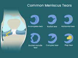
    Meniscus tears are either traumatic or non-traumatic. Traumatic tears occur due to a distinct mechanism of injury, whereas non-traumatic tears occur secondary to age-related changes. Meniscus tears are often classified according to location and orientation, including vertical, longitudinal, radial, horizontal, oblique, or complex tears. Vertical or oblique tears that twist and fold over within the joint are termed bucket handle tears.

    download 2023 11 05T205748.264

    In the past, the presence of pain and mechanical symptoms such as clicking, catching, and locking of the knee were thought to be caused by a meniscus tear. However, current scientific literature does not fully support this theory. Researchers have found that mechanical symptoms are equally prevalent in patients with and without a meniscal tear and are common in those with knee problems in general. Therefore, these symptoms have limited utility as an indicator for the presence of meniscal tears or are not useful clues to the diagnosis.

    Meniscus tears are common in asymptomatic individuals. One study found that in 230 uninjured knees, 30 percent had meniscal tears. If tears are prevalent in a non-painful population, it brings into question the efficacy of surgery in reducing symptoms. Surgery to remove or repair the meniscus has not been shown to lead to better outcomes compared to other interventions. In fact, a brand new Cochrane review concluded that arthroscopic surgery for degenerative meniscal tears provides little to no clinical benefit for pain function and quality of life compared to placebo surgery.

    At this moment, current clinical practice guidelines favor conservative management in those with degenerative meniscus tears. Exercise has been shown to be just as effective in the long term for both populations. We have one study that showed exercise led to similar improvements in pain function and quality of life after 12 months in a younger population with mostly traumatic tears and another study that showed no clinically relevant difference in the function after two years in an older population with degenerative tears.

    We want to provide you with a comprehensive rehab program that covers multiple categories aimed at increasing your capacity and tolerance to various stressors within each category. The first category is knee range of motion. Ideally, you should be able to fully straighten your knee and touch your heel to your butt. You should aim to restore extension and flexion that is equal to your uninjured side. We recommend three options for knee extension or straightening and three options for knee flexion or bending.

    The second category is hip, knee, and ankle strength. We recommend a four-step split squat progression, which will help increase your tolerance to weight-bearing knee flexion in deeper positions. We also recommend a single-leg knee extension and hamstring sliders to work on quadriceps and hamstring strength.

    In conclusion, meniscus tears are a common knee injury that can be managed conservatively through exercise. Surgery has not been shown to provide better outcomes compared to other interventions. Our comprehensive rehab program covers multiple categories aimed at increasing your capacity and tolerance to various stressors. We recommend following this program for non-surgical cases. Always talk to your doctor or physical therapist before starting any exercise program.

    Myths About Meniscus Tears

    We often hear that meniscus tears are the cause of knee pain and mechanical symptoms such as clicking, catching, and locking of the knee. However, recent scientific literature does not fully support this theory. Here are three things you should know about meniscus tears:

    1. There is not a simple cause and effect relationship between symptoms and tears. Patients with and without a meniscal tear have been found to have the same mechanical symptoms, which are actually common in those with knee problems in general.
    2. Meniscus tears are common in asymptomatic individuals. In one study, 30% of uninjured knees had meniscal tears, which brings into question the efficacy of surgery in reducing symptoms.
    3. Surgery to remove or repair the meniscus has not been shown to lead to better outcomes compared to other interventions. In fact, surgery can even lead to an increased risk of developing radiographic knee osteoarthritis.

    Given this information, conservative management is favored for those with degenerative meniscus tears. Exercise has been shown to be just as effective in the long term for both younger patients with traumatic tears and older patients with degenerative tears.

    We provide a comprehensive rehab program that covers multiple categories aimed at increasing your capacity and tolerance to various stressors. In the knee range of motion category, we recommend exercises such as seated active quad contractions, propping your heel up on an elevated surface, and banded tke. In the hip, knee, and ankle strength category, we recommend exercises such as the four-step split squat progression and single leg knee extensions for the quadriceps, and standing knee flexion with an ankle weight or hamstring sliders for the hamstrings.

    It is important to note that these exercises are intended for non-surgical cases. If you had surgery, you should talk to your doctor or physical therapist before starting any exercise program.

    Research on Meniscus Tears

    The meniscus is a fibrocartilaginous structure that is located between the femur and tibia in the knee joint. There are two menisci in each knee, the medial and lateral meniscus. They provide shock absorption, force transmission, and stability to the knee joint.

    Meniscus tears can be either traumatic or non-traumatic. Traumatic tears occur due to a distinct mechanism of injury, while non-traumatic tears occur secondary to age-related changes. Tears can be classified according to their location and orientation, such as vertical, longitudinal, radial, horizontal, oblique, or complex. Bucket handle tears are vertical or oblique tears that twist and fold over within the joint.

    In the past, the presence of pain and mechanical symptoms such as clicking, catching, and locking of the knee were thought to be caused by a meniscus tear. Therefore, surgery to repair or remove the meniscus was performed to reduce symptoms. However, current scientific literature does not fully support this theory.

    There is not a simple cause and effect relationship between symptoms and tears. Mechanical symptoms are equally prevalent in patients with and without a meniscal tear, and are actually common in those with knee problems in general. Meniscus tears are also common in asymptomatic individuals, with one study finding that 30 percent of uninjured knees had meniscal tears.

    Surgery to remove or repair the meniscus has not been shown to lead to better outcomes compared to other interventions. A popular study compared a partial meniscectomy to a placebo or sham surgery and found that surgery provided no more benefit for knee symptoms or function than placebo surgery. It even led to an increased risk of developing radiographic knee osteoarthritis. A brand new Cochrane review concluded that arthroscopic surgery for degenerative meniscal tears provides little to no clinical benefit for pain, function, and quality of life compared to placebo surgery.

    Exercise has been shown to be just as effective in the long term for both younger and older populations with meniscus tears. Clinical practice guidelines favor conservative management in those with degenerative meniscus tears.

    We provide a comprehensive rehab program that covers multiple categories aimed at increasing your capacity and tolerance to various stressors. The program includes knee range of motion exercises, hip, knee, and ankle strength exercises, balance and proprioception exercises, and cardiovascular exercise.

    Surgical Interventions

    We have learned that surgery to remove or repair the meniscus has not been shown to lead to better outcomes compared to other interventions. In fact, a recent Cochrane review concluded that arthroscopic surgery for degenerative meniscal tears provides little to no clinical benefit for pain, function, and quality of life compared to placebo surgery. Therefore, current clinical practice guidelines favor conservative management in those with degenerative meniscus tears.

    Instead, we recommend a comprehensive rehab program that covers multiple categories aimed at increasing your capacity and tolerance to various stressors. We will provide multiple exercise options within each category, ultimately giving you two different workouts to follow. These exercises are intended for non-surgical cases. If you had surgery, you should talk to your doctor or physical therapist before starting, as you may have weight-bearing precautions or other considerations to consider.

    The first category is knee range of motion. Ideally, you should be able to fully straighten your knee and touch your heel to your butt. At minimum, you should aim to restore extension and flexion that is equal to your uninjured side. For knee extension or straightening, we recommend seated active quad contractions, propping your heel up on an elevated surface, and banded tke. For knee flexion or bending, we recommend using a stationary bike, heel slides, and quadruped rock backs or tall kneeling rock backs.

    The second category is hip, knee, and ankle strength. For quadriceps, we recommend a four-step split squat progression and a single-leg knee extension. For hamstrings, we recommend standing knee flexion with an ankle weight, machines, or hamstring sliders.

    By following these exercises, you can increase your tolerance to weight-bearing knee flexion in deeper positions and improve your knee symptoms related to a degenerative meniscus tear.

    Non-Surgical Interventions

    We understand that meniscus tears can cause pain and other mechanical symptoms in the knee joint. However, surgery may not always be the best option for treating this condition. In fact, current scientific literature does not fully support the idea that surgery to repair or remove the meniscus leads to better outcomes compared to other interventions.

    Therefore, we recommend a comprehensive rehab program that includes exercise progressions with sets and reps. This program aims to increase your capacity and tolerance to various stressors within each category. We have categorized the exercises into two main categories:

    Category One: Knee Range of Motion

    The first category focuses on restoring knee range of motion. Ideally, you should be able to fully straighten your knee and touch your heel to your butt. If not, you should aim to restore extension and flexion that is equal to your uninjured side.

    Here are three exercises that can help with knee extension:

    1. Seated Active Quad Contractions: Straighten your leg and squeeze your quad for 10 seconds. Repeat this for 10 repetitions, three times a day. If your motion is improving, you can use a towel or strap to pull up on your foot to intensify the stretch.
    2. Heel Prop: Prop your heel up on an elevated surface for 10 to 15 minutes, three times a day. If you need to enhance the stretch, you can add a weight just above your knee. The goal here is low load, long duration.
    3. Banded TKE: Place a band behind the back of your knee, anchor it to an object, and perform the same quad contractions against resistance.

    For knee flexion, here are three exercises that can help:

    1. Stationary Bike: Start with the seat at a higher level, and as your range of motion improves, lower the seat to expose the knee to more flexion.
    2. Heel Slides: Use a slider on carpet or a towel on a hard surface. Start by actively sliding your heel towards your butt for 10 to 15 repetitions. As motion improves, progress by using a towel or strap to pull the knee into more flexion. You should do these often, at least three times a day, every day.
    3. Quadruped Rock Backs or Tall Kneeling Rock Backs: Kneel on a pad or pillow for these exercises. You can even experiment with using a towel behind the knee, which can help patients move into more knee flexion with less discomfort.

    Category Two: Hip, Knee, and Ankle Strength

    The second category focuses on hip, knee, and ankle strength. We recommend a four-step split squat progression that will help increase your tolerance to weight-bearing knee flexion in deeper positions.

    Here are the four levels of the split squat progression:

    1. Body Weight Squat: Perform three sets of 20 repetitions, getting your hips to at least parallel. If you need to, start with hand assistance, such as using a TRX or another object.
    2. Heels Elevated Squat: Elevate your heels two to three inches. This will help keep your torso more upright and allow you to go deeper, moving your knee into more flexion. Perform three sets of 20 repetitions, and again, try to get your hips to at least parallel.
    3. Split Squat: Stand in a split stance and lower down while driving the front knee forward as far as you can. Perform three sets of 15 controlled reps on each leg before progressing.
    4. Front Foot Elevated Split Squat: Elevate your front foot on an object two to four inches high and lower it down, driving the front knee forward. Over time, you can increase depth and add weight.

    For the hamstrings, we recommend standing knee flexion with an ankle weight, using machines, or performing hamstring sliders. Start with double-leg eccentric sliders for three to four sets of 10 to 15 reps on each leg. Your goal is to move through the full range of motion with the weight at a challenging intensity, but to start, you can shorten the range of motion or even perform with no weight if needed.

    In conclusion, exercise has been shown to be just as effective in the long term for treating meniscus tears compared to surgery. Therefore, we recommend a comprehensive rehab program that includes these exercises to help you recover from this condition.

    Exercise as a Treatment Option

    We have seen that surgery for meniscus tears may not be the best option, and that exercise can be just as effective in the long term for both younger and older populations. Therefore, we present a comprehensive rehab program that covers multiple categories aimed at increasing your capacity and tolerance to various stressors within each category.

    Knee Range of Motion

    The first category we will cover is knee range of motion. Ideally, you should be able to fully straighten your knee and touch your heel to your butt. At minimum, you should aim to restore extension and flexion that is equal to your uninjured side.

    For knee extension or straightening, we recommend three options. The first is seated active quad contractions, where you straighten your leg and squeeze your quad for 10 seconds. Repeat this for 10 repetitions, three times a day. If your motion is improving, you can use a towel or strap to pull up on your foot in order to intensify the stretch.

    Another option is propping your heel up on an elevated surface for 10 to 15 minutes, three times a day. If you need to enhance the stretch, you can add a weight just above your knee. The goal here is low load long duration, so while it may feel slightly uncomfortable, it should not be unbearable.

    Finally, once you gain more range of motion, strength, and control, add in a banded tke. Place a band behind the back of your knee, anchor it to an object, and perform the same quad contractions against resistance.

    For knee flexion or bending, we recommend three options. You can use a stationary bike and start with the seat at a higher level. As range of motion improves, lower the seat to expose the knee to more flexion. Another option is heel slides. You can use a slider on carpet or a towel on a hard surface. Start by actively sliding your heel towards your butt for 10 to 15 repetitions. As motion improves, progress by using a towel or strap to pull the knee into more flexion. You should do these often, so at least three times a day, every day.

    Finally, a more advanced option is quadruped rock backs or tall kneeling rock backs. We recommend kneeling on a pad or pillow for these, and you can even experiment with using a towel behind the knee. Anecdotally, we have found this to help patients move into more knee flexion with less discomfort.

    Hip, Knee, and Ankle Strength

    The second category we will cover is hip, knee, and ankle strength. Let’s start with a four-step split squat progression, which will help increase your tolerance to weight bearing knee flexion in deeper positions.

    Level 1 is a bodyweight squat. Your goal is to perform three sets of 20 repetitions, getting your hips to at least parallel. If you need, start with hand assistance such as using a TRX or another object.

    Level 2 is a heels elevated squat. Elevate your heels two to three inches. This will help keep your torso more upright and allow you to go deeper, moving your knee into more flexion. Your goal is three sets of 20 repetitions, and again, try to get your hips to at least parallel.

    Level 3 is a split squat. Stand in a split stance and lower down while driving the front knee forward as far as you can. Your goal is three sets of 15 controlled reps on each leg before progressing.

    Level 4 is a front foot elevated split squat. Elevate your front foot on an object two to four inches high and lower it down, driving the front knee forward. Over time, you can increase depth and add weight.

    For the quadriceps, we recommend a single leg knee extension for three to four sets of 10 to 15 reps on each leg. Your goal is to move through the full range of motion with the weight at a challenging intensity, but to start, you can shorten the range of motion or even perform with no weight if needed.

    For the hamstrings, the first option will emphasize knee flexion. You can perform standing knee flexion with an ankle weight, use machines, or an exercise we like to use called hamstring sliders. Start with double leg eccentric sliders before progressing to single leg eccentric sliders.

    We hope this comprehensive rehab program will help you increase your capacity and tolerance to various stressors, ultimately leading to better knee function and quality of life.

    Rehab Program

    We will now discuss a comprehensive rehab program for meniscus tears, which includes exercise progressions with sets and reps. The meniscus is a fibrocartilaginous structure located between the femur and tibia bones in the knee joint. There are two menisci in each knee, the medial meniscus on the inner portion and the lateral meniscus on the outer portion. The menisci provide shock absorption, force transmission, and stability to the knee joint.

    Meniscus tears can be either traumatic or non-traumatic. Traumatic tears occur due to a specific injury, while non-traumatic tears occur due to age-related changes. Tears can be classified according to location and orientation, such as vertical, longitudinal, radial, horizontal, oblique, or complex. Vertical or oblique tears that twist and fold over within the joint are termed bucket handle tears.

    In the past, pain and mechanical symptoms such as clicking, catching, and locking of the knee were thought to be caused by a meniscus tear. Therefore, surgery to repair or remove the meniscus was performed to reduce symptoms. However, current scientific literature does not fully support this theory.

    There is not a simple cause and effect relationship between symptoms and tears. For example, a 2018 study found that mechanical symptoms were equally prevalent in patients with and without a meniscal tear and are actually common in those with knee problems in general. Other research has concluded that these symptoms have limited utility as an indicator for the presence of meniscal tears or are not useful clues to the diagnosis.

    Meniscus tears are common in asymptomatic individuals. In one study, researchers found that in 230 uninjured knees, 30 percent had meniscal tears. If tears are prevalent in a non-painful population, it questions the efficacy of surgery in reducing symptoms.

    Surgery to remove or repair the meniscus has not been shown to lead to better outcomes compared to other interventions. For example, a popular study compared a partial meniscectomy to a placebo or sham surgery over the course of five years. Surgery provided no more benefit for knee symptoms or function than placebo surgery. It even led to an increased risk of developing radiographic knee osteoarthritis. In fact, a brand new Cochrane review concluded that arthroscopic surgery for degenerative meniscal tears provides little to no clinical benefit for pain, function, and quality of life compared to placebo surgery.

    Exercise has been shown to be just as effective in the long term for both populations. One study showed exercise led to similar improvements in pain, function, and quality of life after 12 months in a younger population with mostly traumatic tears. Another study showed no clinically relevant difference in function after two years in an older population with degenerative tears.

    Therefore, current clinical practice guidelines favor conservative management in those with degenerative meniscus tears. We will now present a comprehensive rehab program that covers multiple categories aimed at increasing your capacity and tolerance to various stressors within each category. We will provide multiple exercise options, ultimately giving you two different workouts to follow.

    Before diving into these exercises, there is one caveat we want to mention. These exercises are intended for non-surgical cases. If you had surgery, you will ultimately follow a similar framework, but before starting, talk to your doctor or physical therapist as you may have weight-bearing precautions or other considerations to consider.

    Category One: Knee Range of Motion

    Ideally, you should be able to fully straighten your knee and touch your heel to your butt. At a minimum, you should aim to restore extension and flexion that is equal to your uninjured side.

    For knee extension or straightening, here are three options:

    1. Seated active quad contractions: Straighten your leg and squeeze your quad for 10 seconds. Repeat this for 10 repetitions, three times a day. If your motion is improving, you can use a towel or strap to pull up on your foot to intensify the stretch.
    2. Propping your heel up on an elevated surface: Elevate your heel on a surface for 10 to 15 minutes, three times a day. If you need to enhance the stretch, you can add a weight just above your knee. The goal here is low load, long duration, so while it may feel slightly uncomfortable, it should not be unbearable.
    3. Banded TKE: Place a band behind the back of your knee, anchor it to an object, and perform the same quad contractions against resistance.

    For knee flexion or bending, here are three options:

    1. Stationary bike: Start with the seat at a higher level and lower it as range of motion improves to expose the knee to more flexion.
    2. Heel slides: Use a slider on carpet or a towel on a hard surface. Start by actively sliding your heel towards your butt for 10 to 15 repetitions.

    Knee Range of Motion Exercises

    We will now discuss knee range of motion exercises that are essential for rehabilitating meniscus tears. The meniscus is a vital structure that provides shock absorption, force transmission, and stability to the knee joint. Meniscus tears can be traumatic or non-traumatic and can be classified based on their location and orientation. The presence of pain and mechanical symptoms such as clicking, catching, and locking of the knee were traditionally thought to be caused by a meniscus tear. However, surgery to repair or remove the meniscus has not been shown to lead to better outcomes compared to other interventions.

    Therefore, we recommend a comprehensive rehab program that covers multiple categories aimed at increasing your capacity and tolerance to various stressors. In the first category, we will focus on knee range of motion exercises. Ideally, you should be able to fully straighten your knee and touch your heel to your butt. At minimum, you should aim to restore extension and flexion that is equal to your uninjured side.

    For knee extension or straightening, we recommend three options. The first is seated active quad contractions, where you straighten your leg and squeeze your quad for 10 seconds, repeat this for 10 repetitions, three times a day. Another option is propping your heel up on an elevated surface for 10 to 15 minutes, three times a day. If you need to enhance the stretch, you can add a weight just above your knee. Finally, once you gain more range of motion, strength, and control, add in a banded tke. Place a band behind the back of your knee, anchor it to an object, and perform the same quad contractions against resistance.

    For knee flexion or bending, we recommend three options. You can use a stationary bike and start with the seat at a higher level. As range of motion improves, lower the seat to expose the knee to more flexion. Another option is heel slides. You can use a slider on carpet or a towel on a hard surface. Start by actively sliding your heel towards your butt for 10 to 15 repetitions. As motion improves, progress by using a towel or strap to pull the knee into more flexion. You should do these often, at least three times a day, every day. Finally, a more advanced option is quadruped rock backs or tall kneeling rock backs. We recommend kneeling on a pad or pillow for these, and you can even experiment with using a towel behind the knee.

    These knee range of motion exercises will help increase your knee’s flexibility and strength. It is essential to perform these exercises regularly to help you recover from your injury.

    Hip, Knee and Ankle Strength Exercises

    We have put together a comprehensive rehab program that covers multiple categories aimed at increasing your capacity and tolerance to various stressors. Within each category, we will provide multiple exercise options ultimately giving you two different workouts to follow.

    Before diving into these exercises, we want to mention that these exercises are intended for non-surgical cases. If you had surgery, you will ultimately follow a similar framework but before starting, talk to your doctor or physical therapist as you may have weight-bearing precautions or other considerations to consider.

    Category One: Knee Range of Motion

    Ideally, you should be able to fully straighten your knee and touch your heel to your butt. At minimum, you should aim to restore extension and flexion that is equal to your uninjured side.

    For knee extension or straightening, here are three options:

    1. Seated active quad contractions: Straighten your leg and squeeze your quad for 10 seconds. Repeat this for 10 repetitions, three times a day. If your motion is improving, you can use a towel or strap to pull up on your foot in order to intensify the stretch.
    2. Propping your heel up on an elevated surface for 10 to 15 minutes, three times a day. If you need to enhance the stretch, you can add a weight just above your knee. The goal here is low load, long duration, so while it may feel slightly uncomfortable, it should not be unbearable.
    3. Banded TKE: Place a band behind the back of your knee, anchor to an object, and perform the same quad contractions against resistance.

    For knee flexion or bending, here are three options:

    1. Stationary bike: Start with the seat at a higher level. As range of motion improves, lower the seat to expose the knee to more flexion.
    2. Heel slides: Use a slider on carpet or a towel on a hard surface. Start by actively sliding your heel towards your butt for 10 to 15 repetitions. As motion improves, progress by using a towel or strap to pull the knee into more flexion. You should do these often, so at least three times a day every day.
    3. Quadruped rock backs or tall kneeling rock backs: Kneel on a pad or pillow for these and experiment with using a towel behind the knee. Anecdotally, we have found this to help patients move into more knee flexion with less discomfort.

    Category Two: Hip, Knee and Ankle Strength

    Let’s start with a four-step split squat progression which will help increase your tolerance to weight-bearing knee flexion in deeper positions.

    Level 1: Body weight squat – Your goal is to perform 3 sets of 20 repetitions, getting your hips to at least parallel. If you need, start with hand assistance such as using a TRX or another object.

    Level 2: Heels elevated squat – Elevate your heels two to three inches. This will help keep your torso more upright and allow you to go deeper, moving your knee into more flexion. Your goal is 3 sets of 20 repetitions, and again try to get your hips to at least parallel.

    Level 3: Split squat – Stand in a split stance and lower down while driving the front knee forward as far as you can. Your goal is three sets of 15 controlled reps on each leg before progressing.

    Level 4: Front foot elevated split squat – Elevate your front foot on an object two to four inches high and lower it down, driving the front knee forward. Over time, you can increase depth and add weight.

    For the hamstrings, the first option will emphasize knee flexion. You can perform standing knee flexion with an ankle weight, use machines, or an exercise we like to use, hamstring sliders. Start with double leg eccentric sliders.

    We have put together a comprehensive rehab program that covers multiple categories aimed at increasing your capacity and tolerance to various stressors. In this section, we will focus on knee range of motion and hip, knee, and ankle strength.

    Knee Range of Motion

    Ideally, you should be able to fully straighten your knee and touch your heel to your butt. In order to restore extension and flexion equal to your uninjured side, we recommend the following exercises:

    1. Seated active quad contractions: straighten your leg and squeeze your quad for 10 seconds. Repeat for 10 repetitions, three times a day. You can use a towel or strap to pull up on your foot in order to intensify the stretch.
    2. Propping your heel up on an elevated surface for 10 to 15 minutes, three times a day. You can add a weight just above your knee to enhance the stretch.
    3. Banded tke: place a band behind the back of your knee and perform the same quad contractions against resistance.

    For knee flexion or bending, we recommend the following exercises:

    1. Stationary bike: start with the seat at a higher level and as range of motion improves, lower the seat to expose the knee to more flexion.
    2. Heel slides: actively slide your heel towards your butt for 10 to 15 repetitions. As motion improves, progress by using a towel or strap to pull the knee into more flexion. Do these at least three times a day every day.
    3. Quadruped rock backs or tall kneeling rock backs: kneel on a pad or pillow and use a towel behind the knee. This will help you move into more knee flexion with less discomfort.

    Hip, Knee, and Ankle Strength

    We recommend the following exercises to increase your tolerance to weight bearing knee flexion in deeper positions:

    1. Four step split squat progression:
      • Level 1: Body weight squat – perform 3 sets of 20 repetitions getting your hips to at least parallel.
      • Level 2: Heels elevated squat – elevate your heels two to three inches to allow you to go deeper. Your goal is 3 sets of 20 repetitions and again try to get your hips to at least parallel.
      • Level 3: Split squat – stand in a split stance and lower down while driving the front knee forward as far as you can. Your goal is three sets of 15 controlled reps on each leg before progressing.
      • Level 4: Front foot elevated split squat – elevate your front foot on an object two to four inches high and lower it down driving the front knee forward. Over time, you can increase depth and add weight.
    2. Single leg knee extension: perform for three to four sets of 10 to 15 reps on each leg. Your goal is to move through the full range of motion with the weight at a challenging intensity.
    3. Hamstring sliders: start with double leg eccentric sliders and progress to single leg. Your goal is to move through the full range of motion with the weight at a challenging intensity.

    By following these exercises, you can increase your capacity and tolerance to various stressors and ultimately improve your overall knee function.

  • Relationship between exercise intensity and risk of infection

    Relationship between exercise intensity and risk of infection

    So far, there is no clarity on how exercise intensity affects the emission and concentration of aerosol particles in exhaled air. Using a new experimental setup, a research team from Munich has shown that aerosol emissions increase exponentially with intensive physical exertion – so that indoor sports activities result in a higher risk of infectious diseases such as COVID.

    Before the study, it was known that tidal volume in untrained people increases from approximately 5-15 liters per minute at rest to more than 100 l/min during exercise. Highly trained athletes even reach levels of 200 l/min. It was also known that many people have become infected with the SARS-CoV-2 virus when they exercise indoors.

    However, it was unclear how exercise intensity was related to the concentration of aerosol particles in exhaled air and the actual amount of aerosols an individual exhaled per minute, and thus to the potential risk of spreading infectious diseases such as SARS-CoV-2. However, this information is urgently needed, for example to design mitigation measures for gyms and other indoor sports facilities, fitness studios or discos to avoid closures in the event of serious infection waves.

    New methodology produces individually measurable aerosol values

    A team led by Henning Wackerhage, professor of exercise biology at the Technical University of Munich (TUM), and Prof. Christian J. Kähler, director of the Institute for Fluid Mechanics and Aerodynamics at the Universität der Bundeswehr Munich, has developed a new research method to study questions. Their experimental equipment initially filtered the aerosols already present in the ambient air. During the subsequent ergometer stress test, the subjects inhaled the purified air through a special mask that covered the mouth and nose. Exercise intensity was gradually increased from rest to the point of physical exhaustion. The mask was connected to a two-way valve that allowed only exhaled air to escape. The amount of aerosol particles emitted per minute was then measured and directly linked to the current performance of the healthy, 18-40 year old subjects.

    Moderate aerosol emissions during moderate exertion

    This allowed the researchers to investigate for the first time how many aerosol particles are exhaled per minute by an individual at different levels of exercise intensity. The result: aerosol emissions during exercise initially increased only moderately, reaching an average load of about 2 watts per kilogram of body weight. However, above that point they increased exponentially. This means that someone weighing 75 kilograms reaches that threshold at an ergometer setting of approximately 150 watts. This equates to moderate exertion for a casual athlete, perhaps comparable to the exercise intensity of moderate jogging.

    The aerosol output of well-trained athletes was significantly higher than that of untrained subjects at maximum exertion, due to their much higher minute ventilation. The researchers found no significant differences in particle emissions between genders.

    Protective measures are important for high-intensity training

    Although the aerosol experiments provide only indirect knowledge about the amount of viruses in exhaled air, the research suggests useful principles for managing indoor activities when a wave of infections combined with a poorly immunized population threatens to overwhelm the healthcare system.

    “Based on our results, we distinguish between moderate endurance training with an intensity of up to 2 watts per kilogram of body weight and training at high to maximum intensity. Due to the sharp increase in aerosol emissions at high-intensity loads above that initial benchmark, special protective measures are necessary if there is a high risk of infections with serious consequences,” says research leader Prof. Wackerhage: “Ideally, that type of training would be moved outside. If that is not possible, testing should be done to ensure that there are no infected persons in the room. Participants must also keep sufficient distance and a highly efficient ventilation system must be running. In addition, infection risks are reduced by training at lower intensity and keeping sessions shorter. It could also be possible for fitness that young athletes should wear masks during training.” At low workloads, such as light to moderate intensity endurance training, less protection is needed, says Prof. Wackerhage, and the risk of infection can be controlled through distancing and ventilation systems.

    The research team is currently conducting experiments to compare aerosol emissions during strength and endurance training and to correlate this with the age and physical characteristics of subjects.

    Source link

  • Breathe to Win: Scientists show it’s important to screen breathing patterns in athletic populations

    Breathe to Win: Scientists show it’s important to screen breathing patterns in athletic populations

    Breathing patterns are an important indicator of an individual’s health. A healthy individual breathes naturally using primary respiratory muscles (e.g., the diaphragm muscle) that produce a rhythmic observable movement of the upper rib cage, lower rib cage, and abdomen. This is known as the diaphragmatic breathing pattern, which is associated with improvements in posture, core stability and functional performance, as well as reductions in musculoskeletal injuries, pain and stress.

    In contrast, individuals with altered or dysfunctional biomechanical breathing patterns are unable to contract their diaphragm to the desired extent and begin to rely on accessory respiratory muscles to breathe. They exhibit superior rib cage movements and shoulder elevation, decreased abdominal movements, and lateral expansion of the rib cage.

    Previous research suggests a strong link between altered biomechanical breathing patterns and the development of musculoskeletal disorders such as low back pain, neck pain, chronic ankle instability and temporomandibular joint disorders.

    Superior physical performance and the prevention of musculoskeletal injuries are critical for athletes to perform their best in competitive sports. Evidence from previous studies suggests that athletes with diaphragmatic breathing patterns exhibit improved physical and psychological performance. But since athletes with altered breathing patterns may be at greater risk of developing musculoskeletal injuries, identifying the prevalence of altered breathing patterns is of paramount importance to prevent them from developing injuries.

    Now a team of researchers led by Dr. Terada of Ritsumeikan University in Japan conducted a new study, published in The Journal of Strength and Conditioning Researchto investigate the prevalence of dysfunctional and diaphragmatic breathing patterns in an athletic population, and to determine the biomechanical dimensions of these breathing patterns.

    The team tested 1,933 competitive athletes from schools in Japan, across multiple sports and ages, in 2017 and 2020 using a Hi-Lo test – a test that identifies an individual’s breathing pattern. Scores for the Hi-Lo test were determined based on the presence or absence of abdominal excursion, anterior-posterior chest expansion, superior rib cage migration, and shoulder elevation. The team further classified these participants into thoracic dominant and abdominal-only breathers based on the presence of abdominal excursion.

    Findings show that an alarmingly high percentage (91%) of athletes showed dysfunctional breathing patterns, while only 9.4% of them showed diaphragmatic breathing patterns. In fact, among athletes who played baseball, there was a higher rate of diaphragmatic breathing pauses than among those who played tennis, basketball, badminton and volleyball. This indicates that athletes’ breathing patterns vary depending on the type of sport they practice, as each sport has different energy needs and limitations.

    Additionally, the team found that the majority of dysfunctional breathers were high school student athletes, followed by elementary school student athletes and high school student athletes. The proportion of collegiate athletes with dysfunctional breathing patterns was slightly lower in comparison.

    Furthermore, among the population identified as having dysfunctional breathers, 61% of athletes were thoracic-dominant breathers, compared to the 39% who performed only abdominal breathing.

    These findings suggest an overall high prevalence of dysfunctional breathing patterns in the athletic population across all age groups, which should be immediately addressed as an important sports medicine problem.

    When asked about the implications of these findings, Dr. Terada: “Clinicians should consider screening breathing patterns and implementing corrective approaches that target specific components of dysfunctional breathing patterns. They should also consider evaluating sport-specific breathing adjustments and implementing sport-specific modifications. breathing training protocols.”

    The findings also highlight the importance of the Hi-Lo test in recognizing the differences between subcategories (thoracic dominant and abdominal only) of breathing patterns. An understanding of these breathing patterns can help develop individualized intervention plans. Dr. Terada says: “Integrating diaphragmatic breathing exercises and techniques may have beneficial effects on restoring optimal recruitment and motor control patterns of respiratory muscles, improving the efficiency of breathing biomechanics, and reducing psychological stress in athletes with dysfunctional breathing patterns.”

    Source link

  • Antibiotics wreak havoc on athletic performance

    Antibiotics wreak havoc on athletic performance

    New research shows that by killing essential gut bacteria, antibiotics affect athletes’ motivation and endurance. The UC Riverside-led mouse study suggests that the microbiome is a big factor that differentiates athletes from couch potatoes.

    Other studies have examined how exercise influences the microbiome, but this study is one of the few to examine the converse: how gut bacteria also influence voluntary exercise behavior. Voluntary exercise involves both motivation and athleticism.

    The researchers’ methods and results are now detailed in the journal Behavioral processes.

    “We believed that an animal’s collection of gut bacteria, its microbiome, would influence digestive processes and muscle function, as well as motivation for various behaviors, including exercise,” said Theodore Garland, UCR evolutionary physiologist in whose laboratory the study was conducted. “Our study reinforces this belief.”

    Researchers confirmed through stool samples that after ten days of antibiotics, gut bacteria were reduced in two groups of mice: some bred for high levels of running, and others not.

    Neither group of mice showed any sign of sickness behavior as a result of antibiotic treatment. So when cycling was reduced by 21 percent in the athletic mice, researchers were confident that damage to the microbiome was responsible. Furthermore, the high runner mice did not recover their running behavior even 12 days after antibiotic treatment was stopped.

    The behavior of the normal mice was not significantly affected during or after treatment.

    “A normal athlete with a minor injury wouldn’t suffer much. But for a world-class athlete, a small setback can be much bigger,” says Monica McNamara, PhD candidate in evolutionary biology at UCR and first author of the paper . “That’s why we wanted to compare the two types of mice.” Disabling the normal gut microbiome can be compared to an injury.

    One way the microbiome may influence exercise in mice or humans is through its ability to convert carbohydrates into chemicals that travel through the body and affect muscle performance.

    “Metabolic end products from bacteria in the gut can be reabsorbed and used as fuel,” Garland said. “Less good bacteria means less available fuel.”

    In the future, the researchers want to identify the specific bacteria responsible for increased athletic performance. “If we can identify the right microbes, there is the potential to use them as a therapeutic agent to help average people exercise more,” Garland said.

    Lack of exercise is known to be a major risk factor for aspects of mental health, including depression, as well as physical health, including metabolic syndrome, diabetes, obesity, cardiovascular disease, cancer and osteoporosis. Many in the public health community would like to promote physical activity, but few have found ways to do so successfully.

    “Although we study mice, their physiology is very similar to that of humans. The more we learn from them, the greater our chances of improving our own health,” Garland said.

    Certain foods can also increase desirable gut bacteria. As research on “probiotics” develops, Garland recommends that those interested in promoting overall health follow a balanced diet in addition to regular exercise.

    “We know from previous studies that the Western diet, which is high in fat and sugar, can have a negative effect on the biodiversity in your gut and probably, by extension, on athletic ability and possibly even motivation to exercise. Garland said.

    Source link

  • Team sports linked to fewer mental health problems for children

    Team sports linked to fewer mental health problems for children

    A large-scale study of American children and adolescents found that participation in a team sport is associated with fewer mental health problems, but that children involved exclusively in an individual sport – such as tennis or wrestling – may face greater mental health problems. health problems than children who do not exercise at all. Matt Hoffmann from California State University, US, and colleagues present these findings in the open access journal PLOS ONE on June 1, 2022.

    Previous research has consistently suggested that youth participation in organized sports can help protect against mental health problems. However, some studies have linked youth sports participation to poorer mental health. Thus, more detailed research is needed to determine which approaches to sport may be most beneficial.

    To shed new light, Hoffmann and colleagues analyzed data on the exercise habits and mental health of 11,235 children aged 9 to 13. Parents and guardians reported on various aspects of the children’s mental health by completing a form known as the Child Behavior Checklist. The researchers looked for any links between the mental health data and the children’s exercise habits, while also taking into account other factors that may influence mental health, such as family income and overall physical activity.

    In line with the researchers’ expectations, the analysis showed that children involved in team sports were less likely to show signs of anxiety, depression, withdrawal, social problems and attention problems.

    The researchers also expected that individual sports would be associated with fewer mental health problems, albeit to a lesser extent than team sports. Instead, however, they found that children who played only individual sports tended to have greater mental health problems than those who played no sports at all. Nevertheless, participation in both team and individual sports for girls was associated with a lower likelihood of rule-breaking behavior than participation in non-sport activities.

    Overall, these findings add to a growing body of evidence that team sports participation is positively associated with the mental health of children and adolescents. The authors suggest that further research could help clarify the link between individual sports and worse mental health problems, and longitudinal observations are needed to investigate any causal relationships between sports participation and mental health.

    The authors add: “Children and adolescents who played exclusively team sports, such as basketball or football, had fewer mental health problems than those who did not participate in any organized sports. But to our surprise, young people who only participated in individual sports, such as gymnastics or tennis, had more mental health problems than those who did not participate in organized sports.”

    Source link

  • Up to 80% of athletes who die suddenly had no symptoms or a family history of heart disease

    Up to 80% of athletes who die suddenly had no symptoms or a family history of heart disease

    Recommendations on how to use gene testing to prevent sudden cardiac death in athletes and enable safe exercise are published today in the journal European Journal of Preventive Cardiology, a journal of the European Society of Cardiology (ESC).

    “Genetic testing for potentially lethal variants is more accessible than ever before and this paper focuses on which athletes should be tested and when,” said author Dr. Michael Papadakis of St George’s, University of London, UK. “Athletes should be informed of the possible outcomes prior to genetic testing as it could mean exclusion or limited play.”

    In most cases, the clinical evaluation will determine whether preventive therapy is needed, such as a defibrillator and advice about exercise and participation in competitive sports. Dr. Papadakis explained: “Even if a genetic abnormality is found, recommendations on treatment and return to play usually depend on how severe the disease is clinically. Does it cause symptoms such as fainting? Is the heart excessively weak or fat? Can we see many irregularities in the heart rhythm (arrhythmias) and do they get worse during exercise? If the answer to any of these questions is ‘yes’, there is a good chance the game will be restricted in some way.”

    One example is an inherited condition that can cause sudden cardiac death in athletes called hypertrophic cardiomyopathy (HCM), in which the heart muscle is abnormally thick. Dr. Papadakis noted: “We used to be very conservative, but now our advice is more liberal. Athletes with HCM should undergo a comprehensive clinical evaluation to assess their risk of sudden cardiac death and then be offered an exercise prescription. Genetic testing for this condition does. in most cases no impact management. Asymptomatic athletes who are considered to be at low risk may be able to participate in competitive sports after an informed discussion with their doctor. Others at higher risk may be limited to moderate-intensity exercise. The training prescription should be as specific as possible and outline how often, how long, at what intensity and which exercise or sport is safe.”

    However, in some cases, genetic testing can dictate management. An example is long QT syndrome (LQTS), an inherited electrical disorder of the heart. Identification of different genetic subtypes (LQT 1-3) can provide insight into the risk of arrhythmias, identify potential triggers to avoid, and aid in targeting medical therapies and planning exercise advice. Dr. Papadakis said: “For example, sudden immersion in cold water is more likely to cause life-threatening arrhythmias in LQT type 1 than in types 2 or 3, so one should be more cautious with swimmers who have the genetic subtype type 1 than runners.”

    The one situation where genetic testing alone can result in exclusion from the game is a heart muscle disorder called arrhythmogenic cardiomyopathy (ARVC). “Even if an athlete has no clinical evidence of the disease but does have the gene for the condition, he or she should refrain from intense and competitive sports,” says Dr. Papadakis.2 “This is because studies show that people with the gene who exercise at a high level tend to develop the disease earlier in life and tend to develop a more severe disease that can cause a life-threatening cardiac arrhythmia during exercise.”

    Genetic counseling should be conducted prior to testing to discuss the implications for athletes and their families. For example, an athlete’s mother is clinically diagnosed with ARVC and has the causal gene. The athlete is then screened and all clinical tests are normal. The athlete has two choices: 1) clinical monitoring, probably annually, to check for signs of disease; or 2) genetic testing. “The athlete should know that if the test is positive, it could mean the end of his or her career, even if there is no clinical evidence of disease,” said Dr. Papadakis. “On the other hand, if genetic testing is refused, the condition may worsen. Post-test counseling is critical given the potential psychosocial, financial and mental health consequences, especially if the athlete is excluded from the game.”

    For child athletes, genetic counseling at an expert pediatric center with assistance from a pediatric mental health specialist may be necessary. Dr. Papadakis pointed out: “The psychological impact of a positive genetic test result can be significant for the child, especially if it leads to exclusion from sport, even in the absence of clinical disease, as with ARVC.”

    In children with a clinical diagnosis of a hereditary condition, genetic testing can confirm the diagnosis and in some cases help predict the risk of sudden death during exercise. For example, having the gene for an electrical disturbance of the heart called catecholaminergic polymorphic ventricular tachycardia (CPVT) can lead to advice for preventive therapies, such as beta-blockers, and dictate decisions about exercise. “This is important because CPVT predisposes to cardiac arrhythmias during exercise and can cause sudden death at a very young age,” said Dr. Papadakis. “In contrast, the timing of genetic testing in children with a family history of HCM is controversial because it rarely causes sudden death in childhood in the absence of clinical symptoms.”

    The scientific statement was prepared by the Sports Cardiology and Exercise Section of the European Association of Preventive Cardiology, the European Heart Rhythm Association, the ESC Working group on myocardial and pericardial Diseases, the ESC Council on Cardiovascular Genomics, the European Society of Human Genetics and the Society for European Pediatric and Congenital Cardiology.

    Source link

  • Fights in pro hockey do not deter greater violence, research shows

    Fights in pro hockey do not deter greater violence, research shows

    Allowing fights between players in the National Hockey Leagues will not deter greater violence in the modern game, a new study has found.

    In fact, teams and players who fight more often are also responsible for a disproportionate number of violent penalties across the league.

    The results refute league officials’ arguments for continuing to fight in the game, said Michael Betz, author of the study and associate professor of humanities at Ohio State University.

    “The issue of fighting is polarizing within the hockey community and for casual fans. As a former hockey player and researcher, I wanted to see if the arguments in support of fighting held up,” said Betz, who played as a college goalie at Ohio State and briefly as a professional in the ECHL (East Coast Hockey League).

    “What I found was that none of the approaches I tried produced any evidence that fighting or even the threat of fighting deters more violent play in the NHL.”

    The study was published today (June 22, 2022) in the journal PLOS ONE.

    The issue is especially important now with the increased understanding of the consequences of traumatic brain injury, Betz said.

    “Fighting increases the risk of brain injury but is not essential to hockey and removing it would not fundamentally change the sport,” he said.

    For the study, Betz examined data on all regular season penalties from 2010 through 2019. He divided penalties into tactical penalties (designed to give a player a strategic advantage) and violent penalties, which are aimed at intimidating or injuring players . an opponent.

    Violent punishments included boarding fines, assaults, elbowing, front bashing and large hurdles. If fighting were a deterrent, it should reduce the number of violent penalties that could injure a player, Betz said.

    Overall, fights in the NHL decreased dramatically during the period of the study, with the 2018-2019 season seeing 65% fewer fights per game than the 2010-2011 season. Much of that decline is attributed to the league having access to faster, experienced players and needing fewer players who rely on intimidation.

    But if fighting is necessary as a deterrent, there should have been an increase in violent punishment as the number of fights decreased. But the exact opposite happened. Although all types of punishment decreased during the study period, violent punishment fell more than twice as fast as tactical punishment (25% versus 12%), the study found.

    Another team-level analysis also showed that fighting did not protect a team’s players from more violent play: in fact, each additional fight a team was involved in was associated with more violent penalties taken against them.

    “If anything, the fighting seemed to encourage more violence against teams involved in brawls,” Betz said.

    Even within games, the results showed similar patterns. Betz found that the number of violent penalties in a match increased instead of decreased after a fight.

    The study also found that a fight between two teams early in a season did not significantly reduce the number of violent penalties in a second match between the teams later that season.

    One possible explanation is that having a top fighter on your team who can take on any opponent in a fight reduces violence against the fighting player’s team. Betz investigated this by looking at the three players who had the most fights in the 2018/19 season (6) and a player who had one (5) fewer fights that year.

    Whether or not these top fighters were in the lineup had no statistically significant effect on the number of violent penalties their opponents imposed on their teams, the results showed.

    If fighting ever deterred more blatant violence against players, this study shows that is no longer the case in the modern NHL, Betz said.

    “The league may have other reasons why they want to keep fighting in the game – there is evidence that more fighting increases the number of fans at matches,” he said.

    “But they just need to get that out there and not hide behind the deterrent effect, because there is no evidence for that.”

    Betz said he is particularly concerned about the junior hockey leagues in the United States and Canada, which serve as the main training ground for players ages 16 to 19 who aspire to play in college and the professional ranks. These junior leagues follow the NHL’s lead and, unlike colleges, allow fighting.

    “These younger players are not getting paid, and their developing brains are more vulnerable to traumatic brain injuries. The evidence shows that fighting does not protect them from other violence, so there is a real ethical issue here if the fighting continues,” he says . said.

    Source link

  • Research shows that the number of consumer product-related traumatic brain injuries in children has increased significantly since 2000

    Research shows that the number of consumer product-related traumatic brain injuries in children has increased significantly since 2000

    Traumatic brain injury, a major public health problem, is the leading cause of death and disability in children aged 0 to 4 years and 15 to 19 years. With an average of 308,000 cases per year in the United States, such accidents are common among school-age children participating in sports and play activities involving equipment (e.g., cycling, soccer, basketball, and football).

    New research in the American Journal of Preventive Medicine, published by Elsevier, examines consumer product-related traumatic brain injury (CP-TBI) in school-age children over a 20-year period by differentiating age groups, education levels and gender and evaluating trends with the time-point regression method. Their findings reveal insights that have implications for effective preventive strategies and policies.

    This serial cross-sectional study used National Electronic Injury Surveillance System – All Injury Program (NEISS-AIP) data for initial emergency department (ED) visits for CP/TBI from January 2000 to December 2019 for 6.2 million children under the age of 5. 18 years.

    The study documents a significant increase in CP-TBI incidents since 2000, accounting for more than 12% of all US hospital emergency room visits by school-age children in 2019, up from 4.5% in 2000. The rate of increase stabilized overall, after peaking in 2012, to an annual level of 3.6% over the entire study period. This may be partly due to widespread media attention and public health policies that have resulted in greater risk awareness regarding contact sports, increased reporting of incidents, and more effective prevention and treatment.

    The incidence of CP-TBI was higher in boys than in girls. However, annual percentage increases since 2013 have been significantly greatest among girls, especially among high school-age girls.

    “While it appears that efforts to reduce TBI in children’s sports have been effective, our findings suggest that more targeted efforts are needed among girls,” said lead investigator Tuan D. Le, MD, DrPH, Department of Epidemiology and Biostatistics, School of Community and Rural Health, The University of Texas at Tyler Health Science Center, Tyler; and Research Directorate, US Army Institute of Surgical Research, JBSA-Fort Sam Houston, TX, USA.

    CP-TBI has a negative impact not only on affected individuals, but also on their families, schools and healthcare systems. “Parents, sports and activities staff and coaches, educators, caregivers and support members, and the children themselves all need more awareness and training about screening and when to seek care for mild and more severe TBI in children. Improved point-of-care screening should be developed and promoted to identify and treat injuries that are not always immediately visible,” explains Dr. Le.

    He added: “Since inactivity in children is also a serious problem, we are faced with a difficult balancing act: how do we develop awareness of how to avoid risky activities without discouraging children from participating in healthy and fun exercise?”

    Source link

  • Professional football players battled fatigue for six weeks after COVID infection, research shows

    Professional football players battled fatigue for six weeks after COVID infection, research shows

    Professional footballers’ matchday performance fell after recovering from COVID-19, with three-quarters battling fatigue for six weeks, a University of Essex study has found.

    The study – published in Physiological reports — examined top football players for the first time and examined the impact of the long Covid-19 crisis on top athletes.

    The study found that 77 percent of those studied battled general fatigue for 37 days and 54 percent battled muscle fatigue for 38 days after the test came back negative.

    GPS data from ten matches after returning to play revealed a four percent drop in match performance, despite no reduction in lung capacity.

    The research was led by Dr. Michele Girardi, who worked with the university’s School of Sport, Rehabilitation, and Exercise Sciences.

    He hopes the research will help improve return-to-play protocols for sports stars recovering from the virus.

    Dr. Girardi said: “This is one of the first studies to look at the impact of COVID-19 on professional footballers.

    “An original aspect is that we studied the metabolic power of players during official matches after the infection.

    “We were surprised to see such an impact on players’ ability to train at high intensity.

    “The study results suggest that fatigue symptoms should be carefully considered for a safe and effective return to sport post-COVID.

    “We were limited in who we could study, but the findings are cause for concern and show that more needs to be done to help players return to the sport.

    “This research also has wider implications as footballers have been in a unique position during the ongoing pandemic and have almost been canaries in the coal mine.

    “The football world was very unusual because when we all had to isolate ourselves from everyone, they continued to train, meet in groups and play.

    “Much is still being learned about the impact of COVID-19 and we hope this research will help clubs support players’ return to play and help inform public health policy on long COVID-19.”

    Dr. Girardi collaborated with colleagues in Italy to study players in the Italian Serie C league for the article entitled ‘COVID-19 illness in professional football players: symptoms and impact on lung function and metabolic power during matches’.

    One anonymous club opened its doors to a team of researchers, including academics from the University of Padua, the University of Rome “Foro Italico”, the University of Verona and University College London.

    Data from 13 players infected with COVID was studied over the course of about six months.

    They had an average age of 24 years, were just under 6 feet tall and weighed about 12 stone.

    It is now hoped that the research will be expanded with more teams taking part to understand the impact of the coronavirus.

    Dr. Girardi added: “Although this is a relatively small sample size, this is crucial data that shows more needs to be done to understand the impact of COVID on young, healthy people.

    “The virus has not disappeared and sports teams are high-risk environments that can act as real vectors for infections.”

    Source link

  • More evidence to stay off the boards

    More evidence to stay off the boards

    Body checking experience and injury rates among ice hockey players aged 15-17 years.

    Eliason PH, Hagel BE, Palacios-Derflingher L, Warriyar V, Bonfield S, Black AM, Mrazik M, Lebrun C, Emery CA. CMAJ. Jun 20, 2022;194(24):E834-E842. doi: 10.1503/cmaj.211718.

    Full text freely available

    Take home message

    More experience with body checking was not associated with a decrease in the number of concussions or injuries in 15- to 7-year-old ice hockey players.

    Background

    Although body checking in ice hockey can lead to an increased risk of injury and health care costs, arguments remain that increasing a hockey player’s experience with body checking could protect him from injury.

    Study aim

    Eliason and colleagues completed a prospective cohort study to determine the association between body checking experience and rates of concussions and injuries in ice hockey players between 15 and 17 years old.

    Methods

    The researchers invited players from ice hockey leagues in three regions in Alberta, Canada, making body checks possible. All participants completed a baseline questionnaire before the season, which the authors used to estimate the number of years of body checking experience. Designated team members (e.g., manager) recorded each match as an exposure, an injury resulting in more than 7 days lost to hockey, and a concussion as defined in the Consensus Statement on Concussion in Sport.

    Results

    A total of 186 teams (941 players with 1,168 player seasons) participated. Players with three or more years of bodychecking experience tended to sustain new injuries and concussions more than 2.5 times as often as players with two or fewer years of experience.

    Viewpoints

    Interestingly, having more experience was not associated with fewer injuries. These findings support the theory that more experience with body checking is not protective. Therefore, these findings support rule changes regarding the removal of body checks in youth ice hockey. It will be useful to see if these findings can be replicated in other age groups and populations. In the meantime, doctors, parents and players can use this data to advocate for rule changes to reduce body checks.

    Clinical implications

    Clinicians and stakeholders should advocate for fewer body checks to reduce injuries.

    Questions for discussion

    What experiences have you had with rules that limit contact in sports? What implementation barriers have you encountered?

    Written by: Kyle Harris
    Review by: Jeffrey Driban

    related posts

    Less body control may protect adolescent ice hockey players
    Abolishing controls can have positive financial consequences
    Cut out the dangerous checking… Check.

    9 EBP CEU courses

    Source link