A new digital headset designed to measure changes in brain function could change decisions about how quickly an athlete is ready to return to play after a concussion. In an evaluation of the device, researchers at UC San Francisco found that it revealed brain changes even in athletes whose concussion symptoms had disappeared, suggesting they may be playing too fast.
Although the device has not yet been approved by the Food and Drug Administration (FDA), it could fill an important niche among athletes, doctors, trainers and coaches concerned about the long-term effects of repeated sports-related concussions. These include chronic traumatic encephalopathy, Alzheimer’s disease and Parkinson’s disease.
The headset — patented by UCSF and licensed by MindRhythm, a medical technology company — recorded changes in what the researchers call “headpulse,” which are subtle forces exerted on the skull as the heart contracts.
The researchers observed how the device performed in 101 young adults who played Australian Rules Football and had suffered 44 concussions. The results appeared on August 11, 2023 JAMA network opened.
On average, the changes detected by the headset lasted twelve days longer than the players’ symptoms.
“We found a mismatch between the symptoms and the changes in biometrics recorded by the device,” said Cathra Halabi, MD, of the UCSF Department of Neurology and the Weill Institute for Neurosciences, the study’s first author. “This raises concerns about relying on symptoms for return-to-play decisions. Delays may be recommended for symptom-free athletes if head pulse abnormalities persist.”
Researchers said the headset should be used in conjunction with medical expertise.
“We believe it can provide crucial objective biometric measurements that can be used by athletes and medical professionals to decide when to return to play,” said senior author Wade S. Smith, MD, PhD, chief of the UCSF Neurovascular Division and co -author. founder of MindRhythm. “The headset is also used to monitor athletes afterwards to ensure measures remain within the normal range.”
Concussion is at risk when physical activity is resumed
Exercising with a concussion puts the brain at increased risk of damage. “There is a rare condition called second impact syndrome, where a second concussion shortly afterward can cause almost immediate brain death,” Smith said.
More commonly, playing sports with a concussion can result in an increased risk of subsequent brain injury, due to symptoms such as slowed reaction time, impaired balance, or impaired vision.
“Recurrent concussions that occur in close succession can lead to more debilitating symptoms that last longer, keeping athletes out of the game,” Halabi said.
Although the headset was tested in young adults, its use may eventually be expanded to minors. MindRhythm hopes to receive FDA approval within a year, says co-founder and CEO John Keane. “The plan is to make the technology available to the medical community, with the most likely areas of interest being sports medicine and concussion clinics,” he said.
Concussed athletes may be able to record their own biometric measurements, the researchers noted. Doctors or sports trainers would monitor the data remotely and provide advice on when it is safe to resume sports and other physical activities.
The clinical trial evaluated the safety and effectiveness of prodrive C Vivo and prodisc C SK system by comparing it to an approved total disc replacement (TDR) product as a check for 2-level indications.
This is the first IDE study to allow surgeons to choose from two different TDR devices to treat each surgical level separately.
The IDE trial involved 431 subjects at 29 locations in the United States.
WEST CHESTER, Pa., Oct. 26, 2023 /PRNewswire/ — Centinel Spine®LLC, (“the Company”), the leading global medical device company addressing cervical and lumbar spine diseases with the world’s most clinically proven total disc replacement (TDR) technology platform (prodisk®), today announced that the Food and Drug Administration (FDA) has accepted the submission of its Premarket Approval (PMA) application submitted for the Investigational Device Exemption (IDE) study evaluating the company’s benefitsdrive C I’m alive and prodisk C SK cervical TDR system. The filing date for the company’s PMA application was September 27, 2023, and the submission is now under substantive review by the FDA.
The prospective, randomized clinical trial was designed to evaluate the safety and effectiveness of the prodrive C I’m alive and prodisk C SK system by comparing it to an approved TDR product as a control for 2-level indications, making it the first and only trial of its kind with two investigational devices and a TDR control. The study completed enrollment in June 2023 and included 431 subjects at 29 US sites and allowed surgeons to select the study device: the prodrive C I’m alive and/or prodisk CS.K—based on patient anatomy, as well as other surgical factors. The ability to individually treat each level of disease at two levels provides surgeons with both more options and opportunities to tailor the intervertebral disc to the patient’s anatomical needs.
One of the lead investigators in the study, orthopedic spine surgeon Dr. Brian Perri of DOCS Health in Los Angeles, CA, commented on this important milestone: “Centinel Spine is once again working to demonstrate innovation, safety and effectiveness in their line of cervical disc replacements through this unique study. I hope for FDA approval soon, which will make Centinel Spine the first company to offer multiple disc designs for two-level procedures and allow the surgeon to select the best cervical total disc replacement for each level treated.
“The proffesionaldiskC Alive and prodisk C SK The 2-level cervical disc clinical trial was unique in two respects,” said Steve Murray, CEO of Centinel Spine. “It is the first cervical study to use total disc replacement devices in both study arms, and it is the only study to offer disc options in the study arm according to surgeon preference based on patient anatomy. This trial is in line with Centinel Spine’s strategy to be the only company providing both cervical and lumbar intraoperative prostate products.disk options for one- and two-level total drive replacement procedures. We look forward to the FDA’s careful review of the PMA submission.”
The proffesionaldrive C I’m alive system has been in clinical use internationally since 2009 and is currently one of the most implanted TDR devices in the world. The device has a keelless fixation and combines a unique anatomically designed superior endplate with lateral spikes to optimize fit and provide immediate fixation. The proffesionaldisk C SK The device features a flat end plate designed for optimized implant positioning, allowing surgeons to explore the individual patient’s anatomy – with a low-profile central keel that provides immediate fixation and allows for a streamlined keel preparation technique.
More information about the clinical trial can be found at www.clinicaltrials.gov using the identifier NCT04012996.
About Centinel Spine, LLC Centinel spine®LLC is the leading global medical device company addressing cervical and lumbar spine diseases with the world’s most clinically proven total disc replacement (TDR) technology platform (prodisk®). The company’s prodisk technology is the most studied and clinically proven TDR system in the world, validated by more than 540 published articles and more than 250,000 implantations worldwide.
Centinel Spine continues to advance its pioneering culture and corporate mission to become a catalyst for change in the spine industry and change the way spine surgery is experienced. The proffesionaldisk platform remains the only technology with multiple motion preservation solutions for both cervical and lumbar anterior column reconstruction.
For more information, please visit the company’s website at www.CentinelSpine.com or contact:
Varun Gandhi Finance Director 900 Airport Road, Suite 3B West Chester, PA 19380 Phone: 484-887-8871 Email: v.gandhi@centinelspine.com
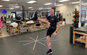
Dynamic versus static stretching are two common methods for warming up and improving flexibility, but they serve different purposes and are best suited for different situations. Understanding the right type of stretching before and after activity is essential to improve performance and prevent injuries. When young athletes engage in physical activity, they are often encouraged by coaches and parents to stretch prior to the activity. The aim is to prepare the muscles for exercise and reduce the risk of injuries.
Preparing the body for physical activity
A thorough warm-up is intended to prepare the body for physical activity by:
Increase in core body temperature
Stimulates blood flow to the arms and legs
Improving coordinated movement
Improving range of motion
Develop body awareness of joint position sense and movement
Using movement to increase the flexibility of muscles and tendons
Dynamic stretching:
Goal: Dynamic stretching involves moving your muscles and joints through a range of motion to increase blood flow, warm up your body, and prepare your muscles and joints for physical activity. It is usually used as part of a warm-up routine before exercise or vigorous exercise.
Technology: Dynamic stretching exercises are performed by actively moving your limbs and muscles without assuming a static position. These stretches mimic the movements you perform during your activity. Examples include leg swings, arm circles, walking lunges, high knees and butt kicks.
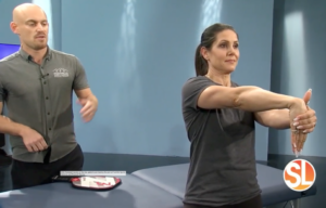
Goal: Static stretching is used to improve overall flexibility and lengthen muscles. It involves holding a stretched position for an extended period of time, usually 15-30 seconds or more, without any bouncing or dynamic movement. It is often used for a post-exercise cooldown or as part of a general flexibility routine.
Technology: Static stretching involves stretching a specific muscle or muscle group to the point of mild discomfort and holding the position without movement. Common static stretches include touching your toes while sitting, stretching your calf against a wall or a standing quad stretch.
Advantages:
Increases flexibility and range of motion.
Helps with muscle relaxation and stress reduction.
Best suited for cooldown or recovery after exercise.
To design dynamic stretching programs:
Exercise continuously, usually in rounds for a total of 10-15 minutes
Vary the program depending on the athlete’s level
Start slow and progress to faster and more advanced movements
Avoid movements that are too intense and tire the muscles.
Take in the whole body and imitate movements used in specific sports
To design static stretching programs:
Stay in one position per muscle group
Hold the stretch for 20-30 seconds
Repeat the stretch 2-3 times per muscle group
Treat all muscle groups used in the specific sport
When should you use dynamic versus static stretching?
Dynamic stretching: Use dynamic stretching as part of your warm-up routine before activities that require strength, speed or agility. It is especially useful for sports such as basketball, football or sprinting, which require explosive movements. Dynamic stretching ensures that your muscles and joints are ready for the demands of such activities.
Static stretching: Reserve static stretching for after your workout or as a separate flexibility routine. It helps improve overall flexibility and can be useful for activities such as yoga or Pilates. Static stretching can also be beneficial for relaxation and stress reduction.
Remember that stretching should be done safely and should not cause pain or discomfort. It is essential to warm up your body before doing static stretches to prevent injuries. Incorporating both dynamic and static stretching into your fitness routine can help you maintain optimal flexibility and reduce the risk of injury during physical activities.
For help designing a stretching program, contact a Foothills Sports Medicine Clinic near you and schedule an appointment.
Dublin, October 26, 2023 (GLOBE NEWSWIRE) – The report “Scoliosis Management – Global Strategic Business Report” has been added to ResearchAndMarkets.com’s to offer.
The global scoliosis treatment market will reach $3.8 billion by 2030
The global scoliosis treatment market, estimated at USD 2.8 billion in the year 2022, is expected to reach a revised size of USD 3.8 billion by 2030, with a CAGR of 4.1% over the analysis period 2022-2030.
This data includes an analysis of the Thoracolumbosacral Orthosis (TLSO) and related orthosis markets worldwide. It includes current, historical, and future annual sales figures in thousands of dollars for the years 2022 through 2030, along with percentage compound annual growth rates (%CAGR).
The analysis is segmented into different product types, including TLSO, Cervical Thoracic Lumbar Sacral Orthosis (CTLSO), Lumbosacral Orthosis (LSO), and Other Product Types. Furthermore, within these product types a distinction is made between the Pediatric and Adult segments.
Thoracolumbosacral orthosis (TLSO), one of the segments analyzed in the report, is expected to register a CAGR of 4.4% and reach $2.9 billion by the end of the analysis period. Growth in the Cervical Thoracic Lumbar Sacral Orthosis (CTLSO) segment is estimated at 3.7% CAGR over the next eight years.
The US market is estimated at $987.6 million, while China is expected to grow at a CAGR of 5%
The US scoliosis treatment market is estimated to reach USD 987.6 million by the year 2022. China, the second largest economy in the world, is expected to reach a projected market size of USD 422.1 million by 2030, at a CAGR of 5% over recent years. analysis period 2022 to 2030.
The data provides a 16-year perspective, breaking down the percentage of value sales for different geographic regions, including the US, Canada, Japan, China, Europe, Asia Pacific and the rest of the world, for the years 2014, 2023 and 2030 Finally, it provides a comprehensive analysis of the scoliosis treatment market, including annual sales figures from 2014 through 2030, and segmented analyzes for different geographic regions.
Other notable geographic markets include Japan and Canada, each expected to grow by 3.1% and 3.4%, respectively, over the 2022-2030 period. Within Europe, Germany is expected to grow at a CAGR of approximately 4.1%.
This comprehensive report also provides detail on the approaches that leading market competitors such as Aspen Medical Products, Bauerfeind AG and Boston Orthotics & Prosthetics are taking, providing invaluable insights that you as an executive can leverage.
What’s new?
Special discussions on the global economic environment and market sentiment
Coverage on global competitiveness and key market shares of competitors
Access to digital archives and a trademarked research platform
Free updates for a year
Access curated YouTube video transcripts of market sentiments shared by CEOs, domain experts and market influencers through interviews, podcasts, press statements and event keynotes
MARKET OVERVIEW
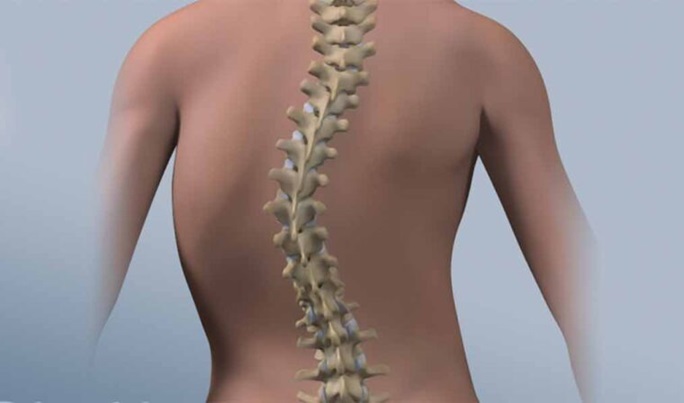
Scoliosis: An abnormal lateral curvature of the spine
Scoliosis Management/Treatment Options
COVID-19 is casting a shadow on the scoliosis treatment market
Competition
Scoliosis Management – Percentage Market Share of Key Competitors Globally in 2023 (E)
Competitive Market Presence – Strong/Active/Niche/Trivial for Global Players in 2023 (E)
Great startup ecosystem
Global market analysis and prospects
The global scoliosis management market will demonstrate steady growth driven by innovations and advancements
North America holds a leading position in the global scoliosis management market
ThoracoLumboSacral Orthosis (TLSO) dominates the market
AIS remains the primary disease type segment
Pediatric/adolescent as an important age group segment
Hospitals and ASCs claim leading share
Technological advancements as pulsating trends drive the scoliosis treatment market forward
Increase R&D activity to drive market expansion
Market restrictions
Market challenges
Recent market activity
GLOBAL BRANDS
MARKET TRENDS & DRIVERS
Rising prevalence of idiopathic and congenital scoliosis drives market growth
Spinal fusion surgery offers great opportunities
The demand for fusionless scoliosis surgery is increasing
Tying the vertebral body provides traction
Schroth therapy – a useful approach to treat scoliosis
Strong focus on minimally invasive spine surgery and increasing use of navigation and robotics in scoliosis surgery to increase prospects
Increasing cases of scoliosis in adults and a growing aging population to propel the adult segment
Increase healthcare spending to stimulate growth
Recent technological advances/innovations in the scoliosis treatment market
New Satellite Rod-Based Sequential Correction for Severe Rigid Spinal Deformities to Reduce Surgical Risks and Other Complications
ApiFix system for correcting moderate scoliosis with single curves
Shriners Hospitals for Children’s The Tether, a device for treating scoliosis, receives FDA approval
Minimally invasive spine surgeries are becoming commonplace, but long-term research into results is essential for wider adoption
Disruptive technologies in the spine space – a review
3D printed braces have potential to improve the treatment of scoliosis
FOCUS ON SELECTED PLAYERS(Total 91 recommended)
Aspen Medical Products, LLC
Bauerfeind AG
Boston Orthoses and prosthetics
Chaneco
Enovis
Fitted, Inc.
Horton’s Orthotics and Prosthetics
Lawall Prosthetics and Orthotics
Optec USA, Inc.
Orthotec
Ottobock SE & Co. KGaA
Spinal Technology Inc.
Real
For more information about this report, visit https://www.researchandmarkets.com/r/ozemky
About ResearchAndMarkets.com ResearchAndMarkets.com is the world’s leading source for international market research reports and market data. We provide you with the latest data on international and regional markets, key industries, top companies, new products and the latest trends.
Post-stroke patients with motor dysfunction who were hospitalized in the Department of Rehabilitation Medicine of our hospital from January 1, 2020 to January 1, 2022, aged 50 to 70 years old, were selected.
Inclusion criteria: [12] ① The patient met the diagnostic criteria for stroke established at the Fourth National Academic Conference on Cerebrovascular Diseases in 1995, [13, 14] and stroke was diagnosed as the primary disease on CT or MRI. ② The time between disease onset and enrollment ranged from two weeks to three months. ③ The patient’s vital signs were stable and the patient was conscious, able to understand the instructions and cooperate with the rehabilitation training. ④ The patient’s score on the Kinesthetic and Visual Imagery Questionnaire (KVIQ) was ≥ 25 points. ⑤ The patient signed the required informed consent form. ⑥ Age between 50 and 70 years.
Exclusion criteria: [14] ① The patient suffered from severe cardiac, hepatic or renal insufficiency, a malignant tumor, etc. ② The patient suffered from impaired consciousness, aphasia, mental disorder or severe cognitive impairment. ③ The patient has had other craniocerebral diseases or traumatic sequelae in the past. ④ The patient has previous severe osteoarticular disorders causing abnormal trunk function.
Finally, a total of 100 patients with stroke and motor dysfunction were included, and they were divided into a control group and a trial group according to the random number table, with 50 cases in each group. There was no significant difference (P > 0.05) in general data such as gender, age, disease course and KVIQ between the two groups, and they were comparable. See Table 1 for details. This study was approved by the local ethics committee (approval number: 2018-ethical review-189) and conducted in accordance with the Declaration of Helsinki. All participants provided written informed consent.
Table 1 Comparison of general data of patients such as gender, age, disease course and lesion site between the two groups
Treatment methods
The patients in the control group underwent routine rehabilitation therapy and remained in the supine position in the same environment for the same amount of time as the combined trunk motor imagery therapy. Meanwhile, the trial group received both routine rehabilitation therapy and combined trunk motor imagery therapy.
Routine rehabilitation therapy
The training included proper limb positioning, neuromuscular promotion techniques, such as the proprioceptive neuromuscular facilitation technique (PNF), Rood’s approach, motor relearning, occupational therapy, daily living training and traditional therapy. The participants received routine rehabilitation therapy for five hours a day, five times a week, for a period of four weeks.
Motor image therapy
The motor imagery therapy training consisted of six steps: [4, 14] ① Illustration of the task: The therapist first demonstrated and explained the content of the imagery training, asking the patients to carefully observe and identify which part of the limb was ‘active’, what kind of movement was to be performed, and the normal movement to master. feeling. ② Preview: Patients were asked to re-imagine the relevant movements. ③ Motor imagery: Patients listened to the motor imagery instruction tape and practiced the imagery. ④ Rehabilitation training: the patients repeatedly practiced the movements of imagery training. ⑤ Problem solving: The patients learned relevant skills through repeated practice. ⑥ Practical application: the patients convert relevant skills into practical skills. Before the motor images, a video of a normal person’s trunk movements was shown, including stable trunk movements with a Bobath ball, and balance movements while sitting, standing, and reaching to move a water cup. The 10-minute video and audio were shown to patients via a computer in a quiet treatment room. During each training session, patients were instructed to close their eyes and sit on a comfortable chair with their bodies relaxed. The patients then imagined the movement of their body based on the specific motor imagery instructions in the video. During the treatment, the therapist occasionally interrupted the patients to ask questions, to see if they could concentrate on the images of the physical movement. At the end of the session, the patients were asked to refocus their attention on their surroundings, after which they were sent back to their room and asked to feel their physical being. The patients were then asked to pay attention to the environmental sounds. Finally, the narrator counted down from 10 to 1, and the patients were asked to open their eyes when the countdown reached 1. A motor imagery video was shown only during the first treatment, after which the patients underwent motor imagery training according to the motor imagery. guidelines for imagery. The motor imagery therapy sessions were conducted for 30 minutes each, with a frequency of five times per week, for a total of four weeks.
Observation indicators and evaluation methods
The evaluation of the patient’s trunk control was performed before treatment and four weeks after treatment using Sheikh trunk control evaluation. The simple Fugl-Meyer assessment (FMA), the Berg rating scale (BBS), and the balance feedback trainer were used to evaluate the motor and balance functions of the patients. In addition, before and after treatment, the sEMG signals of the bilateral erector spinae and rectus abdominis in the maximum flexion and extension range at a uniform speed under the sitting position were measured by sEMG signals. All evaluations were performed in a blinded manner by the same evaluator.
Sheik Hull Check Evaluation
Sheikh is a scale for evaluating the ability to control the trunk. It involves four movements: turning from the supine position to the hemiplegic side, turning to the healthy side, sitting upright from the supine position and maintaining balance in a sitting position on the bed. The scoring method is: 0 points for non-completion, 12 points for completion but needing some assistance (grasping or leaning on an object), and 25 points for normal completion. A higher total score indicates better trunk control.
BBS rating
The balance function is divided into 14 items, from easy to difficult, and each item is scored based on a five-point scale: 0, 1, 2, 3, and 4. The highest score is 4 points and the lowest score is 0 points. . The highest integral score is 56 points, the lowest is 0 points. The higher the score, the better the balance function.
Evaluation of motor functions
FMA is used to evaluate motor function in patients. The highest score is 100. The higher the score, the better the patients’ motor functioning will be.
Evaluation of balance feedback training equipment
The ProKin 254P (PK-254P) balance feedback training device, manufactured by TecnoBody Ltd., Italy, was used to test the postural stability of the patients. Stability tests were performed in standing position with eyes open using the static mode of the PK-254P balancer. The standard standing posture includes: ① Bilaterally symmetrical standing with A1A5 as central axis. ② The patients raise their heads and look straight ahead. ③ Both upper limbs are naturally placed on either side of the body. ④ The medial edges of both feet are 10 cm apart and the highest point of the bilateral arches is on axis A3A5. Observation parameters are as follows: movement length, movement area, mean front-back movement speed, and mean left-right movement speed.
sEMG signal acquisition
While the patients are seated on a square stool, their trunk is subjected to anterior flexion and posterior extension in the maximum range at uniform velocity. The Shanghai NCC 8-channel sEMG signal acquisition system was used to acquire the bilateral erector spinae and rectus abdominis myoelectric signals. The electrodes were taped to the 3 cm lateral opening on the left and right sides of the L3 spinous process (erector spinae) and the 3 cm lateral opening on the left and right sides 3 cm above the navel (rectus abdominis). The conductive diameter of the electrodes was 1 cm and the distance between the electrodes was 2 cm. Dandruff and oil were removed with a fine gauze and alcohol before testing. The root mean square (RMS) of myoelectric signals was then analyzed. The test was repeated three times with an interval of 30 seconds to obtain the average value. The RMS of the bilateral rectus abdominis and erector spinae of the two groups was evaluated before treatment and four weeks after treatment.
static analysis
SPSS software version 16.0 was used to analyze the data. The measurement data is expressed as (\(\bar x \pm s\)). Parametric statistics were applied when the data collected met the assumptions of homogeneity of variance and normal distribution. When these assumptions were not met, non-parametric statistics were used. The paired sample T-test was used for pre- and post-treatment comparison within the same group, while the independent sample T-test was used for between-group comparison, and P< 0.05 indicated that the difference was statistically significant.
Looking for a healthy twist on a breakfast classic?
Here it is!
Our coconut protein pancakes recipe is sure to become a new favorite.
Thanks to the coconut flour base and the addition of collagen, these pancakes are packed with protein and bone-healthy nutrients like potassium, iron, selenium and manganese.
Because this recipe is ready in just ten minutes, you can make this pancake as an energetic start to the day; you can serve it during brunch; or enjoy it as a delicious, bone-boosting dessert.
1 scoop (10 g) collagen peptides (optional for more protein! Add an extra tablespoon of milk)
2 large pasture eggs
1 tablespoon (15 ml) avocado oil
2 tablespoons (30 ml) milk of choice (non-dairy milk for paleo)
1 tablespoon (15 ml) maple syrup (or liquid maple sweetener for keto)
Directions
1) Sift the coconut flour, baking powder and collagen (if using) into a mixing bowl. Add the remaining ingredients and beat everything together until smooth and combined.
2) Heat a frying pan or griddle over medium heat. Once hot, lightly grease the skillet with avocado oil or ghee. Pour the pancake mixture into the frying pan and smooth the batter with a spatula or spoon, as it is quite thick. Cook for 4-5 minutes on each side, or until golden brown.
3) Serve immediately with your favorite nut or seed butter and fruit.
Recipe created by BoneCoach™ Team Dietitian Amanda Natividad-Li, RD & Chef.
Medical disclaimer
The information shared above is for informational purposes only and is not intended as medical or nutritional therapy advice; it does not diagnose, treat or cure any disease or condition; it should not be used as a substitute or substitute for medical advice from physicians and trained medical professionals. If you are under the care of a healthcare professional or are currently taking prescription medications, you should discuss any changes in your diet and lifestyle or possible use of nutritional supplements with your doctor. You should not stop prescribed medications without first consulting your doctor.
Every basketball coach has told his players that free throws win games. A new study from the University of Kansas used innovative, markerless motion capture technology to determine the mechanics of skilled free throw shooters and better understand one of the biggest keys to success in the game.
According to the study, skilled free throw shooters – who could make more than 70% of their shots – performed the shooting motion in a more controlled manner. They had lower knee and center of mass peaks and average angular velocities compared to non-experienced shooters. Also, skilled shooters achieved a greater release height and had less forward torso lean at the point of ball release.
“These findings imply that the movement of basketball shooting is not as simple as some may think. Shooting efficiency cannot simply be attributed to one biomechanical variable. It is based on a mix of multiple segmental body movements performed in a controlled manner,” says Dimitrije. Cabarkapa, lead author of the study and deputy director of the Jayhawk Athletic Performance Laboratory.
The study examined 34 men with at least four years of basketball experience, ranging from recreational to collegiate competitive levels. Each participant attempted 10 free throws with a 10-15 second rest interval between each attempt. A three-dimensional markerless motion capture system developed by the Southwest Research Institute (SwRI Enable, San Antonio, Texas), consisting of nine high-definition cameras (120 fps), was used to capture the biomechanical characteristics of free throw shooting movements and to analyse. .
“We are very interested in analyzing basketball shooting mechanics and what performance parameters distinguish skilled and non-skilled shooters,” Cabarkapa said. “High-speed video analysis is one way we can do that, but innovative technological tools like markerless motion capture systems can allow us to dig even deeper into that. In my opinion, the future of sports science is based on the use of non-invasive and time-efficient testing methods.”
The study, conducted at the Jayhawk Athletic Performance Laboratory, also found that when differentiating between made and missed shots by skilled free throw shooters, an overemphasis on release height could be counterproductive.
“These findings can be metaphorically represented by some healthy habits of daily life. Exercise, drinking water and consuming enough vitamins and minerals are all very beneficial to our health. However, overdoing these things can be harmful in certain cases and can even production of the disease. the opposite effect than expected,” Cabarkapa said.
The study, published in the journal Limits in sports and active life, was co-authored with Damjana Cabarkapa and Andrew Fry of the Jayhawk Performance Athletic Laboratory at KU; Jonathan Miller of KU’s Higuchi Biosciences Center; and Tylan Templin, Lance Frazer and Daniel Nicolella of the Southwest Research Institute.
Using motion capture technology without markers is useful for several reasons, the authors said, because other motion capture systems that use markers to be placed on the skin or clothing have several problems, such as not staying in place and the participant’s awareness of the markers. which can change normal movement patterns. This is crucial when testing in a sport-specific environment, where efficiency is key. The use of markerless motion capture technology enables non-invasive assessment.
Dimitrije Cabarkapa said that, to the authors’ knowledge, this is the first study to use this motion capture system to investigate the biomechanical characteristics of skilled free throw shooters. Previous research has shown that teams with better free-throw skills, especially late in the game, are more likely to win. Although the current study did not include the effects of fatigue on shooting mechanics and accuracy, researchers hope to investigate that factor in upcoming studies, as well as the effect of the presence of a defender on shooting mechanics and accuracy.
The laboratory is part of the Wu Tsai Human Performance Alliance, a consortium of researchers investigating optimal human performance. This alliance includes Stanford University, University of Oregon, Boston Children’s Hospital, Salk Institute, University of California San Diego and KU.
“These findings add to the work we have done in the past and to the body of scientific literature related to basketball shooting performance that we are continually expanding in our laboratory,” said Dimitrije Cabarkapa. “We have found that both the preparation and release phases of the shooting motion are critical to achieving solid levels of shooting efficiency. The implementation of innovative technology can allow us to understand the transition phase of the shooting motion and the kinematic chain in more detail Ultimately, our goal is to have an answer to the question every basketball fan wants to know: ‘Why did Steph Curry miss that shot?’”
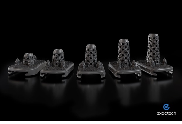
GAINESVILLE, Fla., October 26, 2023–(BUSINESS WIRE)–Exactech, a developer and manufacturer of innovative implants, instrumentation and smart technologies for joint replacement surgery, today announced the 510(k) clearance of its new 3D printed tibial implants, the Vantage® Only 3D and 3D+. This is the second recent approval for the broader Vantage Total Ankle System, following Activit-E™, a new advanced vitamin E polyethylene, which received approval from the Food and Drug Administration last month.
The new implants provide orthopedic surgeons with additional tibial height options for total ankle replacement patients. Both implants are 3D printed and designed with a porous surface that mimics subchondral bone to aid in biological fixation. They are fitted with pointed pegs and a central bone cage, of varying lengths, to achieve initial fixation. The Vantage Ankle 3D builds on the design philosophy of the system’s current tibial offering and is available in a 10mm height. The Vantage Ankle 3D+ is a stem implant with options of 15, 20, 25 and 30 mm.
“As an original member of the Vantage Ankle design team, I am excited about the next generation, state-of-the-art Vantage Ankle 3D and 3D+ tibial components,” said Mark Easley, MD, of Duke Health. “Developed through the collaborative efforts of our extensive team of expert consultant surgeons and experienced engineers, the redesigned tibial implants feature innovative, intuitive surgical technique and surgeon-friendly instrumentation.”
The Vantage Ankle 3D and 3D+ are compatible with Exactech’s patient-specific instruments, the Vantage Ankle PSI cutting guides. 3D Systems (NYSE:DDD), manufacturer of the Vantage Ankle PSI, previously received 510(k) clearance for four additional cutting guides, two of which are specifically made for use with these new tibial implants.
“We are laser-focused on delivering innovative solutions that our surgeons and patients need,” said Devan Carter, Exactech’s foot and ankle marketing director. “With these new additions, the Vantage Ankle now offers solutions from pre-operative planning to complex deformities, bringing us one step closer to providing a complete continuum of care.”
Full market availability for the Vantage Ankle 3D and 3D+ is expected before the end of 2024. For more information, please contact your local Exactech representative.
About Exactech
Exactech is a global medical device company that develops and markets orthopedic implant devices, related surgical instruments and Active Intelligence® platform of smart technologies for hospitals and doctors. Headquartered in Gainesville, Florida, Exactech markets its products in the United States, in addition to more than 30 markets in Europe, Latin America, Asia and the Pacific. Visit www.exac.com for more information and connect with us on LinkedIn, VuMedi, YouTube, Tweet and Instagram. With Exactech by your side you have EXACTLY what you need.
A team of scientists led by KOO Sagang of Seoul National University and the Center for Nanoparticle Research within the Institue for Basic Science Center (IBS), in collaboration with researchers from the Korea Institute of Science and Technology (KIST) and the Seoul National University, developed a new solution for the treatment of rheumatoid arthritis (RA).
RA is a chronic disease that unfortunately cannot be cured. The disease causes a mix of troublesome symptoms, such as inflamed joints, harmful cytokines, and immune system imbalances, which work together to create a relentless cycle of worsening symptoms. While addressing some of these factors can provide short-term relief, others remain unresolved, leading to a frustrating cycle of remission and flare-ups.
One of the biggest hurdles in treating RA is the inability to restore the immune system to a healthy state. This leaves the body unable to control the continued production of harmful substances such as reactive oxygen species (ROS) and inflammatory cytokines, leading to persistent inflammation and discomfort.
Essentially, the ideal treatment for RA should not only provide immediate relief from inflammation and symptoms, but also address the cause by restoring the immune system to its normal, balanced state.
New nanoparticle-based system as a solution
The novel platform involves immobilizing ceria nanoparticles (Ce NPs) on mesenchymal stem cell-derived nanovesicles (MSCNVs). Both components can hinder various pathogenic factors, allowing them to work individually and together to achieve comprehensive treatment.
Ce NPs – can scavenge the overproduction of ROS in RA-induced knee joints. They also cause polarization of M1 macrophages into M2, immediately relieving inflammation and symptoms.
MSCNVs – deliver immunomodulatory cytokines, which convert dendritic cells (DC) into tolerogenic dendritic cells (tDCs). This consequently generates regulatory T cells for long-term immune tolerance.
In short, this approach aims to bridge both innate and adaptive immunity to achieve short-term pain relief, and to convert the tissue environment into an immune-tolerant state to prevent recurrence of symptoms.
Researchers confirmed the efficacy of this approach using a collagen-induced arthritis mouse model. The Ce-MSCNV system was able to comprehensively treat and prevent RA by simultaneously easing and restoring immediate T cell immunity. Supporting data suggests that improvement in conditions can be achieved after only a single dose treatment.
The mice treated with the Ce-MSCNV combination did much better compared to the mice treated with the Ce NP or MSCNV group alone. This clearly demonstrates the synergy between anti-inflammatory agents and immunomodulation and underlines the importance of the combined therapy for effective treatment of RA. Furthermore, administration of Ce-MSCNV prior to booster injection significantly reduced the incidence and severity of symptoms, supporting the prophylactic potential of these nanoparticles.
One of the most difficult decisions in the treatment of intractable diseases is determining how long to continue treatment. For RA, it would not be appropriate to stop treatment just because the target marker has stabilized. A safer indicator should be that the innate and adaptive components of the collapsed immune system are normalized to protect the body.”
Koo Sagang, first author
Koo believes that Ce-MSCNVs’ strategy of working together with different treatment mechanisms offers a unique advantage in this regard. Furthermore, she predicts that a similar approach for this purpose would also be applicable to other refractory, inflammatory and autoimmune diseases. The components within the system can also be changed. For example, depending on the type of disease, other catalysts can be used to generate ROS or other cell-derived nanovesicles. Overall, this study proves the potential of a hybrid nanoparticle system for the comprehensive treatment of autoimmune diseases and modulation of the immune system.
Source:
Institute for Basic Sciences
Magazine reference:
Koo, S., et al. (2023). Ceria-vesicle nanohybrid therapeutic agent for modulation of innate and adaptive immunity in a collagen-induced arthritis model. Nature Nanotechnology. doi.org/10.1038/s41565-023-01523-y.
Alcohol use after injury is associated with prolonged recovery from concussion in NCAA athletes
Chang RC, Singleton M, Chrisman SPD, et al. Alcohol use after injury is associated with prolonged recovery from concussion in NCAA athletes. Clin J Sport Med. Published online 2023:1-8. doi:10.1097/JSM.0000000000001165
Concussions lead to immediate and variable symptoms that affect quality of life. Many collegiate athletes consume alcohol regularly, but there is no consensus regarding the effects of alcohol consumption after a concussion on symptoms.
Study aim
The authors used a prospective cohort to assess whether alcohol consumption after concussion is associated with resolution of concussion symptoms. The authors also evaluated the impact of alcohol consumption on symptom severity.
Methods
The authors used data from the ongoing Concussion Assessment Research and Education (CARE) Consortium. The authors analyzed data from 29 different clinical sites and 484 of 3,518 athletes with concussion. An athlete who reported consuming at least one alcoholic beverage per week during concussion recovery was considered an alcohol user. The authors used the Sport Concussion Assessment Tool 3 (SCAT3) symptom score sheet to track outcomes after concussion.
Results
Athletes who drank alcohol after a concussion took an average of 22 days to return to full participation in sports. This represents a 33% delay in return to play compared to those who did not consume alcohol after injury. On average, those who drank alcohol after a concussion needed about five additional days to return to full-time sports. Additionally, the more alcohol consumed after the concussion, the longer it took for symptoms to resolve. Alcohol consumption had no influence on the severity of symptoms.
Viewpoints
This study clarifies previous research on the relationship between post-concussion alcohol use and delayed symptom resolution. The delay in symptom resolution may contribute to deconditioning among those who have consumed alcohol after a concussion, which could hinder their willingness to contribute to team success. Interestingly, alcohol consumption after an injury does not play a role in the severity of symptoms, according to this study. This suggests that alcohol may slow the healing process rather than causing additional tissue damage. While these results are important, this study did not standardize a “drink.” Therefore, there is a possibility that different types and volumes of alcohol consumed per drink could influence symptom resolution. Furthermore, the authors focused on less than 15% of athletes with a concussion. Therefore, it will be important to replicate these findings to see if they apply to the broader athletic concussion population.
Clinical implications
In practice, physicians should encourage athletes diagnosed with a concussion to reduce alcohol intake or, ideally, to abstain from alcohol consumption until symptoms resolve. Clinicians should also educate athletes about the role alcohol consumption after injury plays in prolonging concussion symptoms.
Questions for discussion
What strategies, if any, do you use to educate athletes about alcohol consumption after a concussion? What changes do you plan to make to patient education based on this research?
related posts
Gender-specific predictors of long-term recovery from concussion
Suggested answer to the most common question about concussions: How many days until I can play?