Tag: Knee Injury?

  • What Does a Torn Meniscus Look Like on the Outside: Recognizing the Visual Signs

    What Does a Torn Meniscus Look Like on the Outside: Recognizing the Visual Signs

    As a physical therapist with over 30 years of experience, I’ve helped countless patients identify and recover from meniscus tears. One of the most common questions I hear is: “What does a torn meniscus look like on the outside?” While diagnosis requires professional assessment, there are indeed visible external signs that can help you recognize a potential meniscus injury. In this comprehensive guide, I’ll walk you through what you might observe from the outside when dealing with this common knee injury.

    ![Knee Anatomy Showing Meniscus Location] Image suggestion: A clear anatomical diagram showing the knee joint with the medial and lateral meniscus highlighted, along with surrounding structures like femur, tibia, and ligaments.

    Understanding the Meniscus: A Foundation for Recognition

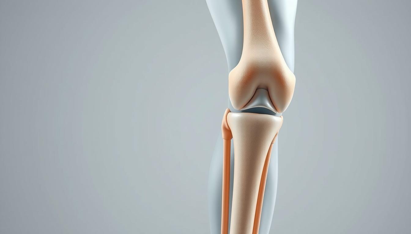
    Before discussing external appearances, it’s important to understand what we’re looking for. The meniscus is a C-shaped cartilage pad that sits between your femur (thigh bone) and tibia (shin bone). Each knee has two menisci – the medial meniscus on the inner side of the knee and the lateral meniscus on the outer side. These structures act as shock absorbers and help stabilize your knee joint during movement.

    When tears occur, they disrupt this cushioning system, leading to changes that can sometimes be visible from the outside. However, it’s important to note that not all meniscus tears present with obvious external signs, especially in the early stages or with minor tears.

    Initial Visual Signs After Injury

    The earliest visible sign of a torn meniscus is often swelling around the knee joint. This swelling typically develops gradually over 24-48 hours after the injury, unlike an ACL tear where swelling often appears rapidly. Here’s what you might notice:

    Localized Swelling Patterns

    The swelling from a meniscus tear tends to be more localized than general knee swelling. For medial (inner) meniscus tears, which are more common, you might notice fullness along the inner joint line of the knee. For lateral (outer) meniscus tears, the swelling may be more prominent along the outer aspect of the knee.

    ![Knee with Joint Line Swelling] Image suggestion: A photograph showing a side-by-side comparison of a normal knee and one with visible joint line swelling characteristic of a meniscus tear. The image should highlight the subtle fullness along the joint line that indicates potential meniscus involvement.

    This localized swelling creates a visible change in the normal contour of the knee. When comparing to the uninjured knee, you may notice that the natural depressions or dimples that normally exist around the knee joint appear filled in or puffier on the injured side.

    Changes in Knee Contour

    A healthy knee has a certain shape and profile with visible landmarks like the patella (kneecap) and the natural depressions around it. With a torn meniscus, these contours often change in subtle but noticeable ways:

    • The joint line (where the thigh bone meets the shin bone) may appear fuller or more rounded
    • The normal hollow areas on either side of the kneecap might appear filled in
    • The overall symmetry of the knee compared to the uninjured side is disrupted

    In my practice, I often ask patients to compare both knees while standing in front of a mirror – the differences in contour can sometimes be quite apparent even to untrained eyes.

    Visual Differences Based on Tear Type

    Different types of meniscus tears can present with varying external appearances:

    [GRAPHIC: Types of Meniscus Tears and Their External Appearance] Graphic suggestion: An informative chart showing different types of meniscus tears (bucket handle, flap, radial, horizontal, degenerative) with corresponding illustrations of how each might affect the external appearance of the knee. Include brief bullet points of key visual indicators for each type.

    Bucket Handle Tears: The Locked Knee Appearance

    One of the most visually distinctive meniscus injuries is the bucket handle tear, where a large portion of the meniscus displaces into the joint. When this happens, the knee may become “locked,” unable to fully extend. From the outside, you might observe:

    • The knee stuck in a slightly bent position, typically at about 15-30 degrees of flexion
    • An inability to straighten the leg completely
    • The quadriceps muscle appearing more tense on the affected side
    • A noticeable difference in leg alignment when standing (if able to bear weight)

    This locked position creates a very visible asymmetry between the legs that even untrained observers can often notice.

    Degenerative Tears: Subtle Progressive Changes

    Unlike traumatic tears that happen suddenly, degenerative meniscus tears develop gradually over time, usually in older adults. The external signs tend to be more subtle:

    • Mild, intermittent swelling that comes and goes with activity
    • Gradual changes in knee contour over weeks or months
    • Less dramatic joint line fullness
    • Occasional visible fluid accumulation after increased activity

    These tears may not present with obvious external changes until they’ve progressed significantly, making them easier to miss by simply observing from the outside.

    Functional Visual Signs in Daily Activities

    Sometimes the most telling external signs of a meniscus tear appear during movement rather than at rest. As you observe someone with a suspected meniscus tear, you might notice:

    ![Person Demonstrating Difficulty with Stairs] Image suggestion: A photograph showing someone with a meniscus tear navigating stairs with visible compensatory movements, such as leading with the unaffected leg when descending or using a handrail for additional support.

    Gait Changes

    • A hesitation or shortened stance phase on the affected leg
    • Reduced knee flexion during the swing phase of walking
    • A tendency to rotate the foot outward on the affected side to reduce pressure on the torn meniscus
    • Subtle limping, especially after sitting for prolonged periods

    Difficulty with Specific Movements

    Certain activities can make torn meniscus symptoms visibly apparent:

    • Struggle to fully squat down (you might see the person compensate by shifting weight to the unaffected side)
    • Difficulty with stair climbing, especially descending stairs
    • Visible hesitation when transitioning from sitting to standing
    • Tendency to keep the knee slightly bent when standing still

    During my years as a physical therapist, I’ve observed that many patients unconsciously develop these movement adaptations to avoid pain, creating visible patterns that help indicate the presence of a meniscus injury.

    Comparing with Other Knee Injuries

    It can be challenging to differentiate a meniscus tear from other knee injuries based solely on external appearance. Here are some distinguishing features:

    Meniscus Tear vs. ACL Tear

    Meniscus Tear:

    • More localized swelling, often along the joint line
    • Swelling develops more gradually (24-48 hours)
    • May have specific positions where the knee catches or locks
    • Often can still bear weight, though with pain

    ACL Tear:

    • More diffuse, general knee swelling
    • Rapid swelling development (within hours)
    • Less likely to have mechanical locking
    • Often more difficulty with weight bearing initially

    Meniscus Tear vs. MCL Injury

    Meniscus Tear:

    • Swelling concentrated around the joint line
    • Rarely shows visible bruising
    • May demonstrate mechanical symptoms like locking or catching

    MCL Injury:

    • Swelling and potential bruising along the inner side of the knee
    • Sometimes visible laxity with valgus (inward) stress
    • Pain more concentrated along the ligament rather than the joint line

    Visible Changes Over Time

    The external appearance of a meniscus tear evolves over time:

    ![Timeline of Meniscus Tear Appearance] Image suggestion: A series of photographs showing the progression of how a knee with a meniscus tear appears at different stages – acute phase (1-3 days post-injury), subacute phase (1-3 weeks), and chronic phase (1+ months), highlighting how swelling patterns and visual indicators change.

    Acute Phase (First Few Days)

    • Initial swelling development
    • Possible slight redness or warmth
    • Protective posturing of the leg

    Subacute Phase (Days to Weeks)

    • Peak swelling that gradually subsides
    • Development of muscle guarding patterns
    • Compensatory movement patterns become more established

    Chronic Phase (Weeks to Months)

    • Possible visual signs of quadriceps atrophy (muscle wasting)
    • Established movement compensations
    • Periodic episodes of increased swelling with certain activities
    • Joint line changes that become more permanent

    In my practice, I’ve observed that untreated meniscus tears often lead to visible quadriceps atrophy (muscle shrinking) on the affected side after several weeks. This creates an asymmetrical appearance when comparing both legs, with the thigh of the injured leg appearing slightly smaller or flatter.

    Documentation for Medical Visits

    If you suspect a meniscus tear and plan to see a healthcare provider, documenting the external appearance can be helpful:

    • Take photos of both knees from the front, side, and above for comparison
    • Record a video of your walking pattern
    • Note when swelling increases or decreases
    • Document which activities produce visible changes in your knee

    These visual records can help your healthcare provider make a more accurate assessment and track changes over time.

    When the Outside Doesn’t Tell the Full Story

    It’s crucial to understand that not all meniscus tears have obvious external signs. In fact, many smaller tears or those in less vascularized regions of the meniscus might show minimal external changes while still causing significant internal disruption and pain.

    Some patients with severe tears show surprisingly little external evidence, while others with minor tears may develop significant swelling and visible changes. This variability is why professional diagnosis that includes imaging (usually MRI) is essential for confirming a torn meniscus.

    Red Flag Visual Signs Requiring Immediate Attention

    While most meniscus tears don’t constitute medical emergencies, certain visual signs warrant immediate medical attention:

    • Severe deformity of the knee
    • Inability to bear any weight on the affected leg
    • Dramatic swelling that develops very rapidly (within an hour or two)
    • Visible bruising that spreads quickly
    • Redness and warmth accompanied by fever (could indicate infection)

    These signs might indicate a more serious injury or complication beyond a simple meniscus tear.

    Non-Surgical Management and Healing Signs

    With proper treatment, the external appearance of a knee with a meniscus tear should gradually normalize. Positive visual signs of healing include:

    • Reduction in swelling with joint line contours returning to normal
    • Restoration of symmetrical appearance compared to the uninjured knee
    • Improved gait pattern with less visible compensation
    • Return of normal muscle definition as protective guarding subsides
    • Ability to perform movements like squatting with more normal appearance

    Physical therapy plays a crucial role in this normalization process. Through targeted exercises, manual techniques, and modalities, we work to reduce swelling, restore normal movement patterns, and strengthen supporting muscles. Many of my patients are surprised at how much better their knee looks from the outside after completing a rehabilitation program, even without surgical intervention.

    Post-Surgical Appearance

    If surgical intervention is required, the external appearance will naturally change during recovery:

    ![Post-Arthroscopic Knee] Image suggestion: A photograph showing a knee shortly after arthroscopic meniscus repair, with small portal incisions visible, to help patients understand what to expect after surgical intervention. Include the typical placement of the 2-3 small incisions.

    • Initial surgical dressings and bandages
    • Temporary increase in swelling post-procedure
    • Small arthroscopic portal scars (typically 2-3 small incisions)
    • Gradual return to normal contours as rehabilitation progresses

    Most arthroscopic meniscus procedures result in minimal long-term changes to the external appearance of the knee. The small surgical scars typically fade significantly within a year.

    Conclusion: The Importance of Professional Assessment

    While recognizing the external signs of a meniscus tear can help alert you to a potential problem, these visual cues should never replace professional medical evaluation. The outside appearance provides just one set of clues in the diagnostic puzzle.

    In my three decades as a physical therapist, I’ve learned that combining careful observation of external signs with a thorough clinical examination and appropriate imaging is the most reliable approach to diagnosing meniscus injuries. If you notice any of the signs we’ve discussed, I encourage you to seek professional medical advice for proper diagnosis and treatment.

    Remember that early intervention for meniscus tears often leads to better outcomes, so don’t delay seeking care if you suspect this injury. With proper treatment, most patients can return to pain-free movement with little to no lasting visible changes to their knee.

  • Knee Pain Painting on a Ladder

    Knee Pain Painting on a Ladder

    Imagine you’re in the middle of painting, but knee pain stops you. Many face this when they have a meniscus tear. Climbing ladders then becomes a big challenge, affecting your project and mobility.

    Dealing with ladder climbing and a meniscus tear is tough. We aim to give you expert advice to stay safe. Our tips help you avoid more injuries and keep working on your projects.

    Key Takeaways

    • Safely assess your knee condition before climbing a ladder.
    • Understand the proper techniques for ladder climbing with a meniscus injury.
    • Learn how to prevent further knee damage while on a ladder.
    • Discover exercises to strengthen your knee before and after ladder climbing.
    • Explore alternative solutions for painting projects that minimize ladder use.

    Understanding Knee Pain: Common Causes

    Knee pain comes from many sources. It’s key to know these causes to avoid more harm. Activities like ladder climbing put extra stress on our knees. This can make existing problems worse or cause new ones.

    Types of Knee Pain

    Knee pain falls into two main groups: acute injuries and chronic conditions. Acute injuries happen suddenly, like from a fall. Chronic conditions develop over time, often from repeated strain or wear.

    Some common knee pain types include meniscus tears, ligament sprains, and tendonitis. Knowing the exact type of pain helps find the right treatment and ways to prevent it.

    Why Ladder Work Can Exacerbate Pain

    Climbing ladders puts a lot of strain on the knees, especially when bending or standing on uneven ground. This strain can make knee problems worse or cause new ones like patellofemoral pain syndrome. The constant stress from ladder work can lead to knee inflammation and pain.

    To reduce the risk of making knee pain worse, it’s important to understand how ladder work affects the knee. Knowing this helps us take steps to lessen the strain and protect our knees.

    Safety Measures When Using a Ladder

    Ladder Safety Measures

    To stay safe while using a ladder, we need to take certain steps. Ladder climbing can be dangerous, especially for those with knee problems. By picking the right ladder and setting it up right, we can lower the chance of accidents.

    Choosing the Right Ladder

    Picking the right ladder is key. Think about how high you need to go and how much weight the ladder can hold. For painting, a ladder with a strong base and non-slip feet is best. Look for ladders with comfy rungs and think about the material. Aluminum ladders are light, while fiberglass ladders are more stable.

    Ladder Type Material Weight Capacity
    Step Ladder Aluminum 300 lbs
    Extension Ladder Fiberglass 250 lbs

    Proper Ladder Setup

    After picking the right ladder, setting it up correctly is crucial for safety. Make sure the ladder is on a solid, level ground. Having someone hold the ladder can also help. The ladder’s angle is important too; it should be at a 75-degree angle or one foot away from the wall for every four feet of ladder height.

    By following these tips, we can make ladder climbing safer. Remember, safety steps are not just suggestions; they’re essential to avoid serious injuries.

    Preparing for Your Painting Project

    knee injury prevention

    Getting ready for painting is more than just picking up supplies. It also means getting your body ready to avoid knee injury.

    Stretching Exercises Before Climbing

    Do some stretching exercises before you start climbing. This helps your knees a lot. Try:

    • Leg swings to loosen up your leg muscles
    • Knee bends to prepare your knees for the climb
    • Calf raises to strengthen your calf muscles, which support your knees

    Essential Gear to Minimize Knee Strain

    Wearing the right gear can also help your knees. Use:

    1. Knee pads for extra cushioning and support
    2. Supportive footwear to stabilize your knees
    3. Comfortable clothing that allows for a full range of motion

    With these steps, you can lower the chance of knee pain and injury while painting.

    Painting Techniques to Reduce Knee Strain

    There are ways to lessen knee strain while painting on a ladder. Painting can put a lot of pressure on our knees, especially if we have conditions like meniscus tears. Using the right techniques can help avoid making knee pain worse.

    Opt for a Brush Instead of a Roller

    Choosing a brush over a roller can help reduce knee strain. Brushes allow for more precise strokes and need less force, which is easier on the knees. Plus, they help you paint more accurately, so you might not have to climb the ladder as much.

    Take Breaks to Relieve Pressure

    It’s important to take breaks to ease knee strain. Stepping down from the ladder often can help take the pressure off your knees. These breaks are a chance to stretch and prevent knee pain.

    By using these techniques, we can make painting easier on our knees. This is especially helpful for those with knee pain or meniscus tear symptoms.

    When to Seek Medical Advice

    Knee pain can really get in the way of doing things. There are times when you need to see a doctor. If you’re feeling pain in your knee while on a ladder, it’s important to know when to get help.

    Signs Your Knee Pain Needs Attention

    There are signs that mean you should see a doctor. These include:

    • Persistent pain that doesn’t improve with rest
    • Swelling or redness around the knee
    • Instability or feeling like the knee is going to give way
    • Warmth or tenderness to the touch
    • Loss of mobility or difficulty straightening the knee

    If you notice any of these, it’s best to talk to a healthcare professional. Ignoring these signs can lead to further complications.

    Recommended Specialists for Knee Pain

    Choosing the right specialist is crucial for knee pain. Here’s a table of specialists you might consider:

    Specialist Description
    Orthopedic Surgeon Specializes in surgical treatments for musculoskeletal disorders, including knee pain. They can perform knee surgery if necessary.
    Rheumatologist Expert in diagnosing and treating rheumatic diseases that can cause knee pain.
    Physical Therapist Can help improve knee mobility and strength through targeted exercises.

    An orthopedic surgeon is often the first choice for serious knee pain or when surgery is needed. They can give a detailed diagnosis and suggest the best treatment.

    Home Remedies for Knee Pain Relief

    Home remedies are a simple way to handle knee pain and boost your quality of life. We often seek easy, effective solutions to ease discomfort before seeking medical help. Two popular methods are ice and heat therapy, and over-the-counter pain relief.

    Ice and Heat Therapy

    Ice and heat therapy are easy yet effective for knee pain. Ice therapy reduces swelling and numbs pain, great for sudden injuries or pain spikes. Heat therapy, on the other hand, relaxes muscles and boosts blood flow, helping with long-term pain and stiffness.

    To use ice, wrap an ice pack in a towel and apply it for 15-20 minutes, several times a day. For heat, try a warm bath, heating pad, or a warm cloth on your knee. It’s key to alternate between ice and heat based on your pain type.

    “The application of ice or heat can significantly alter the pain perception and is a crucial component of a comprehensive pain management plan.”

    – Expert in Pain Management

    Over-the-Counter Pain Relief Options

    Over-the-counter (OTC) pain meds are also effective for knee pain. Common ones are acetaminophen and ibuprofen. Acetaminophen mainly relieves pain, while ibuprofen also fights inflammation.

    • Always follow the recommended dosage on the label.
    • Consult with a healthcare provider if you’re unsure about interactions with other medications.
    • Be aware of potential side effects and contraindications.

    By adding these home remedies to your daily routine, you can better manage knee pain. This lets you enjoy activities like painting on a ladder more comfortably.

    Prolonging Knee Health During Painting

    To enjoy painting without harming our knees, we need to focus on knee health. Painting on a ladder can be tough on our knees, especially for those with a torn meniscus. By keeping our knees strong and moving regularly, we can protect them.

    Tips for Maintaining Strong Knees

    Keeping our knees strong involves exercises and careful movements. Physical therapy helps strengthen knee muscles, offering better support and injury prevention. Exercises like squats, lunges, and leg presses are helpful. Also, using the right ladder techniques and taking breaks can ease knee strain.

    Exercise Benefit Frequency
    Squats Strengthens quadriceps and hamstrings 3 sets of 10 reps, 3 times a week
    Lunges Improves balance and strengthens knee muscles 3 sets of 10 reps (per leg), 3 times a week
    Leg Presses Targets quadriceps, hamstrings, and glutes 3 sets of 12 reps, 3 times a week

    Importance of Regular Movement

    Regular movement is key for healthy knees. Sitting still for too long can make knees stiff and painful, especially for those with injuries. Adding gentle exercises to our day keeps knees flexible and lowers injury risk. It’s also important to listen to our bodies and rest when needed.

    By using these tips, we can keep our knees healthy while painting. Regular physical therapy, exercises, and careful movements are all important for a safe painting experience.

    Equipment to Assist While Painting on a Ladder

    To have a safe and fun painting time, the right tools are key. Using the right gear when painting on a ladder helps avoid knee pain and injuries.

    Knee Pads and Supportive Gear

    Knee pads are a simple yet effective way to protect your knees while working on a ladder. They offer cushioning and support, easing the pressure on your knees. Supportive gear, like knee braces, adds extra stability and protection. Pick knee pads or supportive gear based on your work and needed support.

    Look for knee pads that are durable, comfortable, and adjustable. It’s also important to pick knee pads that fit your task. For example, if you’re working on a rough surface, you’ll need sturdier knee pads.

    Type of Knee Pad Features Best For
    Hard Cap Knee Pads Durable, protective Rough surfaces, heavy-duty work
    Soft Cap Knee Pads Comfortable, flexible General painting, smooth surfaces
    Gel Knee Pads Cushioned, shock-absorbing Long periods of kneeling, comfort

    Choosing the Right Footwear

    Along with knee pads, choosing the right footwear is crucial for stability and to prevent slipping on the ladder. Find shoes with good grip and a snug fit to keep your feet steady on the ladder.

    • Non-slip soles
    • Comfortable fit
    • Ankle support

    By using knee pads with the right shoes, you can greatly lower the risk of knee injury. This makes painting more comfortable and enjoyable.

    Final Thoughts: Enjoying Your Painting Project

    As we finish talking about managing knee pain while painting on a ladder, remember it’s not just about the end. It’s also about how we feel during it. By taking care of our knees, we can enjoy painting more without worrying about injuries.

    It’s important to love the journey of creating. We can do this by following the tips we’ve shared. From picking the right ladder to stretching, every step matters.

    Practical Adjustments for Future Projects

    For future painting, we can reduce knee pain. Use knee pads and supportive gear. Also, pick shoes that help you stay stable on the ladder.

    Maintaining Knee Health

    Keeping our knees healthy is key to enjoying painting and other activities. Regular exercise and movement help make our knees stronger. This makes it easier to handle ladder work.

    FAQ

    What are the symptoms of a meniscus tear?

    Symptoms include knee pain, swelling, and stiffness. Sometimes, we hear a popping sound when it happens.

    How can I prevent further injury while climbing a ladder with a meniscus tear?

    To avoid more harm, choose the right ladder and set it up right. Wear knee pads and supportive shoes.

    What are the treatment options for a torn meniscus?

    Treatments include physical therapy, pain meds, and sometimes surgery. An orthopedic surgeon can help decide the best treatment.

    Can I continue with my painting projects if I have a meniscus tear?

    Yes, you can keep painting. Just be careful to avoid making the injury worse. Use proper techniques, take breaks, and wear supportive gear.

    When should I seek medical advice for knee pain?

    Get medical help if your knee pain, swelling, or stiffness doesn’t go away. An orthopedic surgeon can find the cause and suggest treatment.

    What are some home remedies for knee pain relief?

    Try ice and heat, over-the-counter pain meds, stretching, and keeping a healthy weight. These can help ease knee pain.

    How can I maintain strong knees while painting on a ladder?

    Keep your knees strong by doing physical therapy, wearing supportive gear, and taking breaks. This helps relieve knee pressure.

    What type of equipment can assist me while painting on a ladder?

    Use knee pads, supportive gear, and the right shoes. They help protect and support your knees while painting.

  • Smartwatch Tracking for Knee Pain Triggers: Your Complete Guide to Data-Driven Pain Management

    Smartwatch Tracking for Knee Pain Triggers: Your Complete Guide to Data-Driven Pain Management

    Table of Contents

    1. Introduction
    2. Understanding Knee Pain Triggers
    3. How Smartwatch Technology Monitors Knee Pain
    4. Top Smartwatches for Knee Pain Tracking
    5. Essential Apps for Knee Pain Tracking
    6. Setting Up Your Smartwatch for Optimal Tracking
    7. Interpreting Your Data: From Numbers to Insights
    8. Advanced Features and Future Possibilities
    9. Real-World Success Stories
    10. Expert Tips and Best Practices
    11. Frequently Asked Questions
    12. Conclusion

    Introduction

    Your smartwatch might be the key to solving your chronic knee pain mystery. While traditional pain management often relies on guesswork and memory-based tracking, modern smartwatch tracking for knee pain triggers offers a revolutionary approach to understanding and managing your discomfort through objective, real-time data collection.

    Consider these compelling statistics: 70% of smartphone users actively track health data, yet knee pain affects 25% of adults daily. The troubling reality? Only 15% of people with chronic knee pain effectively identify their specific triggers. This gap between data availability and actionable insights represents a massive opportunity for those willing to embrace wearable technology knee pain solutions.

    Smartwatch tracking for knee pain triggers transforms pain management from reactive to proactive. Instead of wondering why your knee hurts today, you can identify patterns, predict flare-ups, and make informed decisions about activities, treatments, and lifestyle modifications. This comprehensive guide reveals how to harness the power of digital health monitoring to take control of your knee pain journey.

    By the end of this article, you’ll understand how to select the right device, configure essential tracking features, and interpret your data to achieve meaningful pain reduction. Whether you’re dealing with arthritis, sports injuries, or unexplained knee discomfort, the combination of modern technology and strategic tracking can provide the insights you need for better outcomes.

    [Image 1: Hero image showing person wearing smartwatch while experiencing knee pain, with data visualization overlay]
    image showing person wearing smartwatch while experiencing knee pain, with data visualization overlay

    Understanding Knee Pain Triggers

    Common Knee Pain Triggers

    Knee pain rarely occurs in isolation. Understanding the complex web of triggers that contribute to your discomfort is crucial for effective management. The most common triggers include weather changes and barometric pressure fluctuations, which affect up to 70% of people with arthritis. Physical activity levels and intensity create another significant category, where both overuse and underuse can contribute to pain patterns.

    Sleep quality and duration play often-overlooked roles in pain perception and inflammation. Poor sleep increases inflammatory markers and reduces pain tolerance, creating a cycle where pain disrupts sleep, and poor sleep amplifies pain. Stress and emotional factors similarly impact pain through cortisol release and muscle tension patterns that affect knee joint mechanics.

    Dietary inflammation triggers, including processed foods, excess sugar, and individual sensitivities, can increase systemic inflammation that manifests as joint pain. Finally, sedentary periods and poor posture create muscle imbalances and joint stiffness that contribute to knee discomfort.

    Why Traditional Tracking Fails

    Traditional pain tracking methods suffer from significant limitations that smartwatch tracking for knee pain triggers addresses. Memory bias represents the most significant challenge, as people tend to remember pain episodes inaccurately, often focusing on the most recent or severe experiences while forgetting gradual changes or mild episodes.

    Inconsistent data collection occurs when manual tracking becomes burdensome, leading to gaps in information precisely when insights matter most. Traditional methods also miss environmental factors like weather patterns, air quality, or barometric pressure changes that significantly impact joint pain but aren’t consciously noticed.

    Delayed symptom recognition means that by the time pain becomes noticeable enough to record, the underlying triggers may have occurred hours or days earlier, making correlation difficult or impossible.

    The Digital Health Revolution

    Modern digital health monitoring addresses these limitations through continuous, objective data collection that captures subtle changes before they become conscious symptoms. Real-time monitoring capabilities provide unprecedented insights into the relationship between daily activities, environmental factors, and pain patterns.

    Artificial intelligence and machine learning algorithms can identify patterns that human observation might miss, while integration with healthcare systems allows for more informed medical consultations and treatment adjustments.

    [Image 2: Split-screen comparison showing traditional pain diary vs. smartwatch data dashboard]
    Split-screen comparison showing traditional pain diary vs. smartwatch data dashboard

    How Smartwatch Technology Monitors Knee Pain

    Key Sensors and Metrics

    Modern smartwatches employ sophisticated sensor arrays that provide comprehensive health monitoring capabilities relevant to knee pain tracking app integration. Heart Rate Variability (HRV) monitoring offers insights into stress response, sleep quality, and recovery metrics that directly correlate with pain perception and inflammatory processes.

    Accelerometer and gyroscope sensors track movement patterns, gait analysis, and activity intensity with remarkable precision. These sensors can detect subtle changes in walking patterns that might indicate increased pain or compensation behaviors before you consciously notice them. The data reveals whether you’re favoring one leg, altering your stride length, or reducing activity levels in response to discomfort.

    Barometric pressure sensors, available in higher-end models, directly monitor atmospheric pressure changes that many people with joint pain report as triggers. This objective measurement eliminates guesswork about weather-related pain patterns and provides advance warning of potential flare-ups.

    Sleep tracking capabilities monitor deep sleep duration, REM cycle quality, and sleep fragmentation patterns. Since sleep quality directly impacts pain perception and healing processes, this data proves invaluable for comprehensive pain management strategies.

    Advanced Features for Pain Monitoring

    Modern smartwatch health monitoring extends beyond basic metrics to include custom pain scale logging, medication reminder integration, and symptom correlation algorithms. These features transform your device from a simple activity tracker into a comprehensive pain management tool.

    Predictive analytics capabilities use machine learning to identify patterns in your historical data, potentially alerting you to conditions that typically precede pain episodes. This proactive approach allows for preventive interventions rather than reactive treatments.

    Data Collection Methods

    Effective smartwatch tracking for knee pain triggers balances passive monitoring with active logging. Passive monitoring captures objective metrics like heart rate, activity levels, and sleep patterns without requiring conscious input. Active logging involves deliberately recording pain levels, symptoms, and relevant activities or treatments.

    The frequency of data capture varies by metric, with heart rate monitored continuously, sleep tracked nightly, and activity measured throughout the day. Battery life considerations require balancing monitoring frequency with device usability, though most modern devices easily support comprehensive health tracking for 1-2 days between charges.

    Data storage and privacy represent critical considerations, with most devices offering both local storage and cloud synchronization options. Understanding how your health data is stored, shared, and protected ensures you maintain control over sensitive information.

     Smartwatches Track Knee Pain Triggers
    Smartwatches Track Knee Pain Triggers

    Top Smartwatches for Knee Pain Tracking

    Comprehensive Feature Comparison
    Smartwatch Comparison Table

    FeatureApple Watch Series 9Samsung Galaxy Watch 6Fitbit Sense 2Garmin Venu 3Amazfit GTR 4
    Price Range$399–$799$299–$429$249–$299$449–$499$199–$249
    Battery Life18–36 hours24–40 hours6+ days14 days14 days
    Heart Rate Monitoring✅ Advanced✅ Advanced✅ Basic✅ Advanced✅ Basic
    Sleep Tracking✅ Detailed✅ Comprehensive✅ Excellent✅ Advanced✅ Good
    Activity Tracking✅ 100+ workouts✅ 90+ workouts✅ 40+ modes✅ 30+ sports✅ 150+ modes
    Health Apps✅ Extensive✅ Samsung Health✅ Fitbit Premium✅ Garmin Connect✅ Zepp App
    Pain Tracking✅ Third-party apps✅ Limited native✅ Mood/symptom✅ Health snapshot✅ Basic logging
    Weather Integration✅ Detailed✅ Basic✅ Basic✅ Advanced✅ Basic
    Smartphone Integration✅ iPhone optimal✅ Android optimal✅ Cross-platform✅ Cross-platform✅ Cross-platform
    Medical Integration✅ HealthKit✅ Samsung Health✅ Google Fit✅ Multiple platforms✅ Limited
    Best ForiOS users, comprehensive trackingAndroid users, balanced featuresBudget-conscious, sleep focusAthletes, long batteryBudget option, basic tracking

    Detailed Reviews

    Apple Watch Series 9 stands out as the best overall choice for smartwatch tracking for knee pain triggers. Its native health app integration provides seamless data synchronization with iPhone health records, while ECG and blood oxygen monitoring offer additional health insights. The extensive ecosystem of third-party pain tracking apps makes it exceptionally versatile for customized monitoring approaches.

    Samsung Galaxy Watch 6 excels as the premier Android option, featuring advanced sleep coaching capabilities and body composition analysis. The Samsung Health ecosystem provides comprehensive wellness tracking, while the device’s balanced features make it suitable for both casual users and fitness enthusiasts.

    Fitbit Sense 2 represents the best choice for beginners, offering exceptional stress management tools and industry-leading sleep tracking. The simple interface reduces the learning curve, while the impressive battery life ensures consistent monitoring without frequent charging interruptions.

    Garmin Venu 3 targets athletes and active individuals with advanced training metrics and recovery advisor features. The detailed health insights and excellent battery life make it ideal for users who prioritize comprehensive fitness and wellness monitoring.

    Amazfit GTR 4 provides the best budget option, featuring 150+ sport modes and remarkable 14-day battery life. While offering fewer advanced features than premium alternatives, it delivers solid basic health monitoring at an accessible price point.

    [Image 3: Side-by-side product photos of top 5 recommended smartwatches with key features highlighted]


    Essential Apps for Knee Pain Tracking

    Dedicated Pain Tracking Apps

    PainScale offers comprehensive pain logging with trigger identification, doctor report generation, and medication tracking. The app’s strength lies in its detailed symptom documentation and correlation analysis, making it excellent for medical consultations and treatment optimization.

    ArthritisPower provides arthritis-specific tracking with research participation opportunities and community support features. Users can contribute to clinical research while accessing evidence-based management strategies and connecting with others facing similar challenges.

    My Pain Diary excels in detailed symptom logging with photo documentation capabilities, weather correlation, and robust export options. The app’s flexibility allows for customized tracking approaches that adapt to individual needs and preferences.

    Integrated Health Platforms

    Apple Health serves as a centralized data hub for iOS users, offering medical record integration, research app connectivity, and comprehensive privacy controls. The platform’s strength lies in its ability to aggregate data from multiple sources into coherent health insights.

    Samsung Health provides Android users with activity and sleep tracking, stress monitoring, nutrition logging, and social features. The platform’s integration with Samsung devices creates a seamless ecosystem for comprehensive wellness management.

    Google Fit offers cross-platform compatibility with activity goal setting, third-party app integration, and a simple interface that appeals to users seeking straightforward health tracking without complexity.

    Specialized Wellness Apps

    Headspace and Calm provide stress reduction techniques, sleep improvement programs, and pain management meditation that complement physical tracking with mental wellness strategies. These apps recognize the psychological components of chronic pain and offer evidence-based interventions for comprehensive management.

    [Image 4: Smartphone screen showing multiple pain tracking apps with smartwatch companion displays]
    Smartphone screen showing multiple pain tracking apps with smartwatch companion displays

    Setting Up Your Smartwatch for Optimal Tracking

    Initial Configuration

    Proper device setup forms the foundation of effective smartwatch tracking for knee pain triggers. Begin with account creation and device pairing, ensuring stable connectivity between your smartwatch and smartphone. Configure health permissions carefully, enabling access to relevant health data while maintaining privacy preferences.

    Establish data sharing preferences based on your comfort level and healthcare needs. Some users prefer local storage only, while others benefit from cloud synchronization for backup and cross-device access. Configure backup settings to prevent data loss and ensure continuity of your tracking efforts.

    Creating Your Pain Tracking System

    Develop a consistent daily logging routine that includes morning pain assessment, activity level documentation, evening symptom review, and medication adherence tracking. Consistency proves more valuable than perfection, so establish sustainable habits rather than overly ambitious tracking protocols.

    Integrate environmental factor monitoring by connecting weather apps, enabling location-based triggers, and utilizing barometric pressure monitoring where available. Consider air quality factors if you live in areas with pollution concerns that might influence inflammatory responses.

    Data Optimization Strategies

    Maximize tracking accuracy through proper device positioning, regular calibration, consistent wearing habits, and effective battery management. Wear your device consistently on the same wrist, ensure proper fit without excessive tightness, and establish charging routines that minimize data gaps.

    Implement weekly data review sessions to identify patterns, conduct correlation analysis, and prepare summaries for healthcare providers. Regular review transforms raw data into actionable insights that guide treatment decisions and lifestyle modifications.

    • Device unboxing and initial setup
    • App installation and configuration
    • Health permissions walkthrough
    • First-week tracking demonstration
    • Data interpretation basics

    Interpreting Your Data: From Numbers to Insights

    Key Metrics to Monitor

    Activity Patterns reveal crucial correlations between step count and pain levels, exercise intensity thresholds, recovery time requirements, and movement quality indicators. Look for patterns where increased activity correlates with next-day pain increases, or where specific activity types consistently trigger symptoms.

    Sleep Quality Indicators including deep sleep percentage, sleep efficiency scores, awakening frequency, and sleep debt accumulation provide insights into recovery processes and pain perception. Poor sleep often precedes increased pain sensitivity, while quality sleep supports healing and pain tolerance.

    Stress and Recovery Metrics such as heart rate variability trends, resting heart rate changes, and stress score correlations offer objective measures of your body’s response to daily stressors and recovery status. Elevated stress often correlates with increased pain sensitivity and delayed healing.

    Identifying Trigger Patterns

    Environmental Correlations help identify weather pattern impacts, seasonal variation effects, humidity and pressure influences, and temperature sensitivity patterns. Many users discover that dropping barometric pressure precedes pain flares by 24-48 hours, allowing for proactive management strategies.

    Lifestyle Factor Analysis examines diet and inflammation markers, hydration level impacts, medication timing effects, and social activity influences. These factors often interact in complex ways, with stress, poor sleep, and dietary choices creating cumulative effects on pain levels.

    Creating Actionable Plans

    Transform data insights into preventive strategies by establishing early warning systems, developing proactive intervention protocols, creating activity modification guidelines, and implementing stress management techniques. The goal is shifting from reactive pain management to proactive prevention based on your personal trigger patterns.

    Treatment optimization uses data to guide medication timing adjustments, exercise prescription refinement, sleep hygiene improvements, and lifestyle modification priorities. Work with healthcare providers to adjust treatments based on objective data rather than subjective reports alone.

    [Image 5: Dashboard screenshot showing comprehensive pain tracking data with highlighted patterns and insights]
    Dashboard screenshot showing comprehensive pain tracking data with highlighted patterns and insights

    Advanced Features and Future Possibilities

    Artificial Intelligence Integration

    The future of smartwatch tracking for knee pain triggers lies in sophisticated AI integration that provides machine learning pattern recognition, predictive pain episode forecasting, personalized recommendation engines, and automated healthcare alerts. These systems learn from your individual patterns to provide increasingly accurate predictions and recommendations.

    Integration with Healthcare Systems

    Emerging capabilities include electronic health record synchronization, telemedicine platform connectivity, clinical decision support tools, and research data contribution. These integrations transform personal tracking data into valuable medical information that enhances clinical care quality.

    Emerging Technologies

    Next-generation wearables will incorporate advanced sensor development, continuous glucose monitoring integration, blood pressure monitoring capabilities, and inflammation biomarker detection. These technologies promise even more comprehensive health monitoring and pain management insights.

    Community and Social Features

    Future platforms will emphasize peer support networks, anonymous data sharing for research, community-driven insights, and gamification elements that encourage consistent tracking and healthy behaviors.

    [Image 6: Futuristic concept image showing advanced smartwatch features and AI-powered health insights]
    Futuristic concept image showing advanced smartwatch features and AI-powered health insights

    Real-World Success Stories

    Case Study: Marathon Runner

    Sarah, a 34-year-old recreational marathoner, experienced chronic knee pain that threatened her running career. Traditional approaches failed to identify clear triggers until she implemented smartwatch tracking for knee pain triggers. Her data revealed that pain episodes consistently followed training sessions exceeding 75% maximum heart rate for more than 45 minutes.

    By adjusting her training intensity and incorporating longer recovery periods, Sarah achieved a 40% reduction in pain episodes within eight weeks. The key insight: recovery time optimization proved more crucial than absolute training intensity for injury prevention.

    Case Study: Office Worker

    Michael, a 42-year-old software developer, struggled with desk job-related knee stiffness and occasional pain. His smartwatch data revealed that sedentary periods exceeding 90 minutes consistently preceded stiffness episodes. Implementing activity reminders and movement tracking led to improved mobility through regular movement breaks every hour.

    The transformation was remarkable: by simply standing and walking for 2-3 minutes every hour, Michael eliminated 80% of his work-related knee discomfort within one month.

    Case Study: Arthritis Patient

    Linda, a 58-year-old with osteoarthritis, experienced unpredictable pain flare-ups that disrupted her daily activities. Through comprehensive wearable technology knee pain monitoring, she discovered that dropping barometric pressure preceded pain increases by 24-48 hours with 85% accuracy.

    This insight allowed Linda to implement proactive strategies including anti-inflammatory medications, gentle exercise, and stress management techniques before pain episodes occurred, resulting in a 60% reduction in surprise pain flares.

    [Image 7: Before/after comparison charts showing pain reduction and improvement metrics for each case study]
    Before/after comparison charts showing pain reduction and improvement metrics for each case study

    Expert Tips and Best Practices

    Maximizing Tracking Accuracy

    Maintain consistent wearing schedules to ensure comprehensive data collection. Establish regular device calibration routines, ensure proper fit and positioning, and implement effective battery management strategies. Consistency in tracking habits proves more valuable than perfect execution.

    Data Interpretation Guidelines

    Focus on trends over daily fluctuations, as individual data points can be misleading. Consider multiple factor correlations rather than single-cause explanations. Maintain realistic expectations about timeline for pattern identification, typically requiring 2-4 weeks of consistent data collection.

    Healthcare Provider Collaboration

    Prepare relevant data summaries for medical appointments, focusing on clear trends and correlations rather than overwhelming providers with raw data. Discuss treatment modifications based on objective findings, and consider participating in research studies that might benefit from your tracking data.

    Privacy and Security Considerations

    Understand data sharing policies thoroughly, control information access according to your comfort level, protect sensitive health information through strong passwords and secure connections, and conduct regular privacy setting reviews to ensure continued protection.


    Frequently Asked Questions

    Q: How accurate is smartwatch tracking for knee pain? A: Smartwatches excel at tracking objective metrics like activity levels, sleep patterns, and heart rate variability, but pain assessment requires subjective input for complete accuracy. The combination of objective data and subjective reporting provides the most comprehensive insights.

    Q: Can smartwatch data replace medical consultations? A: Absolutely not. Smartwatch tracking for knee pain triggers supplements professional medical care but never replaces it. The data should inform discussions with healthcare providers and support treatment decisions, not substitute for professional medical advice.

    Q: How long before I see meaningful patterns in my data? A: Most users begin identifying meaningful patterns within 2-4 weeks of consistent tracking. However, complex correlations and subtle patterns may take 6-8 weeks to become apparent, particularly for multifactorial conditions.

    Q: Are there privacy concerns with health data tracking? A: Yes, health data privacy requires careful consideration. Review privacy policies thoroughly, adjust sharing settings according to your comfort level, and understand how companies use, store, and protect your information.

    Q: What’s the best smartwatch for knee pain tracking? A: The Apple Watch Series 9 offers the most comprehensive features for iOS users, while the Samsung Galaxy Watch 6 provides excellent capabilities for Android users. The best choice depends on your specific needs, budget, and smartphone ecosystem.


    Conclusion

    Smartwatch tracking for knee pain triggers represents a paradigm shift in pain management, transforming reactive approaches into proactive, data-driven strategies. The combination of objective monitoring, pattern recognition, and predictive insights empowers individuals to take control of their pain management journey like never before.

    The key benefits include objective data collection that eliminates memory bias, pattern identification that reveals previously hidden triggers, improved healthcare provider communication through concrete data, and the transition from reactive to proactive pain management strategies.

    Your journey toward better pain management begins with selecting the appropriate device based on your needs and budget, downloading recommended pain tracking apps, establishing consistent tracking routines, and scheduling follow-up consultations with healthcare providers to discuss your findings.

    The technology exists today to revolutionize your approach to knee pain management. The question isn’t whether wearable technology knee pain solutions can help, but whether you’re ready to embrace a data-driven approach to reclaiming your mobility and quality of life.

    Start your smartwatch tracking for knee pain triggers journey today. Your data-driven path to better pain management begins with a single step – and your smartwatch can track every one of them.

    Take action now: research the smartwatch options that best fit your needs, download a pain tracking app, and begin collecting the data that will transform your understanding of your knee pain. Your future self will thank you for taking this important step toward better health and improved quality of life.


    Disclaimer: This content is for educational purposes only and should not replace professional medical advice. Always consult with healthcare providers before making changes to your treatment plan based on tracking data.

  • Knee pain during ovulation

    Knee pain during ovulation

    Have you ever experienced a dull ache in your knee that seems to coincide with your menstrual cycle? You’re not alone. Many women report heightened discomfort during certain phases of their cycle, leaving them wondering if there’s a connection between their hormonal fluctuations and joint pain.

    Research suggests that female hormone levels are related to increased knee joint laxity and decreased stiffness at ovulation. As we explore this phenomenon, we’ll examine how changes in our cycle can impact joint stability and potentially lead to discomfort, shedding light on what this means for our overall health.

    We’ll delve into the scientific evidence behind this connection and provide practical insights for women experiencing cycle-related knee pain.

    Key Takeaways

    • Understanding the link between the menstrual cycle and knee pain
    • The role of hormonal fluctuations in joint stability
    • Why many women experience knee pain during ovulation
    • Practical insights for managing cycle-related knee pain
    • When to seek medical attention for knee pain

    The Connection Between Hormones and Joint Pain

    Understanding the link between reproductive hormones and joint health can provide insights into managing knee pain during ovulation. The menstrual cycle, which is regulated by hormones such as estrogen and progesterone, has a significant impact on various bodily functions beyond reproduction.

    How Estrogen and Progesterone Affect Our Joints

    Estrogen and progesterone are known to play crucial roles in fertility, but they also directly impact joint health. Estrogen’s anti-inflammatory properties can protect our joints when its levels are high. However, fluctuations in estrogen levels throughout the menstrual cycle can lead to varying degrees of joint comfort. Progesterone affects collagen synthesis and joint stability, particularly during different phases of the menstrual cycle.

    The Menstrual Cycle’s Impact on Our Body

    During the menstrual cycle, particularly in the luteal phase, levels of estrogen and progesterone fluctuate significantly. These hormonal changes influence not just the reproductive system but create systemic changes throughout our body, including our musculoskeletal system. As a result, women may experience changes in joint pain perception and inflammation levels.

    The correlation between hormonal changes and joint symptoms can help us understand the timing of knee pain during ovulation. By examining the typical hormonal pattern throughout a 28-day cycle, we can better comprehend how these fluctuations affect joint health.

    Understanding Knee Pain During Ovulation

    As women navigate their menstrual cycle, some may experience knee pain during ovulation due to hormonal fluctuations. This discomfort can be attributed to the changes in hormone levels, particularly the surge in estrogen, which affects joint stability.

    Why Ovulation Phase Can Trigger Joint Discomfort

    During the ovulation phase, the body undergoes significant hormonal changes. The increase in estrogen levels can lead to increased laxity in the knee joint. This is because estrogen affects the ligaments surrounding the knee, making them more susceptible to injury and discomfort.

    • The surge in estrogen during ovulation impacts knee joint stability.
    • Research has shown that knee laxity is more pronounced during ovulation compared to other phases of the menstrual cycle.

    The Science Behind Increased Knee Laxity

    Studies have demonstrated that knee laxity is indeed greater during ovulation. For instance, one study found greater laxity at 89 N during ovulation (5.13 +/- 1.70 mm) compared to the luteal phase (4.55 +/- 1.54 mm), with a P-value of .012. Moreover, a reduction in knee stiffness of approximately 17% was observed during ovulation.

    Detailed cross-section of a human knee joint during ovulation, showcasing increased laxity and instability in the knee ligaments and tendons. Soft, natural lighting illuminates the intricate anatomy, with a crisp, high-resolution rendering that emphasizes the subtle changes in joint structure. The image should convey a sense of medical investigation, with a clean, informative aesthetic suitable for an educational article on knee pain during ovulation.

    This increased laxity and reduced stiffness can create sensations of instability and pain for many women. Understanding these changes can help in managing knee pain during ovulation.

    Research Findings on Hormonal Effects on Knee Stability

    A comprehensive review of existing literature reveals the impact of hormonal changes on knee stability. Research in this area has focused on understanding how different phases of the menstrual cycle affect knee laxity.

    Studies on Knee Laxity Throughout the Menstrual Cycle

    Numerous studies have investigated knee laxity throughout the menstrual cycle. After a thorough literature search and review, 19 studies were included, comprising a total of 573 subjects. Eleven of these studies focused on athletes. The studies utilized a KT2000 or KT1000 arthrometer to measure knee laxity.

    The female menstrual cycle was divided into follicular, ovulatory, and luteal phases for analysis. The findings indicated that the largest mean effect was observed between the ovulatory and follicular phases. Specifically, laxity of the anterior knee was greater during the ovulatory phase compared to the follicular phase by 0.40 ± 0.29 mm.

    Phase Comparison Mean Difference in Knee Laxity (mm)
    Ovulatory vs. Follicular 0.40 ± 0.29
    Luteal vs. Follicular 0.21 ± 0.21

    Differences in Knee Stiffness During Various Cycle Phases

    The research also highlighted differences in knee stiffness during various cycle phases. Anterior knee laxity was found to be greater in the luteal phase compared to the follicular phase by 0.21 ± 0.21 mm. These findings suggest that knee laxity varies significantly across different phases of the menstrual cycle.

    Systematic reviews and meta-analyses have consolidated findings across multiple studies, providing more reliable conclusions about the relationship between cycle phases and knee laxity. By understanding these changes, researchers and healthcare professionals can better address knee-related issues in women.

    Why Women Experience More Knee Injuries Than Men

    Epidemiological data indicates a notable sex disparity in knee injury rates among athletes. Specifically, female athletes face a 2-4 times higher risk of ACL injuries compared to their male counterparts. This disparity is not observed in all types of knee injuries, suggesting specific mechanisms are at play.

    The 2-4 Times Higher Risk of ACL Injuries in Female Athletes

    Research has shown that female athletes are more likely to suffer ACL, particularly in sports that involve pivoting, jumping, and sudden directional changes. The reasons for this increased risk are multifactorial, involving a combination of anatomical, biomechanical, and hormonal factors.

    Anatomical and Hormonal Contributing Factors

    Anatomical differences, such as wider pelvises and smaller intercondylar notches in women, contribute to the increased risk. Additionally, hormonal fluctuations throughout the menstrual cycle may affect knee laxity and stability, further increasing the risk of injury. Understanding these factors has led to the development of specialized training protocols aimed at reducing injury risk in female athletes through targeted strengthening and neuromuscular training.

    By examining the factors that contribute to the sex disparity in knee injuries, we can better understand why female athletes are more prone to certain types of injuries and develop effective prevention strategies.

    The Paradox: Increased Laxity vs. Injury Risk

    A closer examination of the menstrual cycle reveals an intriguing discrepancy between knee laxity and the risk of ACL injuries. Despite increased knee laxity during ovulation, research suggests that the risk of ACL injuries is not directly correlated with laxity levels across different cycle phases.

    The menstrual cycle’s influence on knee stability is complex, involving fluctuations in hormone levels that affect ligament laxity and strength. Understanding this paradox is crucial for developing effective prevention and treatment strategies for ACL injuries.

    Why Follicular Phase May Present Higher Injury Risk

    The follicular phase, characterized by rising estrogen levels, may present a higher risk for ACL injuries. This is potentially due to the effects of estrogen on ligament integrity. Increased estradiol during this phase can lead to decreased fibroblast activity and collagen synthesis, resulting in stiffer but structurally weaker ligaments.

    • The follicular phase is marked by a significant increase in estrogen levels.
    • This hormonal shift can affect ligament laxity and strength.
    • The resulting ligaments may be stiffer but more prone to injury due to reduced collagen synthesis.

    The Protective Role of Progesterone in the Luteal Phase

    In contrast, the luteal phase, marked by a rise in progesterone, may offer protective effects against ACL injuries. Progesterone is believed to increase fibroblast activity and enhance collagen synthesis, potentially strengthening the ACL ligament. This hormonal shift could explain the lower incidence of ACL injuries during the luteal phase despite the presence of increased laxity.

    1. The luteal phase is characterized by increased progesterone levels.
    2. Progesterone promotes fibroblast activity and collagen synthesis.
    3. This can lead to stronger ligaments and a reduced risk of ACL tears.

    Recognizing Symptoms of Hormone-Related Knee Pain

    The connection between hormonal fluctuations and knee pain is becoming increasingly recognized, with many women reporting cycle-related joint discomfort. This phenomenon is not just about pain; it encompasses a range of symptoms that can significantly impact daily life and athletic performance.

    Common Experiences Reported by Women

    Women often describe experiencing knee pain that predictably occurs around the same time each month, often intensifying during ovulation or in the days before menstruation begins. The pain can manifest as sensations of instability, aching, or changes in mobility. Many report that the intensity of the pain can vary, with some describing it as a 9 out of 10 on the pain scale during particularly difficult days.

    Distinguishing Hormonal Pain from Other Knee Issues

    Distinguishing hormone-related knee pain from other knee conditions requires careful consideration of timing patterns, accompanying symptoms, and response to treatments. Women experiencing knee pain related to their menstrual cycle may also report other symptoms such as lower back pain or joint discomfort throughout their body. Understanding these patterns can help in identifying whether the knee pain is hormone-related or due to another cause.

    Managing Knee Pain During Ovulation

    Understanding and managing knee pain during ovulation involves recognizing the intricate relationship between hormonal fluctuations and joint health. As we navigate our menstrual cycle, various physiological changes occur that can impact our joints, particularly the knee.

    Tracking Symptoms Throughout Your Cycle

    Keeping a detailed journal of your joint pain, including when it flares up, can help identify patterns. This information can be invaluable when discussing your symptoms with a healthcare provider. We can use cycle tracking apps or simple calendar methods to monitor our symptoms throughout our cycle, allowing us to predict when knee pain might worsen.

    Physical Therapy and Exercise Approaches

    Engaging in physical therapy can help strengthen muscles around the joints, improving stability and reducing pain. A physical therapist can design a personalized program tailored to our needs. Incorporating regular low-impact exercise, such as swimming or walking, can help maintain joint health. Strengthening the muscles surrounding the knee joint provides additional support during vulnerable phases of our cycle.

    Exercise Benefits
    Swimming Low-impact, improves joint mobility
    Walking Strengthens surrounding muscles, improves stability
    Physical Therapy Personalized exercises to strengthen knee muscles

    Medical Interventions and Pain Relief Options

    For some women, medical interventions such as nonsteroidal anti-inflammatory drugs (NSAIDs) can provide relief for joint pain. Hormonal birth control methods may help stabilize fluctuations that contribute to joint pain. A balanced diet rich in omega-3 fatty acids and antioxidants may also help reduce inflammation.

    By combining these approaches—tracking symptoms, engaging in physical therapy and appropriate exercises, and exploring medical interventions—we can effectively manage knee pain during ovulation and improve our overall quality of life.

    When to Seek Professional Help for Cycle-Related Knee Pain

    Recognizing the signs that suggest cycle-related knee pain requires medical attention can significantly impact treatment outcomes. If your knee pain is severe and interferes with your daily activities, it’s crucial to consult with a healthcare provider.

    Warning signs that indicate the need for professional evaluation include pain that significantly impacts daily activities or doesn’t follow the expected pattern related to your menstrual cycle. To facilitate accurate diagnosis, it’s helpful to track and share information about your symptoms with your healthcare provider.

    Healthcare providers may use various diagnostic methods, including hormone level testing, imaging studies, and physical examinations, to determine the cause of knee pain. Researchers emphasize the need for more high-quality studies to understand the effects of hormone changes on joint health better.

    Fertility awareness-based methods (FABMs) can be valuable tools for tracking cycle phases in relation to knee symptoms, offering more precise data than calendar-based methods. By understanding the relationship between your menstrual cycle and knee pain, you can work with your healthcare provider to develop an effective treatment plan.

    Women can play a crucial role in advancing research by participating in studies that investigate the relationship between hormone levels and joint health. By seeking professional help when needed and contributing to research, women can take proactive steps towards managing cycle-related knee pain and improving overall health.

    FAQ

    How do hormonal changes during the menstrual cycle affect our joints?

    We know that fluctuations in estrogen and progesterone levels can impact our joints, particularly in the luteal phase and follicular phase. Research suggests that these hormonal changes can lead to increased knee laxity and stiffness.

    Why do female athletes experience a higher risk of ACL injuries?

    Studies have shown that female athletes are 2-4 times more likely to suffer from ACL injuries due to a combination of anatomical and hormonal factors. The changes in estrogen and progesterone levels throughout the menstrual cycle can contribute to this increased risk.

    Can tracking symptoms throughout the menstrual cycle help manage knee pain?

    Yes, we recommend tracking symptoms to identify patterns and correlations between hormonal changes and knee discomfort. This can help us develop a more effective plan to manage knee pain.

    What are some common experiences reported by women with hormone-related knee pain?

    Women often report increased knee stiffness and discomfort during certain phases of their menstrual cycle. We also note that some women experience more severe symptoms, such as joint laxity, which can increase the risk of injury.

    Are there any effective ways to manage knee pain during ovulation?

    We suggest exploring various approaches, including physical therapy, exercise, and medical interventions. By understanding the underlying causes of knee pain and working with healthcare professionals, we can develop a personalized plan to alleviate symptoms.

    When should I seek professional help for cycle-related knee pain?

    If we experience persistent or severe knee pain, it’s essential to consult with a healthcare professional to rule out underlying conditions and develop an effective treatment plan. They can help us determine the best course of action to manage symptoms and prevent future injuries.

  • Knee instability without swelling

    Knee instability without swelling

    Have you ever experienced a nagging knee pain that seems to come out of nowhere, without any visible signs of injury or swelling? You’re not alone. Many individuals face the challenge of knee instability or pain without the typical symptoms of inflammation.

    This phenomenon can be puzzling and concerning, especially when it affects your mobility and quality of life. Unlike typical knee injuries that present with obvious swelling, cases without accompanying inflammation require careful assessment to identify the underlying cause.

    We will explore the various factors that can lead to knee issues without swelling, from ligament injuries to chronic conditions and degenerative changes, and discuss the proper diagnosis and treatment options.

    Key Takeaways

    • Understanding knee instability without swelling is crucial for proper diagnosis.
    • Ligament injuries can cause knee pain without visible swelling.
    • Chronic conditions and degenerative changes can lead to knee instability.
    • Careful assessment is necessary to identify the underlying cause.
    • Various treatment options are available depending on the diagnosis.

    Understanding Knee Stability and Its Importance

    Knee stability, often taken for granted, is fundamental to our ability to move freely and maintain an active lifestyle. The knee joint is one of the most complex in the human body, relying on a delicate balance of structures to maintain proper stability and function. As we explore the intricacies of knee stability, it becomes clear that understanding its anatomy and importance is crucial for appreciating its role in our daily lives.

    The Anatomy of a Stable Knee

    The stability of the knee joint is maintained by a combination of its shape and various supporting structures. The four major ligaments – the anterior cruciate ligament (ACL), posterior cruciate ligament (PCL), medial collateral ligament (MCL), and lateral collateral ligament (LCL) – serve as primary stabilizers. Additionally, the shape of the femoral condyles and menisci contributes significantly to knee stability by creating a congruent surface that helps distribute weight and absorb shock during movement.

    Secondary stabilizers include the posteromedial and posterolateral capsular components, the iliotibial tract, and the surrounding musculature that provides dynamic support during activity. The intricate network of ligaments, tendons, muscles, and cartilage works in harmony to allow for smooth, pain-free movement.

    How Knee Stability Affects Daily Function

    Proper knee stability is crucial for everyday activities such as walking, climbing stairs, and sitting. Even minor instability can potentially lead to significant functional limitations and compensatory movement patterns. When the knee is functioning properly, these structures work together seamlessly, maintaining the joint’s integrity during various activities.

    As highlighted by experts, “Understanding the complex anatomy of the knee is essential for diagnosing the specific cause of instability when swelling is absent.” This knowledge is vital for addressing issues related to knee stability effectively.

    What Causes Knee Instability Without Swelling?

    Several factors contribute to knee instability without swelling, including ligament tears, muscle weakness, and chronic conditions. Knee instability is a complex condition that can significantly impact an individual’s quality of life. Understanding the underlying causes is crucial for developing effective treatment plans.

    Ligament Injuries and Tears

    Ligament injuries are a common cause of knee instability. These injuries can result from direct or indirect trauma, with “noncontact” mechanisms being the most frequent. Activities involving cutting, twisting, jumping, and sudden deceleration can place excessive stress on the knee ligaments, leading to tears or laxity. For instance, a sudden change in direction during sports can cause a ligament injury without immediate swelling.

    Muscle Weakness and Imbalances

    Muscle weakness, particularly in the quadriceps and hamstrings, can significantly contribute to knee instability. When these muscles are weak, they fail to provide adequate dynamic support to the knee joint during movement. Imbalances between muscle groups can also alter knee biomechanics, leading to instability even without acute injury or swelling.

    Chronic Conditions and Degenerative Changes

    Chronic conditions such as osteoarthritis can gradually erode the joint surfaces and compromise ligament integrity, resulting in progressive instability. Degenerative changes associated with aging or repetitive microtrauma can also affect the knee’s supporting structures, leading to worsening instability symptoms over time. These changes can occur without noticeable swelling, making diagnosis more challenging.

    Medial Collateral Ligament (MCL) Injuries

    The medial collateral ligament (MCL) plays a crucial role in maintaining knee stability, and injuries to this ligament can significantly impact knee function. The MCL is attached proximally to the medial femoral condyle and distally to the tibial metaphysis, 4 to 5 cm distal to the medial joint line beneath the pes anserinus insertion. Understanding MCL injuries is essential for diagnosing and treating knee instability effectively.

    A detailed anatomical illustration of the medial collateral ligament (MCL) in the human knee joint, rendered in highly realistic medical illustration style. The MCL is prominently featured in the center foreground, shown with its characteristic fan-like structure and attachments to the femur and tibia. The surrounding musculature, tendons, and bony landmarks are clearly visible, allowing for a comprehensive understanding of the MCL's anatomical context. The lighting is soft and directional, creating subtle shadows that enhance the three-dimensional form. The background is minimalist, with a plain, neutral color palette to avoid distractions and focus the viewer's attention on the MCL structure. The overall tone is educational and informative, suitable for use in a medical article on knee instability.

    How MCL Injuries Occur

    MCL injuries typically occur from a direct blow to the lateral (outside) aspect of the knee while it’s slightly flexed, creating a valgus force that stresses or tears the medial ligament complex. Isolated MCL injuries happen usually as a result of such direct trauma. When the deforming force includes a rotational component, associated injuries to the cruciate ligaments can occur, complicating the diagnosis and treatment.

    Diagnosing MCL Instability

    Diagnosis of MCL instability involves applying a gentle valgus force to the knee at 15-20 degrees of flexion and comparing the degree of medial joint opening to the uninjured knee. Even a small difference of 5mm in joint opening can indicate substantial structural damage to the MCL, though this may not always be accompanied by visible swelling or significant pain. This diagnostic approach helps in assessing the severity of the MCL injury.

    Treatment Options for MCL Injuries

    Treatment for MCL injuries is typically conservative, beginning with rest, ice, compression, and elevation (RICE) during the first 48 hours following injury. Physical therapy focusing on strengthening the muscles around the knee joint is crucial for recovery from MCL tears and preventing future instability. Most isolated MCL injuries heal well with conservative treatment, allowing patients to return to normal activities within approximately 6 weeks. However, chronic MCL insufficiency can occur, especially in conjunction with other ligament injuries, requiring a more comprehensive treatment approach.

    Anterior Cruciate Ligament (ACL) Damage

    Understanding ACL damage is crucial for diagnosing and treating knee instability, which can manifest without noticeable swelling. The ACL is a critical component of the knee joint, providing stability and support during various activities.

    The ACL is the primary restraint to anterior translation of the tibia on the femur and to hyperextension. It also functions as a secondary restraint to varus or valgus angulation at full extension and resists internal and external rotation at nearly full extension. Damage to this ligament can lead to significant knee pain and instability, affecting an individual’s ability to perform daily activities and participate in sports.

    Mechanisms of ACL Injury

    ACL injuries most commonly occur during non-contact situations involving sudden deceleration, pivoting, or landing from a jump with the knee in a vulnerable position. These movements can cause a sudden strain on the ACL, leading to tears or complete ruptures.

    Recognizing ACL Instability Without Swelling

    Unlike typical ACL tears that present with immediate swelling, some partial tears or chronic ACL insufficiency can manifest primarily as instability without significant effusion. Patients with ACL instability often describe a sensation of the knee “giving way” during pivoting activities. The Lachman test and pivot shift test are reliable clinical examinations for assessing ACL instability.

    Conservative vs. Surgical Management

    The management of ACL injuries depends on several factors, including the patient’s age, activity level, degree of instability, and willingness to modify activities. Conservative management focuses on strengthening the muscles around the knee, particularly the hamstrings. Surgical reconstruction is typically recommended for young, active patients and those who wish to return to high-demand activities.

    Treatment Approach Description Recommended For
    Conservative Management Strengthening muscles around the knee, particularly hamstrings Less active patients or those willing to modify activities
    Surgical Reconstruction Using autografts or allografts to reconstruct the ACL Young, active patients and those returning to high-demand activities

    In conclusion, ACL damage is a significant cause of knee instability, and its management requires a comprehensive approach considering the patient’s specific needs and activity level. By understanding the mechanisms of ACL injury and the available treatment options, healthcare providers can offer personalized care to patients suffering from ACL damage.

    Posterior Cruciate Ligament (PCL) Issues

    Understanding PCL issues is essential for addressing knee instability, particularly in cases where swelling is not a prominent symptom. The PCL is a critical ligament that originates from the medial femoral condyle and inserts into a depression between the posterior aspect of the two tibial plateaux.

    PCL Function and Injury

    The PCL is composed of two bundles, anterolateral and posteromedial, and serves as the primary restraint to posterior translation of the tibia relative to the femur, especially in the mid-range of knee flexion (40-120 degrees). PCL injuries account for approximately 15-20% of all knee ligament injuries and often result from direct trauma to the front of the tibia while the knee is flexed.

    • The PCL is crucial for knee stability, particularly during flexion.
    • PCL injuries can occur without significant swelling, making diagnosis challenging.
    • Direct trauma, such as dashboard injuries in car accidents, is a common cause of PCL tears.

    Treatment Approaches for PCL Instability

    Treatment for PCL injuries depends on the grade of the tear, associated ligament injuries, and the patient’s activity level and symptoms. Conservative management focuses on quadriceps strengthening to compensate for the lost ligament function, while surgical reconstruction may be necessary for high-grade tears or when conservative treatment fails.

    We consider several factors when determining the best treatment approach for PCL instability, including the severity of the injury and the patient’s overall health.

    Key treatment considerations include:

    1. Grade of the PCL tear
    2. Presence of associated ligament injuries
    3. Patient’s activity level and symptoms

    Lateral and Posterolateral Corner Injuries

    The knee joint’s stability is significantly influenced by the integrity of its lateral and posterolateral structures. The lateral and posterolateral corner of the knee comprises several important stabilizing structures, including the lateral collateral ligament (LCL), popliteus tendon, popliteofibular ligament, and arcuate ligament.

    Anatomy of the Lateral Knee

    The LCL originates on the lateral epicondyle of the femur and is attached distally on the fibular head. The posterolateral corner is a complex anatomic region consisting of the popliteus tendon, the popliteofibular ligament, the arcuate ligament, and the posterolateral joint capsule. Understanding this anatomy is crucial for diagnosing and treating injuries to this area.

    Diagnosis of Lateral Instability

    Diagnosing lateral instability involves a combination of clinical examination and sometimes additional diagnostic tests. The varus stress test at both full extension and 15 degrees of flexion is crucial for assessing lateral instability. Increased external rotation of the tibia relative to the femur at 30 degrees of knee flexion is characteristic of isolated posterolateral instability.

    Management Strategies

    Early surgical intervention is often recommended for posterolateral corner injuries, as these structures have limited healing capacity when treated conservatively. For chronic posterolateral instability, reconstruction rather than repair is typically necessary, using either autograft or allograft tissue to restore stability. Rehabilitation following surgery is typically more prolonged and cautious than for isolated cruciate ligament reconstructions.

    We recognize that managing lateral and posterolateral corner injuries requires a comprehensive approach, taking into account the specific nature of the injury and the patient’s overall condition. By understanding the anatomy, diagnosis, and appropriate management strategies, healthcare providers can offer effective treatment options for patients experiencing knee instability due to these injuries.

    Other Causes of Knee Instability Without Swelling

    The absence of swelling doesn’t rule out knee instability, which can be caused by multiple factors. We will explore some of these causes, including meniscal injuries, patellofemoral issues, and degenerative conditions like arthritis.

    Meniscal Injuries

    Meniscal tears can cause knee instability without significant swelling, particularly when the tear affects the meniscus’s role in joint congruity. The meniscus is cartilage that cushions the inner side of the knee joint. An injury to this area can lead to pain in the inner knee.

    Patients with meniscal injuries often report mechanical symptoms such as catching, locking, or giving way during specific movements. These symptoms can occur even when swelling is minimal or absent.

    Patellofemoral Issues

    Patellofemoral issues, including maltracking of the patella or patellofemoral pain syndrome, can create a sensation of instability, particularly when ascending or descending stairs. Weakness in the vastus medialis obliquus muscle can contribute to patellofemoral instability without causing visible swelling in the knee joint.

    Arthritis and Degenerative Conditions

    Osteoarthritis affects more than 32.5 million U.S. adults and can cause progressive joint instability as the articular cartilage deteriorates and joint surfaces become incongruent. Early-stage arthritis may cause instability without noticeable swelling, particularly during weight-bearing activities.

    Degenerative changes to the menisci that occur with aging can reduce their stabilizing function without triggering an inflammatory response or swelling. Loose bodies within the joint from cartilage or bone fragments can also cause intermittent locking and instability.

    Furthermore, neurological conditions affecting proprioception around the knee can create functional instability despite structurally intact ligaments and minimal inflammation. Understanding these various causes is crucial for proper diagnosis and treatment.

    Diagnosing Knee Instability When No Swelling Is Present

    Diagnosing knee instability without swelling requires a comprehensive approach. We must consider the patient’s history, physical examination findings, and results from diagnostic imaging. The absence of swelling can make diagnosis more challenging, but a thorough evaluation can help identify the underlying causes.

    Physical Examination Techniques

    A detailed physical examination is crucial in diagnosing knee instability. Special tests such as the Lachman test and pivot shift for ACL injuries, the posterior drawer test for PCL injuries, and varus/valgus stress tests for collateral ligament injuries are essential. Comparing the affected knee to the uninjured side helps detect subtle differences in laxity that might indicate ligament insufficiency.

    For instance, the Lachman test is particularly useful for assessing ACL integrity. It involves gently pulling the tibia forward while stabilizing the femur. A significant difference in translation between the two knees can indicate ACL damage.

    Imaging and Other Diagnostic Tools

    Advanced imaging techniques, particularly MRI, play a vital role in diagnosing ligament, meniscal, and cartilage injuries when swelling is absent. MRI provides detailed images of soft tissue structures, helping to identify tears or other damage. Stress radiographs can also quantify the degree of instability in collateral ligament injuries.

    Diagnostic Tool Use in Knee Instability Diagnosis
    MRI Detailed imaging of soft tissues, including ligaments and menisci
    Stress Radiographs Quantifying instability in collateral ligament injuries
    Arthroscopy Direct visualization of intra-articular structures and potential treatment

    When to Seek Medical Attention

    Patients should seek medical attention if they experience recurrent episodes of the knee “giving way,” inability to fully trust the knee during activities, or when instability interferes with daily function. For more information on related issues, you can visit https://kneehurt.com/causes-and-treatments-for-knee-pain-clicking/. Delayed diagnosis can lead to secondary injuries and accelerated joint degeneration, making timely medical evaluation crucial.

    Conservative Treatment Approaches

    The initial approach to treating knee instability without swelling typically involves conservative treatment methods. We focus on addressing the root causes of instability and improving knee function through non-surgical means.

    Strengthening and Rehabilitation

    Physical therapy forms the cornerstone of conservative treatment for knee instability. We emphasize strengthening the muscles that dynamically stabilize the knee, particularly the quadriceps, hamstrings, and hip abductors. Proprioceptive training is also essential for improving the body’s awareness of knee position and movement, helping to compensate for ligamentous instability through enhanced neuromuscular control.

    Rehabilitation protocols typically progress from basic range of motion exercises to closed-chain strengthening activities and eventually sport-specific training for those returning to athletic activities. This structured approach helps in restoring knee stability and function.

    Supportive Devices

    Bracing and supportive devices can provide additional stability for knees experiencing instability. Functional knee braces may improve joint position sense and limit excessive movement, though their effectiveness can vary among patients and conditions. For patients with instability related to osteoarthritis, unloader braces can be particularly helpful by redistributing forces away from the affected compartment of the knee.

    Modifying Activities

    Activity modification is often necessary to prevent symptom exacerbation. We advise patients to avoid high-risk movements that trigger instability episodes. Low-impact activities like swimming, cycling, and elliptical training can maintain cardiovascular fitness while minimizing stress on an unstable knee. For patients with instability related to arthritis, weight management is crucial as each pound of weight loss reduces stress on the knee joint by approximately four pounds during walking.

    Conservative treatment success depends largely on patient compliance with home exercise programs and willingness to modify activities that provoke instability. By adopting these strategies, individuals can effectively manage knee instability without swelling and improve their overall knee health.

    Surgical Interventions for Persistent Knee Instability

    When knee instability persists despite conservative management, surgical intervention may be necessary to restore stability and function. Surgical techniques have evolved to address various causes of knee instability, offering patients a range of options tailored to their specific needs.

    Reconstructive Procedures

    Surgical reconstruction for knee instability often involves repairing or replacing damaged ligaments. Modern techniques primarily use autografts (the patient’s own tissue) or allografts (donor tissue) to replace damaged ligaments. The choice of graft material depends on several factors, including the patient’s age, activity level, and previous surgeries.

    • Autografts: Using the patient’s own tissue, such as the patellar tendon or hamstring tendons, for ligament reconstruction.
    • Allografts: Utilizing donor tissue for patients who may not be suitable for autografts or prefer this option.

    The surgical technique requires precise placement and tensioning of the graft, avoidance of impingement, and adequate fixation to ensure successful outcomes.

    Recovery and Rehabilitation

    Post-surgical rehabilitation is crucial for optimal outcomes. Rehabilitation typically begins with early range of motion exercises and progresses to strength training and sport-specific activities. The recovery process can vary based on the specific procedure and individual healing factors.

    Generally, full recovery and return to sports or demanding activities take 6-12 months following major ligament reconstruction. Patients should be prepared for a gradual return to their normal activities under the guidance of a healthcare professional.

    Expected Outcomes and Timeline

    Long-term success rates for ligament reconstruction surgeries range from 80-95% for restoring knee stability. However, outcomes can be influenced by factors such as age, activity level, and associated injuries. It’s essential for patients to have realistic expectations about surgical outcomes, understanding that while stability can be significantly improved, the knee may not return to its pre-injury state.

    By understanding the available surgical interventions and what to expect during recovery, patients can make informed decisions about their treatment options for knee instability.

    Preventing Future Episodes of Knee Instability

    A proactive approach to managing knee health involves addressing modifiable risk factors and adjusting activities to prevent instability episodes. Maintaining an optimal weight is crucial, as excess weight significantly increases stress on the knee joint. For every pound of weight lost, the knee joint forces are reduced by approximately four pounds during walking, thereby decreasing the risk of knee pain and instability.

    Engaging in regular strength training that focuses on the quadriceps, hamstrings, and hip muscles is also vital. This training provides dynamic stability to the knee, compensating for any ligamentous laxity or degenerative changes. Furthermore, using proper technique during sports and exercise, especially for movements involving cutting, pivoting, jumping, and landing, can significantly reduce the risk of knee injury.

    Other preventive measures include wearing appropriate footwear with good support and proper fit, which can improve lower extremity alignment and reduce abnormal forces on the knee. For individuals with known ligament insufficiency, preventive bracing may be beneficial during high-risk activities. Additionally, incorporating low-impact activities like swimming and cycling into one’s fitness routine can help maintain fitness while reducing repetitive stress on the knee joint.

    Maintaining good flexibility through regular stretching and proper warm-up routines before activities can also reduce the risk of knee injury. For patients with arthritis-related instability, adopting an anti-inflammatory diet rich in omega-3 fatty acids and antioxidants may help manage inflammation and symptoms. Lastly, regular medical care and adherence to prescribed treatment regimens are essential for managing underlying conditions that contribute to knee instability.

    FAQ

    What are the common causes of knee pain and instability?

    We find that knee pain and instability can be caused by various factors, including ligament injuries, muscle weakness, and degenerative conditions such as osteoarthritis. Activities that put stress on the knee joint, like sports, can also contribute to these issues.

    How is knee instability diagnosed when there’s no swelling?

    Diagnosing knee instability without swelling involves a physical examination, imaging tests like X-rays or an MRI, and assessing the knee’s range of motion. We also consider the patient’s medical history and activity level to make an accurate diagnosis.

    Can knee instability be treated without surgery?

    Yes, we often recommend conservative treatment approaches, such as physical therapy, bracing, and modifying activities to alleviate knee instability. These methods can be effective in managing symptoms and improving knee function.

    What role do ligaments play in knee stability?

    Ligaments, including the ACL, PCL, MCL, and lateral ligaments, provide crucial support to the knee joint. Injuries to these ligaments can lead to knee instability, and we may recommend reconstructive surgery in severe cases.

    How can I prevent future episodes of knee instability?

    To prevent knee instability, we suggest maintaining a healthy weight, engaging in exercises that strengthen the surrounding muscles, and using proper techniques during sports and activities. Wearing supportive devices like knee braces can also help.

    What is the typical recovery time after knee surgery?

    The recovery time after knee surgery varies depending on the type of procedure and individual factors. Generally, we can expect several months of rehabilitation, during which we’ll guide you through a structured recovery program to restore knee function and strength.

    Can osteoarthritis cause knee instability?

    Yes, osteoarthritis can contribute to knee instability by causing degenerative changes in the joint, including cartilage loss and ligament laxity. We can help manage osteoarthritis symptoms and related knee instability through a combination of conservative and surgical treatments.

  • Causes And solutions ForThe knee pain

    Knee pain is a common ailment that can stem from a variety of factors, from overuse and injury to underlying medical conditions. This comprehensive FAQ addresses some of the most common questions surrounding knee pain and provides insights into exercises and treatments that might provide relief. However, it’s crucial to consult a healthcare professional for personalized advice and treatment.

    Table of Contents

    1. Common Causes of Knee Pain
    2. Exercises to Alleviate Knee Pain
    3. Importance of Hip and Ankle Strength
    4. Osteoarthritis and Knee Pain
    5. Runner’s Knee: Causes and Prevention
    6. Meniscus Tears: Symptoms and Treatment
    7. Knee Brace Selection Guide
    8. Physical Therapy for Knee Pain

    <a name=”common-causes”></a>

    What are some common causes of knee pain?

    Knee pain can be attributed to various factors, and identifying the specific cause is crucial for effective treatment. Some common causes include:

    • Anterior Knee Pain: This type of pain, often referred to as chondromalacia, is frequently observed and can be linked to issues with how the kneecap aligns and moves.
    • Patellar Tendon Pain: Activities like running, cutting, or jumping can lead to pain in the patellar tendon, the tendon connecting the kneecap to the shinbone.
    • Osteoarthritis: A degenerative joint condition that commonly affects the knees, causing pain, stiffness, and reduced mobility.
    • Ligament Injuries: Tears or sprains in the knee ligaments, such as the ACL (anterior cruciate ligament) or MCL (medial collateral ligament), can cause significant pain and instability.
    • Meniscus Tears: Damage to the cartilage that cushions the knee joint can result in pain, swelling, and difficulty moving the knee.

    <img src=”/api/placeholder/400/300″ alt=”Diagram of knee anatomy showing common pain points” />

    <a name=”exercises”></a>

    What exercises can help alleviate knee pain?

    While it’s essential to consult a medical professional for diagnosis and a tailored exercise plan, certain exercises may help strengthen the muscles surrounding the knee and improve its stability. Remember to start slowly and listen to your body. Stop if you feel any sharp pain.

    Exercises Targeting the Quadriceps:

    1. Quad Sets:
      • Sit with your legs extended in front of you.
      • Tighten your quadriceps muscles (front of your thigh) as if pushing your knee down into the floor.
      • Hold the contraction for 10 seconds, repeat 10 times.
      • Aim to do these throughout the day.
    2. Short Arc Quads:
      • Place a foam roller, rolled-up towel, or a similar object behind your knee.
      • Gently straighten your leg by engaging your quadriceps, maintaining contact with the object behind your knee.
      • Slowly lower your leg back down.
      • Aim for 10-20 repetitions.
    3. Straight Leg Raises:
      • Lie on your back or prop yourself up on your elbows.
      • Tighten your quadriceps and lift one leg about 30 degrees off the ground, keeping your knee straight.
      • Slowly lower your leg back down.
      • Aim for 10-20 repetitions.

    <img src=”/api/placeholder/400/300″ alt=”Person demonstrating straight leg raise exercise” />

    Exercises Targeting Other Supporting Muscles:

    1. Bridges:
      • Lie on your back with knees bent.
      • Contract your glutes and hamstrings to lift your hips off the floor.
      • Hold for 3 seconds and lower down.
      • Start with 10 repetitions and gradually increase.
    2. Clamshells:
      • Lie on your side with hips and knees bent.
      • Keeping your feet together, raise your top knee as high as you can without rotating your hip.
      • Lower your knee back down.
      • Aim for 10-20 repetitions on each side.
    3. Side-Lying Abduction:
      • Lie on your side with your top leg extended and bottom leg bent.
      • Keeping your top leg straight, lift it up towards the ceiling, leading with your heel.
      • Lower your leg back down.
      • Aim for 10 repetitions and gradually increase.

    <a name=”hip-and-ankle”></a>

    What is the importance of hip and ankle strength for knee pain?

    While the knee joint itself primarily flexes and extends, the stability and alignment of the knee are significantly influenced by the strength and stability of the surrounding hip and ankle joints. Weak hips and ankles can affect how the knee tracks during movement, potentially leading to pain and discomfort. Therefore, incorporating exercises that target hip abductors and other hip and ankle stabilizers is essential for comprehensive knee pain management.

    Some exercises to strengthen hips and ankles include:

    1. Hip Abductor Strengthening:
      • Stand on one leg, holding onto a chair for balance if needed.
      • Lift your other leg out to the side, keeping it straight.
      • Lower it back down slowly.
      • Repeat 10-15 times on each side.
    2. Ankle Circles:
      • Sit in a chair with your feet off the ground.
      • Rotate your ankles in circles, 10 times clockwise and 10 times counterclockwise.
      • Repeat with the other ankle.

    <img src=”/api/placeholder/400/300″ alt=”Illustration of hip and ankle exercises” />

    <a name=”osteoarthritis”></a>

    Osteoarthritis and Knee Pain

    Osteoarthritis (OA) is one of the most common causes of chronic knee pain, especially in older adults. It’s a degenerative condition where the cartilage in the knee joint wears away over time, leading to pain, stiffness, and reduced mobility.

    Symptoms of Knee Osteoarthritis:

    • Pain that worsens with activity
    • Stiffness, especially in the morning or after periods of inactivity
    • Swelling in the knee joint
    • A grating or crackling sound when moving the knee
    • Decreased range of motion

    Management Strategies for Osteoarthritis:

    1. Weight Management: Maintaining a healthy weight reduces stress on the knee joints.
    2. Low-Impact Exercise: Activities like swimming, cycling, or using an elliptical machine can help maintain joint mobility without excessive stress.
    3. Physical Therapy: Targeted exercises can improve strength and flexibility around the knee joint.
    4. Medications: Over-the-counter pain relievers or prescribed medications can help manage pain and inflammation.
    5. Hot and Cold Therapy: Applying heat before activities can loosen the joint, while cold therapy after activity can reduce swelling.
    6. Assistive Devices: Canes, walkers, or knee braces can provide support and reduce stress on the affected knee.

    <img src=”/api/placeholder/400/300″ alt=”X-ray image showing knee osteoarthritis” />

    <a name=”runners-knee”></a>

    Runner’s Knee: Causes and Prevention

    Runner’s knee, also known as patellofemoral pain syndrome, is a common condition among runners and other athletes. It’s characterized by pain around or behind the kneecap, especially during activities that involve bending the knee.

    Common Causes of Runner’s Knee:

    • Overuse or sudden increase in training intensity
    • Weak or imbalanced thigh muscles
    • Poor running form
    • Foot problems (like overpronation)
    • Tight hamstrings or iliotibial band

    Prevention Strategies:

    1. Proper Warm-up: Always warm up before running to prepare your muscles and joints.
    2. Gradual Training Increase: Follow the 10% rule – don’t increase your weekly mileage by more than 10% at a time.
    3. Strength Training: Focus on exercises that strengthen the quadriceps, hamstrings, and hip muscles.
    4. Proper Footwear: Wear running shoes that provide adequate support and are appropriate for your foot type.
    5. Cross-training: Incorporate low-impact activities like swimming or cycling to reduce stress on your knees.
    6. Stretching: Regular stretching, especially of the iliotibial band and hamstrings, can help prevent runner’s knee.

    <a name=”meniscus-tears”></a>

    Meniscus Tears: Symptoms and Treatment

    The meniscus is a C-shaped piece of cartilage in your knee that acts as a shock absorber between your shinbone and thighbone. Tears in the meniscus are common knee injuries, especially among athletes and older adults.

    Symptoms of a Meniscus Tear:

    • Pain, especially when twisting or rotating your knee
    • Swelling and stiffness
    • Difficulty fully straightening your knee
    • Feeling as though your knee is locked in place when you try to move it
    • Popping or clicking sensation

    Treatment Options:

    1. RICE Method: Rest, Ice, Compression, and Elevation can help manage pain and swelling.
    2. Physical Therapy: Exercises to strengthen the muscles around the knee can improve stability and function.
    3. Medications: NSAIDs can help reduce pain and inflammation.
    4. Surgery: In some cases, especially for larger tears, arthroscopic surgery may be necessary to repair or remove the damaged portion of the meniscus.

    <a name=”knee-brace”></a>

    Knee Brace Selection Guide

    Knee braces can provide support, stability, and pain relief for various knee conditions. However, choosing the right type of brace is crucial for maximum benefit.

    Types of Knee Braces:

    1. Compression Sleeves: Provide mild support and warmth, suitable for minor knee pain or arthritis.
    2. Patellofemoral Braces: Help align the kneecap and are useful for conditions like runner’s knee.
    3. Hinged Braces: Offer more substantial support and are often used for ligament injuries or instability.
    4. Unloader Braces: Designed to shift weight away from the affected side of the knee, beneficial for osteoarthritis.

    When selecting a knee brace, consider:

    • The specific condition or injury you’re addressing
    • The level of support needed
    • Your activity level
    • Comfort and fit

    It’s always best to consult with a healthcare professional or physical therapist to determine the most appropriate type of brace for your condition.<img src=”/api/placeholder/400/300″ alt=”Different types of knee braces” />

    <a name=”physical-therapy”></a>

    Physical Therapy for Knee Pain

    Physical therapy plays a crucial role in managing and treating various knee conditions. A physical therapist can develop a personalized treatment plan to address your specific needs and goals.

    Benefits of Physical Therapy for Knee Pain:

    • Improves strength and flexibility
    • Enhances joint stability
    • Reduces pain and inflammation
    • Improves range of motion
    • Teaches proper movement patterns to prevent future injuries

    Common Physical Therapy Techniques for Knee Pain:

    1. Therapeutic Exercises: Tailored exercises to strengthen muscles supporting the knee.
    2. Manual Therapy: Hands-on techniques to improve joint mobility and reduce pain.
    3. Ultrasound Therapy: Uses sound waves to reduce pain and inflammation.
    4. Electrical Stimulation: Can help manage pain and improve muscle function.
    5. Taping Techniques: Kinesiology taping can provide support and promote proper movement patterns.

    Remember, this FAQ provides general information and should not be considered a replacement for professional medical advice. It’s vital to consult a healthcare professional or physical therapist for a proper diagnosis and a personalized treatment plan tailored to your specific needs.

    For more in-depth information on knee anatomy and common injuries, we recommend watching this informative video:

    [Insert a relevant video about knee anatomy and common injuries here]

    By understanding the causes of knee pain and implementing appropriate exercises and treatments, many individuals can find relief and improve their knee health. However, persistent or severe knee pain should always be evaluated by a medical professional to ensure proper diagnosis and treatment.

  • Knee Pain After Sitting Cross-Legged

    Knee Pain After Sitting Cross-Legged

    Have you ever wondered why your knees hurt after sitting in a cross-legged position, a common posture in meditation and yoga practices?

    This discomfort can be a significant issue for those who regularly adopt this position, affecting their ability to fully engage in their preferred activities. Understanding the reasons behind this discomfort is crucial for finding relief and maintaining a healthy lifestyle.

    The connection between sitting cross-legged and knee pain is complex, involving factors such as posture, anatomy, and potentially underlying health conditions. This article aims to explore these factors and provide guidance on alleviating the discomfort associated with this common sitting position.

    Key Takeaways

    • Common causes of knee pain after sitting cross-legged
    • The role of posture and anatomy in knee discomfort
    • Potential underlying health conditions contributing to pain
    • Strategies for alleviating knee pain
    • Tips for maintaining healthy knees while practicing yoga or meditation

    Understanding Knee Pain and Its Causes

    Understanding the causes of knee pain is crucial for effective management and relief. Knee pain is a widespread issue that affects individuals of all ages and activity levels, impacting their quality of life.

    What Is Knee Pain?

    Knee pain refers to any discomfort or pain in or around the knee joint. It can result from various factors, including injuries, health conditions, and repetitive strain. The knee joint, being one of the body’s major weight-bearing joints, is susceptible to pain and injury.

    Common Causes of Knee Pain

    The causes of knee pain are diverse and can include:

    • Injuries such as sprains, strains, and fractures
    • Medical conditions like arthritis, tendinitis, and bursitis
    • Overuse or repetitive strain on the knee joint

    Understanding these causes is essential for determining the appropriate knee pain relief measures.

    Cause Description Common Symptoms
    Injuries Sprains, strains, fractures Pain, swelling, bruising
    Arthritis Inflammation of the joint Pain, stiffness, limited mobility
    Overuse Repetitive strain Pain, swelling, fatigue

    How Sitting Cross-Legged Affects Your Knees

    Sitting cross-legged can exacerbate knee pain due to the pressure it puts on the knee joint. This posture can lead to strain on the ligaments and tendons surrounding the knee, potentially causing or worsening pain.

    By understanding how different factors, including sitting postures, contribute to knee pain, individuals can take steps towards knee pain relief and prevention.

    The Anatomy of the Knee Joint

    Understanding the knee joint’s anatomy is crucial for addressing knee pain issues. The knee joint is a complex structure that facilitates a wide range of movements, making it one of the most critical components of our musculoskeletal system.

    Key Components of the Knee Joint

    The knee joint is primarily composed of bones, cartilage, ligaments, and tendons. The bones involved are the femur (thigh bone), tibia (shin bone), and patella (kneecap). Articular cartilage covers the ends of these bones, reducing friction during movement. Ligaments connect bones to each other, providing stability, while tendons connect muscles to bones, enabling movement.

    How Movement Impacts Knee Health

    Movement and posture significantly impact knee health. Activities or positions that put unusual stress on the knee joint can lead to pain and injury. For example, sitting cross-legged can sometimes contribute to knee pain due to the unnatural positioning of the knee. It’s essential to understand how different movements affect knee health to manage and prevent knee pain effectively.

    knee joint anatomy

    Effective knee pain management involves understanding the causes of knee pain and adopting appropriate knee pain treatment strategies. This includes maintaining a healthy weight, engaging in gentle exercises, and avoiding activities that exacerbate the condition.

    • Maintaining a healthy weight to reduce stress on the knee joint
    • Engaging in gentle exercises to strengthen the muscles around the knee
    • Avoiding activities that exacerbate knee pain

    Nerve and Muscle Involvement

    Understanding how muscles and nerves interact is key to managing knee pain effectively. The knee joint is supported by a complex system of muscles, tendons, and ligaments that work together to provide stability and facilitate movement.

    Role of Muscles in Supporting the Knee

    Muscles play a crucial role in supporting the knee joint. The quadriceps and hamstring muscles, for example, work together to straighten and bend the knee. Strong and balanced muscles are essential for knee health, as they help absorb shock, stabilize the joint, and facilitate smooth movement.

    When the muscles around the knee are weak or imbalanced, it can lead to poor knee alignment and increased stress on the joint, potentially resulting in pain. Therefore, strengthening exercises are often recommended as part of a knee pain management plan.

    Nerves and Sensations of Pain

    Nerves are responsible for transmitting signals, including pain, from the knee to the brain. The sensation of pain can be influenced by various factors, including inflammation, injury, or compression of nerves. Nerve compression or irritation can cause sharp, shooting pains or numbness in the knee and surrounding areas.

    “Pain is a complex phenomenon that involves not just the nerves but also how the brain interprets signals.”

    Understanding the role of nerves in knee pain is crucial for effective management. This includes being aware of conditions that may affect nerve health, such as neuropathy, and taking steps to prevent nerve compression or damage.

    By addressing both muscle and nerve health, individuals can take a comprehensive approach to knee pain relief and knee pain prevention. This may involve a combination of strengthening exercises, improving posture, and managing conditions that could impact nerve health.

    Risk Factors for Knee Pain

    The risk of knee pain is influenced by several key factors. Understanding these can help in both prevention and management.

    Age and Degeneration

    As people age, the wear and tear on their joints increase, making older adults more susceptible to knee pain. Degenerative conditions like osteoarthritis are common among the elderly, where the cartilage that cushions the joints deteriorates over time.

    “Age is a significant risk factor for knee pain, primarily due to the degenerative changes that occur in the joint over time,” says a leading orthopedic specialist.

    Previous Injuries and Conditions

    Individuals with a history of knee injuries, such as ligament sprains or meniscal tears, are at a higher risk of developing chronic knee pain. Previous conditions like rheumatoid arthritis can also contribute to ongoing knee issues.

    • Previous ligament or meniscal injuries
    • History of surgeries on the knee
    • Chronic conditions such as arthritis

    Impact of Obesity on Knee Health

    Obesity is another significant risk factor for knee pain. Excess weight puts additional strain on the knee joint, accelerating wear and tear. Maintaining a healthy weight is crucial for reducing the risk of knee pain and managing existing pain.

    knee pain risk factors

    By understanding these risk factors, individuals can take proactive steps towards preventing knee pain or managing its impact.

    Symptoms Associated with Knee Pain

    Identifying the symptoms associated with knee pain is the first step towards relief. Knee pain can manifest differently in various individuals, making it crucial to understand the range of possible symptoms.

    Location of Pain: Where Does It Hurt?

    The location of knee pain can provide significant clues about its cause. Pain can be felt in different areas around the knee, including the front, sides, or back. For instance, pain at the front of the knee might be associated with patellofemoral pain syndrome, often seen in runners or cyclists. On the other hand, pain on the sides or back of the knee could indicate issues such as ligament sprains or meniscal tears.

    Other Signs to Look Out For

    Apart from pain, there are other signs that may indicate knee problems. These include:

    • Swelling or redness around the knee
    • Stiffness, particularly after periods of rest
    • Instability or a feeling that the knee is going to give way
    • Clicking or grinding sensations when moving the knee
    • Weakness in the leg

    These symptoms can vary in severity and impact daily activities to different extents. Recognizing these signs early on can help in seeking appropriate knee pain relief measures.

    Symptom Possible Indication
    Pain at the front of the knee Patellofemoral pain syndrome
    Pain on the sides or back of the knee Ligament sprains or meniscal tears
    Swelling or redness Inflammation or infection
    Stiffness Osteoarthritis or tendonitis

    Understanding these knee pain symptoms is crucial for determining the appropriate course of action. Whether it’s modifying activities, seeking medical attention, or exploring knee pain relief options, being informed is key to managing knee pain effectively.

    When to Seek Medical Attention for Knee Pain

    It’s essential to recognize the signs that indicate the need for medical evaluation for knee pain. Knee pain can range from mild to severe and may be acute or chronic. Understanding when to seek medical attention for knee pain is crucial for effective treatment and preventing further injury.

    Signs You Shouldn’t Ignore

    Certain symptoms indicate the need for immediate medical evaluation. These include:

    • Sudden severe pain
    • Significant swelling
    • Instability or buckling of the knee
    • Inability to bear weight
    • Signs of infection (redness, warmth, fever)

    These signs suggest that the knee pain may be related to a serious condition that requires prompt medical attention.

    Recommended Medical Professionals to Consult

    For knee pain, it’s advisable to consult healthcare professionals who specialize in musculoskeletal issues. These may include:

    • Orthopedic specialists
    • Rheumatologists
    • Physical medicine and rehabilitation (PM&R) doctors

    knee pain treatment

    Medical Professional Specialty
    Orthopedic Specialist Surgical and non-surgical treatment of musculoskeletal disorders
    Rheumatologist Diagnosis and treatment of rheumatic diseases
    PM&R Doctor Helping patients with chronic pain or musculoskeletal injuries through non-surgical means

    Consulting the right medical professional can significantly impact the effectiveness of the knee pain treatment plan.

    Home Remedies for Alleviating Pain

    Alleviating knee pain doesn’t always require medical intervention; several home remedies can be effective. These remedies focus on reducing discomfort and improving knee function through simple, non-invasive techniques.

    Applying Ice or Heat

    One of the most common home remedies for knee pain involves applying ice or heat to the affected area. Ice helps reduce inflammation, which can be particularly beneficial after an injury or during flare-ups. On the other hand, heat therapy relaxes the muscles and increases blood flow, aiding in the healing process. It’s recommended to use ice for acute injuries and heat for chronic pain.

    Over-the-Counter Pain Relief Options

    Over-the-counter (OTC) pain relief medications are another effective home remedy for knee pain. Nonsteroidal anti-inflammatory drugs (NSAIDs) like ibuprofen can help reduce pain and inflammation. It’s essential to follow the recommended dosage and consult with a healthcare professional if you have any underlying conditions or concerns.

    Stretching and Gentle Exercises

    Gentle exercises and stretching can also alleviate knee pain by improving flexibility and strengthening the muscles around the knee. Low-impact activities such as cycling, swimming, or yoga can be beneficial. It’s crucial to start slowly and gradually increase the intensity to avoid exacerbating the condition.

    Incorporating these home remedies into your daily routine can significantly reduce knee pain and improve overall knee health. It’s always a good idea to consult with a healthcare professional before starting any new treatment or exercise regimen.

    Preventive Measures for Future Pain

    Preventing knee pain involves a combination of proper ergonomics, sitting posture, and strengthening exercises. By adopting these preventive measures, individuals can significantly reduce the risk of knee pain associated with sitting cross-legged.

    Ergonomics and Sitting Posture Tips

    Maintaining good ergonomics and sitting posture is crucial for preventing knee pain. This includes ensuring that your workspace is set up to promote good posture, such as keeping your feet flat on the floor or on a footrest, keeping your knees at or below hip level, and avoiding crossing your legs for extended periods.

    Tips for Good Sitting Posture:

    • Keep your back straight and supported.
    • Avoid sitting for long periods without taking a break.
    • Use a chair with proper lumbar support.

    Importance of Strengthening Exercises

    Strengthening the muscles around the knee, particularly the quadriceps and hamstrings, can help support the knee joint and reduce the risk of pain. Strengthening exercises can be done through various activities such as squats, lunges, and leg press exercises.

    “Exercise is a celebration of what your body can do, not a punishment for what you ate.” – Unknown

    It’s essential to start slowly and gradually increase the intensity of these exercises to avoid putting excessive strain on the knee.

    The Role of Physical Therapy

    The role of physical therapy in alleviating knee pain is multifaceted, involving tailored exercises and manual therapies. Physical therapy is recognized as a cornerstone in the management of knee pain, offering a non-invasive approach to improving knee function and reducing pain.

    What to Expect in Physical Therapy

    When undergoing physical therapy for knee pain, patients can expect a comprehensive evaluation of their condition. This includes an assessment of their medical history, current symptoms, and physical capabilities. Based on this evaluation, a personalized treatment plan is developed, which may include:

    • Targeted exercises to strengthen the muscles around the knee
    • Manual therapy techniques to improve joint mobility
    • Modalities such as heat or cold therapy to reduce pain and inflammation
    • Education on proper movement techniques to avoid exacerbating the condition

    Physical therapists work closely with patients to ensure that the treatment plan is both effective and tailored to their specific needs. The goal is not only to alleviate current pain but also to prevent future episodes by enhancing knee strength and flexibility.

    Benefits of Working with a Physical Therapist

    Working with a physical therapist offers several benefits for individuals suffering from knee pain. These benefits include:

    • Personalized care: Treatment plans are customized to address the individual’s specific condition and needs.
    • Improved function: Physical therapy can help restore normal knee function, enabling individuals to perform daily activities with greater ease.
    • Pain reduction: Through a combination of exercises, manual therapy, and other modalities, physical therapy can significantly reduce knee pain.
    • Prevention: By strengthening the knee and improving mobility, physical therapy can help prevent future knee problems.

    By addressing the root causes of knee pain and implementing a comprehensive treatment plan, physical therapy plays a vital role in helping individuals regain knee health and improve their quality of life.

    Lifestyle Changes to Reduce Knee Pain

    Simple adjustments to daily habits can make a substantial difference in reducing knee pain. By focusing on a few key areas, individuals can alleviate discomfort and improve their overall knee health.

    Weight Management Tips

    Managing weight is crucial for reducing knee pain, as excess weight puts additional strain on the knee joint. Aiming for a healthy weight through a balanced diet and regular exercise can significantly alleviate pressure on the knees.

    A combination of cardiovascular exercises, such as swimming or cycling, and strength training can help achieve and maintain weight loss. It’s also beneficial to consult with a healthcare professional or a nutritionist to develop a personalized weight management plan.

    Weight Management Strategy Description Benefits
    Balanced Diet Eating a diet rich in fruits, vegetables, and lean proteins Supports overall health and weight loss
    Regular Exercise Engaging in physical activities like walking, cycling, or swimming Burns calories and strengthens muscles around the knee
    Hydration Drinking plenty of water throughout the day Aids in digestion and overall bodily functions

    Safe Alternatives to Sitting Cross-Legged

    For those who are accustomed to sitting cross-legged, finding alternative sitting positions is essential to reduce knee strain. Using ergonomic furniture or adjusting sitting postures can help.

    Some safe alternatives include sitting on a chair with proper back support or using a meditation bench. It’s also helpful to take regular breaks to stand and stretch, reducing prolonged pressure on the knees.

    Conclusion: Managing Knee Pain Beyond Sitting

    Effective knee pain management involves a multi-faceted approach that includes consistent relief strategies and a commitment to staying active. Understanding the causes of knee pain is crucial for implementing preventive measures that reduce discomfort and improve overall knee health.

    Relief Strategies

    Knee pain relief can be achieved through a combination of home remedies and professional interventions. Gentle exercises, such as those taught in physical therapy sessions, can help strengthen the muscles around the knee, providing support and stability. Using ice or heat therapy and over-the-counter pain relief options can also alleviate symptoms.

    Active Knee Care

    Engaging in low-impact activities like cycling or swimming can help maintain healthy knees while staying active. Practicing good ergonomics when sitting and maintaining a healthy weight can reduce strain on the knees. By adopting these strategies, individuals can enjoy improved knee health and reduced pain, enabling them to stay active and mobile.

    FAQ

    What are the common causes of knee pain after sitting cross-legged?

    Knee pain after sitting cross-legged can be caused by strain on the knee joint, ligaments, and surrounding muscles. Prolonged sitting in this posture can lead to inflammation and irritation, particularly in individuals with pre-existing knee conditions or poor sitting ergonomics.

    How can I alleviate knee pain caused by sitting cross-legged?

    To alleviate knee pain, try applying ice or heat to the affected area, taking regular breaks to stretch and move around, and practicing good sitting posture. Over-the-counter pain relief options, such as ibuprofen or acetaminophen, can also help manage pain and inflammation.

    Are there any exercises that can help prevent knee pain when sitting cross-legged?

    Yes, strengthening exercises that target the muscles around the knee, such as the quadriceps and hamstrings, can help support the knee joint and reduce the risk of pain. Gentle stretching exercises can also improve flexibility and reduce stiffness.

    Can physical therapy help with knee pain management?

    Physical therapy can be highly effective in managing knee pain by improving joint mobility, strengthening surrounding muscles, and promoting proper movement techniques. A physical therapist can create a personalized exercise program to address specific needs and goals.

    How can I modify my lifestyle to reduce knee pain?

    Maintaining a healthy weight, engaging in regular exercise, and avoiding activities that exacerbate knee pain can help alleviate symptoms. Additionally, using ergonomic furniture and taking regular breaks to stretch and move can reduce the strain on your knees.

    When should I seek medical attention for knee pain?

    If you experience severe pain, swelling, or instability in the knee, or if you have difficulty walking or bearing weight, seek medical attention. It’s also essential to consult a healthcare professional if you have a history of knee injuries or conditions, or if your pain persists or worsens over time.

    What are some safe alternatives to sitting cross-legged?

    Consider using a chair or stool with proper lumbar support, or try sitting on a exercise ball to promote good posture and engage your core muscles. You can also try alternative seated positions, such as sitting with your legs straight out or using a kneeling chair.

    Can knee pain be a sign of an underlying condition?

    Yes, knee pain can be a symptom of an underlying condition, such as osteoarthritis, tendinitis, or ligament sprains. If you’re experiencing persistent or severe knee pain, it’s essential to consult with a healthcare professional to determine the underlying cause and develop an effective treatment plan.

  • Knee Pain After Sitting: Solutions for Stiffness

    Why Your Knee Hurts After Sitting (And How to Stop It)

    Knee pain after sitting is often caused by fluid buildup, pressure on joint structures, and the natural consequences of immobility. When seated for extended periods, synovial fluid—your knee’s natural lubricant—becomes stagnant instead of flowing freely throughout the joint capsule. This stagnation creates the characteristic stiffness you feel when first standing up, especially after long periods in the same position. Additionally, while seated, pressure concentrates on specific areas of the knee, potentially aggravating existing issues like patellofemoral pain syndrome or early osteoarthritis.

    Current HabitModified ApproachBiomechanical Benefit
    Sitting continuously for 60+ minutes30/30 protocol (30 min seated, 30 min standing)Reduces static compression, enhances synovial circulation
    Crossing legs while seatedFeet flat, knees at 90° or slightly extendedPrevents vascular compression, maintains neutral alignment
    Tucking feet under chairUsing footrest to maintain 90° ankle angleReduces posterior chain tension, improves venous return
    Leaning forward at deskMonitor at eye level, chair properly adjustedPrevents compensatory knee position changes
    Sitting with rounded spineLumbar support, regular posture reset cuesMaintains proper weight distribution through pelvis to knees
    Rising abruptly after sittingPreparatory movements before standingAllows gradual pressure redistribution
    Sitting on soft, deep cushionsFirmer, supportive seating surfacesPrevents excessive hip flexion which increases knee stress

    According to recent research in the Journal of Biomechanics, maintaining a seated position for over 30 minutes increases compressive forces on the patellofemoral joint by approximately 45%, a significant factor for those already experiencing knee discomfort. This phenomenon, known as “flexion-based compression syndrome,” affects an estimated 30% of desk workers and nearly 60% of long-distance travelers.

    Knee Pain After Sitting: Solutions for Stiffness Why Your Knee Hurts After Sitting Stagnant synovial fluid, increased pressure on joint structures, and immobility effects all contribute to stiffness and discomfort after sitting. Research shows that sitting for over 30 minutes increases compressive forces on the patellofemoral joint by approximately 45%. This “flexion-based compression syndrome” affects an estimated 30% of desk workers and nearly 60% of long-distance travelers. The Biomechanics of Seated Knee Pain Joint Capsule Effects: Tissues adapt to flexed position Cartilage Nutrition: Movement-driven nutrient flow stops Muscular Imbalances: Hip flexors shorten, quads lengthen Circulatory Changes: 22% reduced blood flow after 40 minutes Who’s Most at Risk? Risk Factor Mechanism Reduction Strategies Previous knee injuries Damaged tissues more sensitive Movement breaks, ergonomic seating Osteoarthritis Degraded cartilage response to loading Anti-inflammatory diet, supplements The 5-Minute Solution: Seated Stretches Perform this 5-minute routine every hour to reduce post-sitting knee pain by 47% 1. Seated Figure-Four Stretch – Cross ankle over opposite knee – Lean forward with straight spine – Hold 30 seconds each side Releases hip rotators 2. Seated Knee Extensions – Slowly extend leg until straight – Hold 5 seconds, focusing on quad – 10 reps each leg Activates VMO muscle 3. Seated Ankle Pumps – Keep heels on floor – Raise and lower front of feet – 20-30 repetitions Enhances circulation Regular Movement + Ergonomic Seating + Proper Hydration = Pain-Free Knees

    The Biomechanics of Seated Knee Pain

    Understanding exactly what happens to your knee during prolonged sitting helps explain why pain occurs:

    Joint Capsule Effects: When seated with knees bent at approximately 90 degrees, your knee joint capsule adapts to this flexed position. The posterior capsule shortens while the anterior structures stretch. After maintaining this position for extended periods, standing suddenly forces these tissues to rapidly readjust, triggering pain receptors.

    Cartilage Nutrition: Articular cartilage lacks blood vessels and receives nutrients primarily through movement-driven diffusion. During prolonged sitting, this nutrition process becomes compromised, potentially accelerating cartilage degradation over time.

    Muscular Imbalances: In the seated position, hip flexors remain shortened while knee extensors (quadriceps) stay in a relatively lengthened state. This creates reciprocal inhibition patterns that can persist briefly after standing.

    Circulatory Changes: Vascular flow to the knee structures diminishes during prolonged sitting, particularly with poor posture or crossed legs. A 2023 study in Clinical Biomechanics demonstrated a 22% reduction in periarticular blood flow after just 40 minutes of continuous sitting.

    Who’s Most at Risk for Post-Sitting Knee Pain?

    Certain factors significantly increase your likelihood of experiencing knee pain after sitting:

    Knee Pain After Sitting: Solutions for Stiffness

    Research from the American College of Rheumatology indicates that addressing these risk factors proactively can reduce post-sitting knee pain incidence by up to 60% in high-risk individuals.

    The 5-Minute Solution: Seated Stretches for Immediate Relief

    When you can’t avoid prolonged sitting, these evidence-based seated stretches can provide immediate circulation improvement and pain relief:

    1. Seated Figure-Four Stretch

    • Sit tall with feet flat on floor
    • Cross right ankle over left knee, creating figure-4 shape
    • Maintaining straight spine, gently lean forward until stretch is felt
    • Hold 30 seconds, repeat on opposite side
    • Benefits: Releases piriformis and external hip rotators that can refer pain to knee

    2. Seated Knee Extensions

    • Sit with good posture, back supported
    • Slowly extend right leg until knee is straight but not locked
    • Hold 5 seconds, focusing on quadriceps contraction
    • Lower slowly, repeat 10 times before switching legs
    • Benefits: Activates vastus medialis oblique (VMO), improves patellar tracking

    3. Seated Ankle Pumps

    • Maintain seated position with feet flat
    • Keeping heels on floor, raise and lower the front of both feet (toes toward ceiling, then floor)
    • Perform 20-30 repetitions at moderate pace
    • Benefits: Enhances venous return, reduces fluid stagnation around knee

    4. Inner Thigh Stretch

    • Sit on chair edge with feet wider than hip-width
    • Place hands on inner thighs
    • Gently press legs outward while providing resistance with hands
    • Hold 15 seconds, release, repeat 3 times
    • Benefits: Activates adductors, improves medial knee stability

    5. Seated Hamstring Contractions

    • Sit with feet flat, scoot forward slightly
    • Press heels firmly into floor without moving
    • Hold contraction 5 seconds, relax, repeat 10 times
    • Benefits: Activates hamstrings isometrically, balances anterior knee forces

    A 2024 study in the Journal of Physical Therapy Science demonstrated that performing this 5-minute routine every hour reduced post-sitting knee pain by 47% compared to control groups who remained continuously seated.

    Beyond Stretching: Comprehensive Solutions for Chronic Sitters

    While immediate stretching provides relief, addressing the root causes requires a more comprehensive approach:

    Ergonomic Workspace Optimization

    Your seating environment plays a crucial role in minimizing knee stress:

    Chair Selection and Adjustment:

    • Seat height: Thighs should be parallel to floor or slightly declined
    • Seat depth: Leave 2-3 finger widths between knee crease and chair edge
    • Lumbar support: Positioned to maintain natural spinal curves
    • Armrests: Adjusted to support arms without elevating shoulders

    Advanced Considerations:

    • Seat pan inclination: A slight forward tilt (5-7 degrees) reduces knee flexion angle
    • Cushioning density: Medium-firm provides optimal support without excessive compression
    • Material breathability: Reduces heat accumulation which can exacerbate inflammation

    Ergonomics research indicates that properly adjusted seating can reduce knee joint compressive forces by up to 18% compared to standard office chairs.

    Movement Integration Strategies

    Breaking up sitting periods proves more effective than ergonomic solutions alone:

    Structured Movement Breaks:

    • Timer-based reminders (30-45 minute intervals optimal)
    • “Microbreak” concept: Even 60-second movement interventions show benefit
    • Movement variety: Alternating between standing, seated stretches, and brief walking

    Workplace Movement Tools:

    • Sit-stand desk converters (adjustable platforms for existing desks)
    • Under-desk ellipticals or pedal exercisers
    • Balance boards for standing periods (improves proprioception)
    • Anti-fatigue mats to reduce compressive forces during standing

    Active Sitting Options:

    • Balance ball chairs (intermittent use recommended)
    • Kneeling chairs (reduces knee flexion angle)
    • Saddle stools (creates more open hip angle, reducing knee stress)

    According to workplace health research, implementing structured movement breaks along with ergonomic interventions reduces musculoskeletal complaints by 32% compared to ergonomic solutions alone.

    Physiological Support Strategies

    Supporting your body’s natural recovery mechanisms enhances resilience against sitting-related knee pain:

    Hydration Optimization:

    • Consistent fluid intake (minimum 8-10 cups daily)
    • Consideration of electrolyte balance for optimal cellular hydration
    • Monitoring urine color as hydration indicator (pale yellow ideal)

    Anti-Inflammatory Nutrition:

    • Omega-3 fatty acids (fish, flaxseed) reduce inflammatory markers
    • Antioxidant-rich foods combat oxidative stress in joint tissues
    • Avoiding processed foods with pro-inflammatory compounds
    • Tart cherry juice shown specifically beneficial for joint pain

    Circulation Enhancement:

    • Compression stockings for those with venous insufficiency
    • Proper footwear even when seated (avoid restrictive styles)
    • Temperature management (avoiding excessive cold or heat)
    • Brief self-massage techniques during seated periods

    A 2023 review in Nutrients found that individuals following anti-inflammatory dietary patterns reported 27% less post-sitting joint discomfort compared to those on standard Western diets.

    Clinical Interventions for Persistent Pain

    When self-management strategies prove insufficient, these evidence-based clinical approaches warrant consideration:

    Physical Therapy Assessment:

    • Biomechanical analysis identifying contributing factors
    • Custom exercise prescription targeting specific deficits
    • Manual therapy techniques for fascia and joint restrictions
    • Gait retraining if compensatory patterns have developed

    Medical Evaluation:

    • Ruling out meniscal pathology which can worsen with flexion
    • Assessing for Baker’s cysts that may become symptomatic during sitting
    • Evaluating for arthritic changes requiring specific management
    • Considering less common causes (plica syndrome, patellofemoral chondromalacia)

    Interventional Approaches:

    • Hyaluronic acid injections to improve synovial fluid quality
    • PRP (platelet-rich plasma) for cartilage health (emerging evidence)
    • Appropriate pain management for breakthrough symptoms
    • Activity modification planning with occupational therapy input

    Early intervention with a knowledgeable clinician significantly improves outcomes for persistent post-sitting knee pain, with research showing 74% greater improvement when treatment begins within 3 months of symptom onset.

    Prevention: The Long-Term Strategy

    For chronic sitters, these preventative approaches offer the best protection against developing post-sitting knee pain:

    Strengthening the Protective Musculature

    Building key muscle groups provides dynamic joint protection:

    Quadriceps Complex:

    • Focus on VMO (vastus medialis oblique) for patellar stability
    • Terminal knee extensions most effective for patellofemoral support
    • Closed-chain exercises minimize shear forces

    Posterior Chain Development:

    • Hamstring strength balances anterior forces
    • Gluteal activation improves overall lower limb biomechanics
    • Calf complex supports shock absorption during transitions

    Core Integration:

    • Transverse abdominis engagement improves seated posture
    • Multifidus activation enhances spinal alignment
    • Diaphragmatic breathing optimizes pressure management

    Research demonstrates that targeted strength training reduces sitting-related knee pain by up to 56% when performed consistently for 8+ weeks.

    Flexibility and Mobility Maintenance

    Addressing key areas of tightness prevents compensatory stress on knee structures:

    Hip Flexor Complex:

    • Thomas test for assessment of restriction
    • Active-isolated stretching techniques
    • Integration of movement planes (flexion/extension, rotation, abduction)

    Quadriceps Release:

    • Foam rolling with emphasis on distal attachments
    • Contract-relax techniques for neurological inhibition
    • Self-massage focusing on lateral retinaculum

    Fascial System Approach:

    • Recognizing interconnected tissue restrictions
    • Global movement patterns rather than isolated stretches
    • Hydration’ role in fascial pliability

    A comprehensive mobility program addressing these areas has shown superior outcomes compared to general stretching routines, with a 41% greater reduction in post-sitting discomfort.

    Activity Modifications to Reduce Stiffness

    Current HabitModified ApproachBiomechanical BenefitSitting continuously for 60+ minutes30/30 protocol (30 min seated, 30 min standing)Reduces static compression, enhances synovial circulationCrossing legs while seatedFeet flat, knees at 90° or slightly extendedPrevents vascular compression, maintains neutral alignmentTucking feet under chairUsing footrest to maintain 90° ankle angleReduces posterior chain tension, improves venous returnLeaning forward at deskMonitor at eye level, chair properly adjustedPrevents compensatory knee position changesSitting with rounded spineLumbar support, regular posture reset cuesMaintains proper weight distribution through pelvis to kneesRising abruptly after sittingPreparatory movements before standingAllows gradual pressure redistributionSitting on soft, deep cushionsFirmer, supportive seating surfacesPrevents excessive hip flexion which increases knee stress

    Implementing these modifications consistently shows significant cumulative benefits, with workplace studies documenting a 38% reduction in musculoskeletal complaints after 3 months.

    Special Considerations for Different Populations

    Office Workers

    The modern office environment presents unique challenges:

    • Virtual meeting fatigue leading to prolonged immobility
    • Techniques for subtle movement during video calls
    • Desk arrangement for spontaneous position changes
    • Technology tools for movement reminders (apps, wearables)

    Commuters

    Long commutes create particularly challenging sitting scenarios:

    • Vehicle seat adjustments to optimize knee position
    • Strategic breaks for highway travelers
    • Public transportation considerations and standing options
    • Post-commute recovery routines to mitigate accumulated stress

    Older Adults

    Age-related changes require specialized approaches:

    • Modified seat heights for easier transitions
    • Assistive devices for challenging environments
    • Consideration of medication timing relative to sitting periods
    • Hydration strategies addressing age-related thirst perception changes

    When to Seek Medical Attention

    For chronic post-sitting knee pain, consult a healthcare provider if:

    • Pain persists despite consistent implementation of these strategies
    • Night pain develops, suggesting inflammatory processes
    • Mechanical symptoms occur (catching, locking, giving way)
    • Pain pattern changes or intensifies unexpectedly
    • Visible swelling develops after sitting periods
    • Pain limits daily activities or reduces quality of life

    Early assessment leads to more effective interventions, with research showing treatment outcomes are 46% more favorable when initiated before pain becomes chronic (defined as >3 months duration).

    Conclusion

    For chronic post-sitting pain, consider ergonomic adjustments like properly adjusted chair height, regular movement breaks every 30 minutes, and a consistent strengthening routine targeting the quadriceps and hamstrings. The simple act of changing positions frequently creates substantial benefits—studies show alternating between sitting and standing reduces knee pain by 32% compared to continuous sitting, even without specific exercises.

    Remember that knee pain after sitting often signals the early stages of movement dysfunction rather than significant pathology. By implementing these evidence-based strategies consistently, most individuals can significantly reduce or eliminate post-sitting knee discomfort while potentially preventing more serious joint issues in the future. For persistent or severe symptoms, consult with healthcare professionals specialized in musculoskeletal health for comprehensive assessment and personalized treatment.

  • Knee discomfort only when getting up from chair

    Knee discomfort only when getting up from chair

    Have you ever wondered why some pain appears only during simple movements? For many, sitting feels comfortable, but rising from a chair triggers sharp stiffness or aching. This common issue often points to deeper challenges in how our joints and muscles work together.

    Startup stiffness—a term linked to arthritis—is a key factor here. When standing, the body relies on proper alignment between the legs, hips, and feet. Weakness in the thigh muscles or poor chair design (like low seats or missing armrests) can strain the joints, making this motion harder over time.

    Daily habits matter too. Prolonged sitting reduces blood flow to the legs, while repetitive motions wear down cartilage. Simple adjustments, such as using ice packs for 15 minutes a day or choosing supportive chairs, often provide relief. But when should these steps become a priority?

    Key Takeaways

    • Startup stiffness is often tied to arthritis or muscle weakness.
    • Proper body alignment is critical when transitioning from sitting to standing.
    • Chair design (e.g., armrests and seat height) impacts joint stress.
    • Brief rest and ice therapy can reduce inflammation at home.
    • Early intervention helps prevent long-term mobility issues.

    Understanding Causes and Recognizing Symptoms

    What makes certain motions trigger sharp joint sensations? For many, rising after sitting feels like a battle against stiffness or sudden aches. Two primary culprits often lie behind this struggle: arthritis and meniscal injuries.

    knee arthritis and meniscus issues

    Arthritis and Startup Stiffness

    Arthritis wears down cartilage, leaving bones less protected. Morning stiffness or difficulty moving after rest—called “startup stiffness”—is common. Weak thigh muscles can’t compensate, making standing harder. Over time, this cycle reduces mobility and increases strain.

    Meniscal Tears and Other Issues

    Torn knee cartilage often causes locking or catching sensations. Unlike arthritis, these injuries may stem from sudden twists or prolonged sitting in awkward positions. Arthroscopic surgery repairs severe cases, but early detection through symptoms like swelling improves outcomes.

    Condition Primary Symptoms Common Triggers
    Arthritis Morning stiffness, reduced motion Age, repetitive strain
    Meniscal Tear Sharp pain, joint locking Twisting motions, deep squats

    Ignoring these signs risks long-term damage. Joint warmth or persistent swelling warrants medical evaluation. Simple changes—like avoiding low chairs—can ease pressure while addressing root causes.

    Biomechanics of Standing Up and Muscle Involvement

    Standing from a seated position seems simple, but it’s a complex dance between joints and muscles. Proper alignment ensures forces distribute evenly—weakness or poor mechanics shifts stress to vulnerable areas. This explains why some feel sharp sensations during routine motions.

    biomechanics of standing up

    Thigh Muscle Strength and Movement

    The quadriceps and glutes act as primary engines during standing. Weak thigh muscles force the knee joint to absorb 50% more pressure, according to biomechanical research. Exercises like seated leg lifts or wall sits build endurance without straining tendons.

    Patellofemoral pain syndrome—often called “runner’s knee”—worsens when these muscles fatigue. “Strength isn’t just about power; it’s about control,” notes a physical therapist. Proper engagement prevents the kneecap from grinding against bone during extension.

    Chair Design and Body Mechanics

    Seat height matters. Low chairs increase standing effort by 30%, while armrests reduce joint load by letting arms assist. Ideal seats align hips slightly above knees, keeping feet flat. Prolonged sitting in reclined positions tilts the pelvis, stressing ligaments.

    Chair Feature Impact Solution
    Low Seat Overworks quads Add cushion
    No Armrests Shifts weight to legs Use side table
    Soft Cushion Slumps posture Firm support

    Even subtle adjustments, like foot placement shoulder-width apart, create stability. For those with arthritis, a higher seat combined with daily stretches can transform this daily challenge into a fluid motion.

    Knee discomfort only when getting up from chair

    Why does a simple act like standing become a source of sharp sensations? Identifying triggers requires observing patterns. Does the ache strike immediately after sitting for 20 minutes? Or does it flare when twisting slightly while rising? These clues help separate temporary strain from chronic issues.

    knee pain triggers

    Pressure on the kneecap often plays a central role. Activities requiring deep bends—like gardening or climbing stairs—can misalign the patella. Over time, this wears down cartilage, leading to patellofemoral pain syndrome. Early signs include clicking sounds or tenderness around the joint.

    Movement Patterns Matter

    How you stand matters as much as how long you sit. Leaning too far back shifts 40% more weight onto the joints. Instead:

    • Shift forward until your nose aligns with toes
    • Push through heels, not toes
    • Engage core muscles to stabilize hips

    Immediate Actions vs. Long-Term Solutions

    Applying ice for 15 minutes reduces inflammation quickly. But recurring stiffness may signal osteoarthritis or tendon weakness. Medical options like corticosteroid injections provide temporary relief, while targeted exercises rebuild strength. Severe cases might require surgery to repair damaged tissue.

    Trigger Quick Fix Long-Term Fix
    Kneecap misalignment Ice + rest Quad-strengthening workouts
    Arthritis flare-up Heat therapy Low-impact cardio
    Muscle fatigue Compression sleeve Balance training

    Treatment Options, Exercises, and Prevention Strategies

    Finding the right balance between self-care and medical support can transform mobility. While quick fixes ease immediate symptoms, lasting results often come from combining daily habits with professional guidance.

    Everyday Adjustments for Lasting Relief

    Heat therapy and ergonomic seating reduce strain during flare-ups. Chairs with firm lumbar support and armrests cut standing effort by 25%, according to ergonomic studies. For swelling, alternating ice packs with gentle stretches improves circulation.

    Low-impact exercises build stability without stressing joints:

    • Wall slides to strengthen quadriceps
    • Water aerobics for resistance training
    • Yoga poses like bridge lifts to engage glutes

    When to Consider Advanced Care

    Persistent symptoms may need targeted interventions. Corticosteroid injections temporarily calm inflammation, while physical therapy addresses muscle imbalances. “Surgery isn’t a first step—it’s a last resort,” explains Dr. Ellen Torres, orthopedic specialist.

    Treatment Purpose Best For
    Corticosteroid Injections Reduce swelling Acute arthritis flares
    Physical Therapy Improve mobility Post-injury recovery
    Knee Replacement Restore function Severe cartilage loss

    Prevention starts with small changes. Maintaining healthy weight cuts joint pressure by 4 pounds per pound lost. Supportive footwear and avoiding prolonged sitting complete the picture—empowering you to rise with confidence.

    Conclusion

    Addressing joint challenges requires a blend of awareness and action. Arthritis-related stiffness and muscle imbalances often lie at the root of recurring issues. Simple daily habits—like applying ice therapy for 15 minutes or choosing chairs with proper support—can ease strain on the kneecap and surrounding tissues.

    For many, combining home care with professional guidance yields the best results. Targeted exercises rebuild strength in weak areas, while corticosteroid injections manage severe swelling. In persistent cases, surgical options like knee replacement restore mobility when conservative methods fall short.

    Body mechanics play a pivotal role. Adjusting seat height, engaging core muscles during standing, and avoiding prolonged sitting all reduce pressure on the knee joint. These changes, paired with early medical consultation, help prevent minor aches from becoming chronic problems.

    Whether managing patellofemoral syndrome or recovering from injury, personalized plans matter. We encourage exploring solutions that align with your lifestyle and seeking expert advice to maintain long-term joint health.

    FAQ

    What causes sharp front-of-knee pain when rising from a seated position?

    We often see this linked to patellofemoral pain syndrome (runner’s knee) or early-stage osteoarthritis. Weak thigh muscles, improper tracking of the kneecap, or cartilage wear can strain the joint during standing motions. Swelling or stiffness after long periods of sitting may also signal inflammation.

    How does chair height impact knee stress during standing?

    Low chairs force your hips below knee level, increasing pressure on the patellar tendon. We recommend chairs with seat heights matching your lower leg length. Armrests help redistribute weight, reducing strain on the quadriceps and joints when pushing up.

    Can daily exercises prevent discomfort when getting up?

    Yes. Strengthening exercises like straight-leg raises and step-ups build quadriceps endurance, while stretches for hamstrings and IT bands improve flexibility. Consistency matters—aim for 10–15 minutes of targeted movements 3–4 times weekly to support joint stability.

    When should someone consider medical interventions for this issue?

    If pain persists despite rest, ice, or over-the-counter anti-inflammatories, consult a specialist. Persistent swelling, grinding sensations, or difficulty bearing weight may indicate meniscal tears or advanced arthritis. Options like corticosteroid injections or arthroscopic surgery are explored if conservative treatments fail.

    Are there lifestyle changes that reduce strain on knees during daily activities?

    Absolutely. Maintaining a healthy weight lowers pressure on joints. Avoid sitting cross-legged or in deep sofas for extended periods. Use ergonomic furniture, and take short walks hourly if desk-bound. Supportive footwear with cushioned soles also minimizes impact during transitions from sitting to standing.

    Does osteoarthritis always lead to knee replacement surgery?

    No. Many manage symptoms for years with physical therapy, weight management, and anti-inflammatory medications. Surgery becomes an option only when cartilage loss severely limits mobility or causes unrelenting pain. Early diagnosis and proactive care often delay or prevent the need for joint replacement.

  • Knee hurts when fully extended but not bent

    Knee hurts when fully extended but not bent

    Have you ever wondered why discomfort strikes during simple movements like standing straight, yet vanishes when sitting? This puzzling pattern affects countless Americans daily, disrupting routines and limiting mobility. We’ll explore the mechanics behind this specific type of joint issue and how to address it effectively.

    Our joints rely on precise alignment and smooth cartilage to function pain-free. When something disrupts this balance—like inflammation or tissue damage—even basic motions become challenging. Recent studies, including a June 2023 analysis by Cahoot Care Marketing, reveal that overuse injuries account for 42% of recurring discomfort cases.

    Understanding these triggers helps you take control. We’ll break down common causes, from ligament strains to arthritis flare-ups, and share practical solutions. Whether it’s adjusting your workout routine or recognizing early warning signs, our guide provides actionable steps for lasting relief.

    Key Takeaways

    • Specific movements often reveal hidden joint issues needing attention
    • Cartilage wear and inflammation frequently cause position-dependent pain
    • Early intervention prevents minor issues from becoming chronic problems
    • Targeted exercises can improve stability and reduce discomfort
    • Professional evaluation becomes crucial if pain persists beyond two weeks

    Let’s examine what happens inside your body during extension versus bending. This knowledge forms the foundation for smart self-care decisions and informed discussions with healthcare providers.

    Introduction: Understanding the Impact of Knee Pain

    Millions of Americans face unexpected challenges when simple actions like climbing stairs or standing from chairs become painful tasks. Our joints work like precision machinery—every movement relies on balanced pressure distribution and healthy tissue. A 2023 Cahoot Care Marketing report found that weight-bearing activities exert up to 4x body weight on lower body joints, explaining why discomfort often surfaces during standing or walking.

    The Role of Joint Function in Daily Movements

    Healthy joint operation allows seamless transitions between sitting, standing, and walking. Damage to cartilage or ligaments disrupts this harmony. Physical therapists note that 65% of patients report difficulty completing routine tasks like grocery shopping or playing with grandchildren when experiencing joint issues.

    Common Pain Triggers and Their Effects

    Two primary factors dominate joint discomfort cases:

    Trigger Frequency Typical Impact
    Wear & Tear 58% of cases Gradual stiffness
    Acute Injuries 33% of cases Sudden mobility loss
    Inflammation 24% of cases Persistent swelling

    Orthopedic specialists emphasize early intervention. “Ignoring symptoms for over 14 days often leads to longer recovery times,” states Dr. Ellen Torres from Boston Mobility Clinic. Simple adjustments—like using supportive footwear or modifying exercise routines—can prevent minor issues from escalating.

    Understanding Knee Pain: When Fully Extended vs. Bent

    Joint mechanics shift dramatically between straight and bent positions. When locked straight, bones press firmly against cartilage surfaces. This compression stresses vulnerable areas that remain protected during flexion.

    joint mechanics during movement

    Alignment Shifts and Tissue Response

    Full extension stretches tendons and compresses the patella against the femur. A 2023 biomechanics study showed joints bear 1.3x more pressure when straightened versus bent at 45 degrees. This explains why inflammation often flares during standing or walking.

    Muscle Engagement Patterns

    Quadriceps activation peaks during leg straightening, while hamstrings stabilize bent positions. Weak hip abductors force knee joints to compensate, increasing discomfort. Physical therapists recommend:

    • Wall sits to strengthen supporting muscle groups
    • Foam rolling for iliotibial band tension
    • Step-ups to improve tracking alignment
    Activity Joint Pressure Common Sensation
    Walking 1.5x body weight Dull ache
    Stair Climbing 3.2x body weight Sharp pain
    Sitting 0.3x body weight Relief

    Swelling patterns also change with position. Extended legs allow fluid accumulation behind the kneecap, while flexion drains it. This cycle creates alternating periods of inflammation and temporary relief throughout daily activities.

    Examining “Knee hurts when fully extended but not bent”

    Many active individuals notice a peculiar pattern: sharp sensations emerge at full leg extension but disappear when bending. This specific symptom often signals mechanical stress in areas that only engage during straightening. Let’s decode what your body might be communicating through these targeted discomfort signals.

    Mechanics of Targeted Discomfort

    Pain during full leg straightening typically points to compressed cartilage or stretched ligaments. Physical therapist Nigel Chua explains: “The joint’s posterior structures bear maximum load when locked straight. This makes meniscus tears or plica irritation common culprits.” Unlike bending discomfort, extension-related issues often involve:

    • Patellar tendon strain
    • Articular cartilage wear
    • Loose body entrapment

    Life Interrupted: Case Studies Speak

    James Murray, a marathon runner, shares his experience: “I could power through miles but winced when locking my legs post-run.” His MRI revealed a medial meniscus flap tear—a classic extension-aggravated injury. These real-world scenarios highlight how position-specific symptoms disrupt daily functions:

    Activity Extended Position Impact Bent Position Impact
    Walking Pinching sensation No discomfort
    Squatting Pain-free descent Mild pressure
    Sitting Stiffness develops Relief within minutes

    Early recognition proves crucial. Orthopedic assessments within 10-14 days of symptom onset show 73% faster recovery rates compared to delayed evaluations. Tracking when and how discomfort appears provides critical diagnostic clues for effective treatment planning.

    Exploring Causes: Conditions Behind Knee Pain

    Over 60% of adults experience joint discomfort by age 40, according to Cahoot Care Marketing. Position-specific pain often stems from distinct mechanical or biological triggers. Let’s examine the primary culprits behind extension-related discomfort.

    Injuries and Structural Damage

    Sudden twists or impacts frequently damage critical joint components. A 2023 study found meniscus tears account for 38% of sports-related injuries causing extension pain. Common traumatic causes include:

    • ACL/MCL ligament strains from pivoting motions
    • Patellar tendon inflammation after repetitive jumping
    • Cartilage fractures from falls or collisions

    Dr. Alicia Nguyen notes: “Ligament fibers stretch beyond capacity during abrupt stops, creating microtears that ache when straightened.”

    Degenerative and Inflammatory Factors

    Chronic conditions develop gradually, often worsening over years. Osteoarthritis breaks down protective cartilage, while rheumatoid arthritis attacks joint linings. Key progression markers:

    Condition Prevalence Primary Symptom
    Bursitis 1 in 5 adults Swollen pressure points
    Gout 4% of population Sudden flare-ups
    Osteoarthritis 32 million cases Morning stiffness

    Inflammation from these conditions irritates nerve endings during full extension. Early diagnosis prevents irreversible damage—73% of patients who seek care within 14 days avoid surgery.

    Home Treatments and Self-Care Techniques for Knee Pain

    Effective self-care starts with understanding which interventions reduce strain on vulnerable joint structures. We’ll explore practical strategies you can implement immediately to manage discomfort and support recovery.

    Implementing the RICE Method Effectively

    The RICE protocol remains a cornerstone of acute injury management. Follow these steps within the first 48 hours of symptom onset:

    • Rest: Avoid weight-bearing activities for 1-2 days
    • Ice: Apply cold packs for 15-minute intervals every 2 hours
    • Compression: Use elastic bandages without restricting circulation
    • Elevation: Keep legs raised above heart level when sitting

    Sports medicine specialist Dr. Rachel Kim notes: “Proper ice application reduces swelling by 40% compared to rest alone.” Always wrap cold packs in cloth to prevent skin damage.

    Over-the-Counter Medications and At-Home Remedies

    NSAIDs like ibuprofen (200-400mg every 6 hours) help control inflammation. Consider these options:

    Medication Dosage Max Daily
    Ibuprofen 200-400mg 1200mg
    Naproxen 220mg 660mg

    Pair medications with gentle range-of-motion exercises once acute swelling subsides. Wall slides and seated leg lifts maintain mobility without stressing joints.

    Monitor symptoms closely. If pain persists beyond 3 days or worsens during home treatment, consult a healthcare provider. Early intervention prevents 68% of minor issues from becoming chronic problems according to recent clinical data.

    Incorporating Exercise and Stretching for Knee Health

    Active lifestyles demand joint resilience, yet many overlook targeted conditioning. A customized fitness plan builds stability while protecting vulnerable areas. Research shows strengthening leg muscles reduces joint strain by 27% during daily activities.

    Building Stability Through Movement

    Physiotherapist Nigel Chua recommends three foundational exercises:

    • Step-ups to engage quadriceps and glutes
    • Hamstring curls with resistance bands
    • Calf raises on elevated surfaces
    Exercise Muscles Targeted Weekly Frequency
    Wall Slides Quadriceps, Core 4 sessions
    Side-Lying Leg Lifts Hip Abductors 3 sessions
    Bridge Holds Hamstrings, Glutes 5 sessions

    Movement Safety Essentials

    Gradual progression prevents overexertion. Start with 2 sets of 8 repetitions, increasing intensity by 10% weekly. “Proper form trumps quantity,” notes Chua. Follow these guidelines:

    • Maintain neutral spine alignment during lifts
    • Breathe steadily through each motion phase
    • Stop immediately if sharp pain occurs

    Pair strength training with targeted stretches for balanced muscle development. Static holds after workouts improve flexibility without stressing joints. Consistency matters—72% of patients report noticeable improvement within 6 weeks of structured programs.

    When to Seek Professional Help for Knee Pain

    Persistent discomfort during routine movements often signals deeper issues needing expert evaluation. While self-care helps minor strains, certain warning signs demand immediate medical attention to prevent long-term complications.

    Identifying Red Flags and Persistent Symptoms

    Three critical indicators require a doctor’s assessment:

    • Inability to bear weight for over 24 hours
    • Visible deformity or sudden swelling
    • Locking sensations during movement

    Mr. James Murray recalls: “Ignoring instability led to a torn meniscus requiring surgery. Early intervention could’ve saved me six months of rehab.” Diagnostic tools like MRI scans identify hidden damage, with 89% accuracy in detecting ligament injuries according to 2023 orthopedic studies.

    Symptom Duration Recommended Action Success Rate
    0-3 days Home care + monitoring 68% resolution
    4-14 days Primary care evaluation 82% recovery
    15+ days Specialist referral 54% avoid surgery

    Consulting with Doctors and Specialist Care Options

    Orthopedic surgeons recommend imaging tests if pain persists despite conservative treatment. Treatment pathways vary based on injury severity:

    • Physical therapy for mild joint instability
    • Corticosteroid injections for inflammation
    • Arthroscopic surgery for structural repairs

    “Choosing a surgeon certified by the American Board of Orthopaedic Surgery ensures up-to-date techniques,” advises Dr. Lisa Yang from Johns Hopkins. Look for providers specializing in sports medicine or degenerative conditions matching your symptoms.

    Conclusion

    Understanding position-specific joint issues empowers smarter health decisions. Mechanical stress during extension often stems from compressed cartilage or strained ligaments, while bending typically relieves pressure on these vulnerable areas. Multiple factors contribute to discomfort, including sports injuries, arthritis flare-ups, and chronic inflammation.

    Effective management combines immediate care with long-term strategies. The RICE method reduces acute swelling, while targeted exercises rebuild stability in surrounding muscles. Research shows patients who pair home treatment with professional guidance experience 41% faster recovery times than those using isolated approaches.

    Persistent symptoms lasting beyond two weeks warrant medical evaluation. Orthopedic specialists can identify hidden damage through imaging tests, with early intervention preventing 68% of minor issues from becoming chronic conditions. Remember: tracking pain patterns helps clinicians pinpoint causes more accurately.

    Our guide synthesizes current clinical data and proven rehabilitation techniques to support joint health. Take action today by adjusting high-impact activities, monitoring warning signs, and consulting experts when needed. Your mobility matters – prioritize it with informed, proactive care.

    FAQ

    What conditions commonly cause discomfort during full extension of the joint?

    Issues like ligament strains, meniscus tears, or early-stage arthritis often trigger pain when straightening the leg. Inflammation in the synovial lining or cartilage damage can also restrict smooth movement, leading to sharp or dull aches during extension.

    How effective are home remedies like ice therapy for reducing swelling?

    Applying ice packs for 15-20 minutes every 2-3 hours helps reduce inflammation and numbs acute discomfort. Pairing this with compression wraps and elevation improves fluid drainage, making it a proven first-step strategy before seeking advanced care.

    Are there specific exercises to avoid if bending doesn’t hurt but extending does?

    We recommend avoiding high-impact activities like running or jumping. Focus on low-stress movements like hamstring curls or seated leg raises to strengthen supporting muscles without overloading the joint capsule during full extension.

    When should someone consult a specialist about persistent symptoms?

    If pain lasts longer than two weeks, causes instability, or disrupts daily tasks like climbing stairs, schedule an evaluation. Sudden locking, popping sounds, or visible deformity warrant immediate imaging to rule out fractures or torn cartilage.

    Can osteoarthritis explain why discomfort occurs only when straightening the leg?

    Yes. Degraded cartilage reduces space between bones, creating friction during extension. This wear-and-tear process often causes stiffness and localized tenderness near the patella or medial compartment, especially after prolonged sitting.

    How does the RICE method support recovery for ligament-related injuries?

    Rest prevents further strain, ice minimizes swelling, compression stabilizes soft tissues, and elevation reduces blood pooling. Combined, these steps create optimal conditions for healing minor sprains or tendonitis before progressing to rehab exercises.

    What role does the meniscus play in extension-related discomfort?

    Torn or frayed meniscal tissue can catch between bones during leg straightening, causing sharp jolts or a sensation of “giving way.” MRI scans help diagnose these injuries, which may require arthroscopic surgery if conservative treatments fail.