Your knees shouldn’t dictate your training destiny. Many experienced lifters face a frustrating dilemma when knee pain threatens to derail years of hard-earned progress. The good news is that you don’t have to choose between building powerful legs and protecting your joints.
Heavy lifters require specialized approaches to leg training. Standard advice often falls short because it doesn’t account for the unique demands of moving serious weight. This guide presents proven knee-friendly lower body workouts specifically designed for lifters who refuse to compromise on strength development.
You’ll discover exercises that reduce knee stress while maintaining the training stimulus necessary for continued gains. Each movement has been selected based on biomechanics, load capacity, and real-world effectiveness for experienced strength athletes dealing with knee concerns.
Free 4-Week Knee-Friendly Heavy Lifting Program
Get your complete downloadable program designed specifically for heavy lifters with knee concerns. Includes exercise progressions, load recommendations, and form coaching cues.
- Progressive 4-week training split
- Exercise substitution guide
- Video form demonstrations
- Load progression calculator
Why Heavy Lifters Need Knee-Friendly Alternatives
Heavy lifting creates unique demands on your knee joints. The forces transmitted through your knees during maximal strength work exceed those in standard fitness training by substantial margins. Understanding these mechanical realities helps you make smarter training decisions.
Knee pain doesn’t always mean structural damage. Many experienced lifters develop discomfort from accumulated training stress rather than acute injury. The repetitive nature of heavy leg training can create inflammation and tracking issues even with perfect form.
Continuing to train through knee pain often leads to compensation patterns. Your body shifts load to other joints and muscles when your knees hurt. This creates a cascade of issues affecting your hips, lower back, and overall movement quality.
The Heavy Lifter’s Knee Challenge
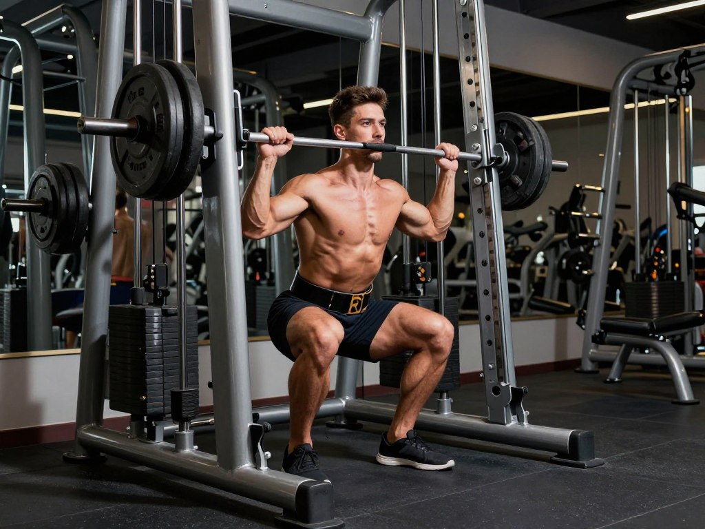
Traditional lower body exercises place significant shear force on the knee joints. Barbell back squats, while excellent for strength development, create substantial anterior knee stress. This becomes problematic when you’re moving weights exceeding double your body weight regularly.
The deeper you squat, the more your knees travel forward over your toes. This forward knee translation increases the moment arm at the joint. Heavy loads combined with this mechanical disadvantage create the perfect storm for knee irritation.
Key Point: Knee-friendly doesn’t mean easy or ineffective. The exercises in this guide allow you to train heavy while redistributing forces away from vulnerable knee structures. You’ll maintain training intensity while giving your knees the break they need to recover.
When to Modify Your Lower Body Training
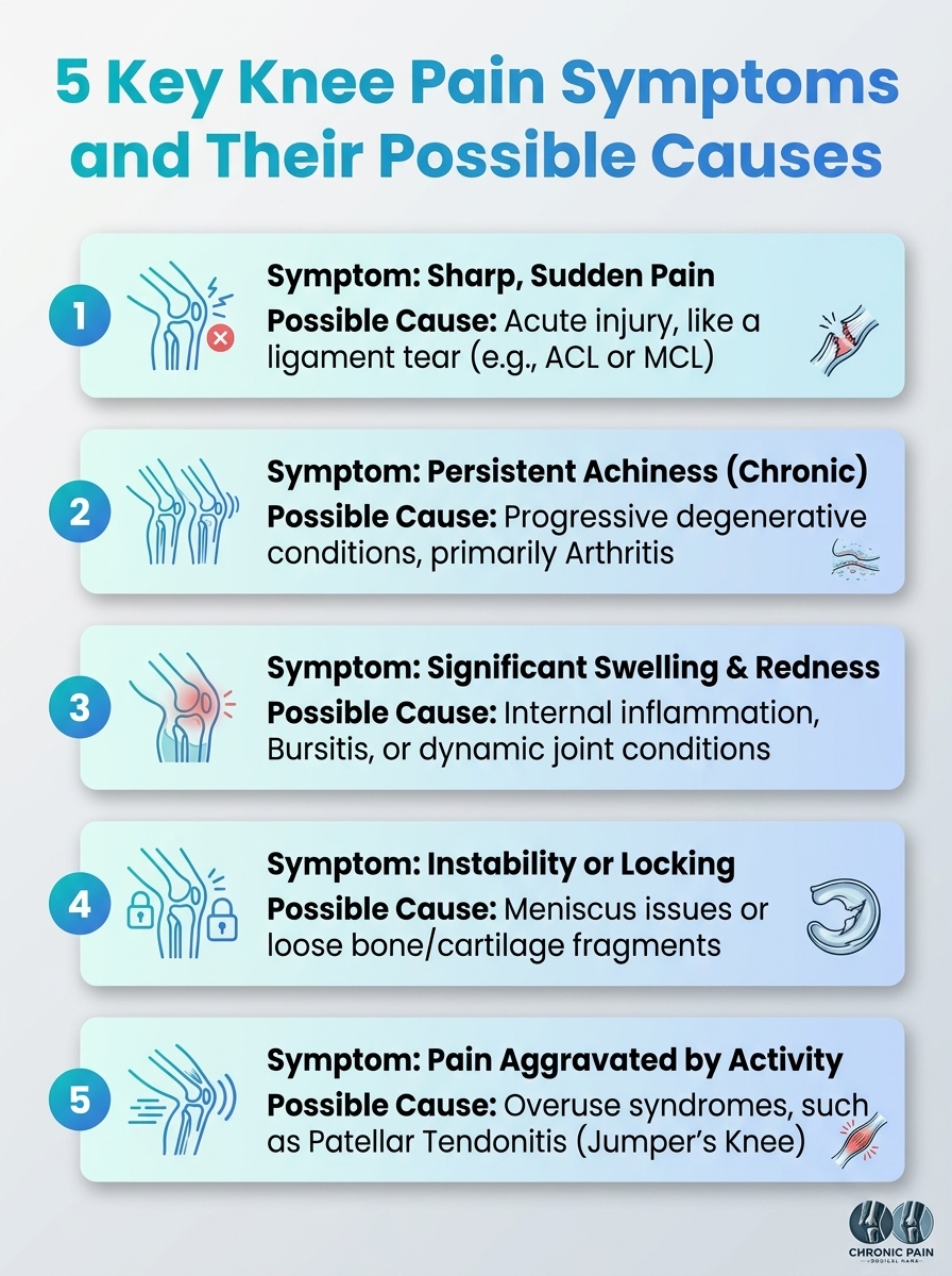
Several signs indicate you need knee-friendly alternatives. Sharp pain during or after leg training signals immediate concern. Swelling around the knee joint suggests inflammatory processes that require attention. Grinding sensations or clicking sounds often indicate tracking problems or cartilage issues.
Morning stiffness lasting more than thirty minutes points to joint inflammation. Difficulty descending stairs reveals eccentric loading problems. Pain that worsens throughout your training session indicates you’re exceeding your knee’s current capacity.
- Anterior knee pain during or after squatting movements
- Swelling that persists beyond normal training soreness
- Limited range of motion compared to your baseline
- Compensatory movement patterns developing in other joints
- Decreased training performance despite adequate recovery
Addressing these symptoms early prevents minor issues from becoming chronic problems. The exercises that follow provide effective alternatives while your knees heal. Many lifters find these movements become permanent fixtures in their training even after knee pain resolves.
Exercise 1: Belt Squat – The Ultimate Knee-Friendly Heavy Movement
The belt squat represents the gold standard for knee-friendly lower body work. This movement allows you to load your glutes and hamstrings heavily while dramatically reducing knee stress. The belt attachment point eliminates axial spine loading while maintaining the squat pattern.
Your torso stays more upright during belt squats compared to barbell variations. This positioning reduces forward knee travel significantly. The result is substantial quad, glute, and hamstring stimulation with minimal patellofemoral compression forces.
How to Perform the Belt Squat Correctly
Position yourself on the belt squat platform with feet shoulder-width apart. Attach the loading belt securely around your hips, positioned just below your iliac crest. Grip the handles lightly for balance but don’t support your body weight through your arms.
Initiate the descent by pushing your hips back slightly while maintaining an upright torso. Keep your chest tall and core braced throughout the movement. Descend until your thighs reach parallel or slightly below while maintaining neutral spine position.
Drive through your entire foot to return to the starting position. Focus on spreading the floor apart with your feet to engage your glutes maximally. Maintain consistent tempo throughout each repetition to maximize muscle tension.
Key Form Cues
- Keep your torso vertical throughout the entire range of motion
- Drive your knees outward to track over your toes
- Maintain even weight distribution across your foot
- Brace your core as if preparing for a punch
- Control the eccentric portion for three seconds
Common Mistakes to Avoid
- Allowing excessive forward knee travel
- Leaning forward excessively with your torso
- Bouncing out of the bottom position
- Supporting body weight through your arms
- Using momentum instead of muscular control
Why Belt Squats Are Knee-Friendly
The loading vector in belt squats differs fundamentally from barbell squats. Weight hangs below your center of mass rather than compressing from above. This changes the force distribution throughout your lower body dramatically.
Your knee joint experiences primarily vertical compression rather than anterior shear force. The reduced forward knee travel decreases the moment arm at your knee joint. These mechanical advantages allow you to train your legs heavily without aggravating knee pain.
Research shows belt squats reduce patellofemoral joint stress by approximately forty percent compared to back squats. Your quadriceps still receive substantial training stimulus. The difference lies in how forces transmit through your knee structure.
Programming Recommendations for Belt Squats
Start with moderate loads to establish proper movement patterns. Use three to four sets of eight to twelve repetitions initially. This rep range builds muscle while allowing your body to adapt to the new movement pattern.
Progress load conservatively by adding five to ten pounds weekly. Your strength on belt squats will increase rapidly as you master the technique. Within four to six weeks, most lifters move impressive loads that rival their back squat numbers.
Advanced Programming: For maximum strength development, work up to sets of three to five reps with heavy loads. Include lighter sets of fifteen to twenty reps weekly to promote blood flow and joint health. This combination builds both strength and muscular endurance while keeping your knees healthy.
Perform belt squats once or twice weekly depending on your overall training volume. Place them early in your leg training when you’re fresh. This exercise can serve as your primary lower body movement or as a valuable assistance exercise.
| Training Phase |
Sets |
Reps |
Load |
Frequency |
| Adaptation (Weeks 1-2) |
3 |
12-15 |
Light to Moderate |
1-2x per week |
| Hypertrophy (Weeks 3-6) |
4 |
8-12 |
Moderate to Heavy |
2x per week |
| Strength (Weeks 7-10) |
5 |
4-6 |
Heavy |
1-2x per week |
| Maintenance |
3-4 |
6-10 |
Moderate Heavy |
1x per week |
Exercise 2: Trap Bar Deadlift – Heavy Pulling Without Knee Stress
The trap bar deadlift offers heavy lifters a powerful pulling variation that minimizes knee stress. This movement allows you to load your posterior chain substantially while maintaining a more knee-friendly position than conventional deadlifts.
Your starting position in trap bar deadlifts places less demand on knee flexion. The neutral grip and centered load distribution create optimal pulling mechanics. This exercise builds tremendous strength in your glutes, hamstrings, and lower back while being gentler on your knees.
Proper Trap Bar Deadlift Execution
Step inside the trap bar with your feet positioned hip to shoulder-width apart. Grip the handles with a neutral grip while keeping your arms fully extended. Your shins should be relatively vertical with minimal forward lean.
Set your back in a neutral position by engaging your lats and bracing your core. Your hips should be positioned between your knees and shoulders, not extremely low or high. This middle position optimizes force production while protecting your knees.
Drive through your entire foot simultaneously while maintaining back tension. Think about pushing the floor away rather than pulling the weight up. Keep the bar path vertical and close to your body throughout the entire range of motion.
Pro Tip: The trap bar deadlift requires less ankle dorsiflexion than conventional deadlifts. This means your knees stay more vertical throughout the movement. The reduced knee travel forward decreases compression forces on your patellofemoral joint significantly.
Why This Movement Protects Your Knees
The trap bar’s design allows you to maintain a more vertical shin angle. Your knees travel forward minimally during the pull. This positioning reduces the moment arm at your knee joint compared to conventional deadlifts.
The centered load distribution eliminates the need to pull the bar around your knees. Conventional deadlifts require your knees to clear backward as the bar passes. This creates shear forces that can aggravate knee pain. The trap bar eliminates this mechanical issue entirely.
Your quadriceps contribute to the movement without bearing excessive load. The emphasis shifts toward your posterior chain muscles. This distribution allows heavy training without overloading knee extensors that may already be irritated.
Programming the Trap Bar Deadlift for Maximum Results
Treat trap bar deadlifts as a primary strength movement. Program them early in your training session when your nervous system is fresh. This allows you to handle maximum loads safely and effectively.
Use sets of three to six repetitions for pure strength development. The trap bar allows most lifters to handle significant weight quickly. Many experienced lifters exceed their conventional deadlift numbers within several weeks of focused trap bar work.
- Begin each training session with two warm-up sets using just the bar and light weight
- Progress to three working sets in your target rep range with appropriate load
- Add one backoff set with eighty percent of your top weight for higher reps
- Increase load by five to ten pounds weekly when you complete all prescribed reps
- Deload every fourth week by reducing volume by thirty to forty percent
Include trap bar deadlifts one to two times weekly depending on your overall deadlift volume. If you’re still performing conventional deadlifts, use trap bar variations as your secondary pulling movement. For lifters with knee concerns, trap bar deadlifts can completely replace conventional variations.
Exercise 3: Pendulum Squat – Controlled Resistance for Knee Safety
The pendulum squat machine provides a fixed movement path that optimizes knee joint positioning. This guided resistance allows you to train your lower body heavily while eliminating instability that might cause pain. The arc-shaped movement pattern mimics natural squatting biomechanics.
Your back remains supported throughout the entire range of motion on a pendulum squat. This support removes axial loading from your spine while allowing focused leg work. The shoulder pads distribute forces evenly without creating compression through your knee joints.
Executing the Pendulum Squat
Position yourself on the machine with your back flat against the pad. Place your feet on the platform shoulder-width apart with toes pointed slightly outward. Your foot position should feel natural and allow full depth without heel lifting.
Release the safety mechanism and control your descent along the machine’s path. The pendulum arc naturally guides your knees through an optimal trajectory. Descend until your thighs reach parallel or slightly below while maintaining complete back contact with the pad.
Drive powerfully through your midfoot and heel to return to the starting position. Focus on feeling your glutes and hamstrings engage throughout the movement. The machine’s design ensures consistent tension on your muscles without placing excessive stress on your knee structures.
Joint-Friendly Benefits of Pendulum Squats
The fixed path of the pendulum squat removes instability variables. Your body doesn’t need to balance the load or control multiple planes of motion. This allows you to focus purely on generating force without worrying about knee stability issues that might cause pain.
The arc pattern of pendulum squats creates a natural movement flow. Your knees track through space along an optimal path determined by the machine’s engineering. This consistency reduces aberrant forces that might irritate your knee joints during free-weight movements.
Most pendulum squat machines position your torso at a slight angle. This positioning reduces forward knee translation compared to vertical squatting. Your knees stay more aligned over your feet throughout the movement, decreasing anterior knee stress significantly.
Sets, Reps, and Progression Guidelines
Use pendulum squats as either a primary or secondary lower body movement. The machine’s safety allows you to push close to failure without spotters. This makes it ideal for hypertrophy-focused training with moderate to high repetitions.
Start with three sets of ten to fifteen repetitions to establish your baseline capacity. Focus on smooth tempo and full range of motion rather than maximum load initially. As your movement quality improves, gradually increase resistance while maintaining perfect form.
Hypertrophy Protocol
- Four sets of twelve to fifteen reps
- Tempo: three seconds down, one second pause, one second up
- Rest ninety seconds between sets
- Add weight when you complete all reps with perfect form
Strength Protocol
- Five sets of six to eight reps
- Tempo: two seconds down, explosive up
- Rest two to three minutes between sets
- Increase load by five percent weekly
Advanced lifters can use pendulum squats for brutal finisher sets. After your primary strength work, load the machine with moderate weight and perform one to two sets of twenty to thirty repetitions. These high-rep sets create massive metabolic stress while keeping your knees safe due to the controlled movement pattern.
Get Your Personalized Knee-Safe Training Plan
Work with experienced strength coaches who specialize in heavy lifting programming for athletes with knee concerns. We’ll design a complete training system tailored to your specific goals, experience level, and joint health needs.
4.9
Based on 200+ lifters
Program Effectiveness
4.8
What You’ll Receive:
- Comprehensive movement assessment and program design
- Weekly check-ins with load and volume adjustments
- Video form analysis with personalized corrections
- Exercise library with knee-friendly alternatives
- Nutrition guidance for recovery and performance
- Direct coach access via messaging platform
Exercise 4: Spanish Squat – Therapeutic Strength Building
The Spanish squat combines therapeutic benefits with legitimate strength training stimulus. This movement originated in rehabilitation settings but has gained popularity among heavy lifters seeking knee relief. The band setup creates a posterior force that off-loads your knee joints while still challenging your quadriceps.
Your knees receive decompression during Spanish squats due to the band tension. The backward pull reduces pressure on your patellofemoral joint surfaces. This allows you to train your leg muscles intensely while actually helping your knees feel better rather than worse.
Setting Up and Performing Spanish Squats
Loop a heavy resistance band around a sturdy anchor point at knee height. Step into the band and position it in the crease behind your knees. Walk forward until the band creates substantial tension pulling your knees backward.
Stand with your back against a wall for additional support. Your feet should be positioned twelve to eighteen inches from the wall. The band tension should be significant enough that you feel it pulling your knees backward even in the standing position.
Descend into a squat while maintaining contact between your back and the wall. The band pulls your knees backward as you squat down, creating a shearing force opposite to normal squatting. This reverse shear actually benefits your knee joints by decompressing the joint surfaces.
Hold the bottom position for three to five seconds on each repetition. This isometric component builds tremendous quad strength while the band continues decompressing your knees. Control your ascent back to the starting position while maintaining band tension throughout.
The Unique Benefits for Knee Health
Spanish squats create what physical therapists call posterior tibial translation. The band pulls your shin bone backward relative to your thigh bone. This creates space within your knee joint that reduces compression on irritated structures.
The wall support allows you to focus purely on your quadriceps without balance concerns. You can push to muscular failure safely since you’re supported and can simply step out of the band when finished. This makes Spanish squats ideal for high-rep finisher work.
Many lifters report immediate knee pain reduction after Spanish squat sessions. The decompression effect can provide temporary relief that makes your subsequent training more comfortable. Regular use often leads to long-term improvements in knee health.
Programming Spanish Squats for Strength and Recovery
Use Spanish squats as an accessory movement or as part of your warm-up routine. Perform three to four sets of fifteen to twenty repetitions with longer hold times. The goal is muscular fatigue and joint decompression rather than maximum load.
Include Spanish squats two to three times weekly on lower body training days. Perform them either before your main lifts as activation work or after as a therapeutic finisher. Both approaches provide benefits depending on your specific needs.
Recovery Application: On active recovery days, perform five sets of twenty reps with sixty seconds rest. Use moderate band tension and focus on the decompression sensation. This protocol promotes blood flow and joint health without creating excessive training stress.
Progress Spanish squats by increasing hold time rather than resistance. Work up to ten-second holds in the bottom position. You can also add light dumbbells held at your sides once bodyweight becomes too easy. The band decompression remains effective even with additional load.
Exercise 5: Reverse Sled Drag – Zero Impact Lower Body Power
Reverse sled drags build incredible lower body strength without any eccentric loading. This unique characteristic makes sleds ideal for knee-friendly training. Your joints experience no impact forces or rapid deceleration that typically aggravate knee pain.
The continuous tension of sled work challenges your muscles differently than traditional resistance training. There’s no resting point during the movement. Your glutes, hamstrings, and quadriceps work constantly to move the load, creating substantial training stimulus without joint stress.
Executing Reverse Sled Drags Properly
Load the sled with appropriate weight based on your strength level and training surface. Grass and turf create more resistance than smooth concrete. Start conservatively and adjust as you learn how different surfaces affect difficulty.
Grab the sled straps or handles and walk backward while maintaining an athletic posture. Keep your torso upright with a slight forward lean. Your arms should be relatively straight, allowing your lower body to generate all the pulling force.
Take controlled steps backward while driving through your entire foot. Keep your knees tracking in line with your toes throughout each step. Maintain consistent tension on the sled rather than allowing jerky movements that might stress your joints.
Cover distances of twenty to fifty yards per set depending on available space and training goals. The key is maintaining good posture and consistent speed throughout each drag. If you start leaning back excessively or taking choppy steps, reduce the load.
Why Sled Work Protects Your Knees
The absence of eccentric muscle actions eliminates a primary source of knee stress. Traditional exercises require your muscles to lengthen under load. This eccentric component creates muscle damage and joint inflammation. Sled work involves only concentric actions, dramatically reducing tissue trauma.
Your knee joints never experience impact forces during sled drags. Each step involves smooth transitions without rapid deceleration. This consistent, low-impact nature allows you to accumulate high training volumes without aggravating irritated knee structures.
The backward motion emphasizes your posterior chain while reducing quad dominance. Your glutes and hamstrings bear the primary workload. This force distribution takes pressure off knee extensors that might already be overworked from other exercises.
Programming Sled Drags for Maximum Benefit
Use sled drags as either primary strength work or conditioning finishers. For strength development, load the sled heavy and perform shorter distances of fifteen to twenty-five yards. Rest three to four minutes between sets to allow full recovery.
For conditioning and metabolic work, use moderate loads for longer distances of fifty to one hundred yards. Reduce rest periods to ninety seconds or less. This approach builds work capacity while maintaining knee-friendly loading patterns.
| Training Goal |
Load |
Distance |
Sets |
Rest |
| Maximum Strength |
Very Heavy |
15-20 yards |
6-8 |
3-4 minutes |
| Hypertrophy |
Heavy |
25-40 yards |
4-6 |
2-3 minutes |
| Conditioning |
Moderate |
50-100 yards |
4-8 |
60-90 seconds |
| Active Recovery |
Light |
100+ yards |
2-4 |
60 seconds |
Include sled work two to four times weekly depending on your overall training volume. Sled drags recover faster than traditional leg exercises due to the lack of eccentric damage. This allows higher frequency training without excessive fatigue accumulation.
Exercise 6: Safety Bar Box Squat – Controlled Depth with Reduced Knee Stress
The safety bar box squat combination creates one of the most knee-friendly loaded squatting variations available. The safety bar’s cambered design shifts the load’s center of mass. This allows you to maintain a more upright torso position, reducing forward knee travel significantly.
Adding a box provides a depth gauge and teaches proper hip loading mechanics. You learn to sit back into your hips rather than driving your knees forward. This movement pattern protects your knees while building tremendous posterior chain strength.
Setting Up Your Safety Bar Box Squat
Position a box or bench at a height that places your thighs parallel to the ground when seated. The exact height depends on your limb proportions and mobility. Start slightly higher if you have knee issues, lowering the box as your tolerance improves.
Load the safety bar and position it across your upper back. The cambered arms rest against your torso while the pad sits on your traps. This weight distribution eliminates the need to hold the bar with your hands, reducing upper body tension.
Set up with the box directly behind you. Your stance should be slightly wider than normal squatting, typically outside shoulder-width. This wider stance allows better hip engagement and reduces knee stress further.
Initiate the descent by pushing your hips back toward the box. Your knees should track over your toes but travel forward minimally. Think about sitting back onto the box rather than squatting straight down. This hip-dominant pattern protects your knee joints.
The Box Touch Technique
As your glutes contact the box, maintain muscle tension throughout your body. Don’t fully relax or collapse onto the box. Think of the box as a depth gauge rather than a resting place. Your muscles should remain engaged even at the bottom position.
Pause briefly on the box without rocking or using momentum. This eliminates the stretch reflex and forces your muscles to generate force from a dead stop. The pause also gives you time to reset your position if needed between repetitions.
Drive explosively off the box by pushing through your entire foot simultaneously. Think about spreading the floor apart with your feet to maximize glute activation. Maintain your torso angle as you rise, preventing excessive forward lean that might stress your knees.
Why This Combination Works
The safety bar’s forward weight distribution encourages an upright torso position automatically. You don’t need to fight to stay vertical like with a straight barbell. This natural positioning reduces the moment arm at your knee joints substantially.
The box provides instant feedback on your depth consistency. You know exactly when you’ve reached your target depth on every repetition. This consistency helps you avoid going too deep, which might cause knee pain, while ensuring adequate range of motion for muscle development.
The pause on the box eliminates momentum and eccentric loading transitions. Your knee joints don’t experience rapid direction changes that often cause pain. Each repetition starts from a controlled position, reducing impact forces on your joint structures.
Loading Parameters and Progression
Safety bar box squats respond well to various loading schemes. Use lower reps of three to five for maximum strength development. The safety bar typically handles ten to fifteen percent less load than straight bar squats, but this difference disappears with practice.
Include one to two box squat sessions weekly as either primary or secondary lower body work. Alternate box heights every few weeks to train different ranges of motion. Higher boxes emphasize your posterior chain while protecting your knees maximally. Lower boxes increase quad involvement as your knee tolerance improves.
Advanced Technique: Perform accommodating resistance box squats using bands or chains. This method loads the top portion of the movement more heavily while reducing load at the bottom where your knees are most vulnerable. Bands or chains represent ten to thirty percent of the total load at the top position.
- Weeks 1-3: Focus on technique with moderate loads, five sets of five reps
- Weeks 4-6: Increase intensity to eighty-five percent of max, four sets of three reps
- Weeks 7-9: Include heavy triples and doubles, working up to ninety percent
- Week 10: Deload with reduced volume and intensity
- Weeks 11-12: Test new maximums or begin new progression cycle
Exercise 7: Lying Leg Curl – Isolated Hamstring Development
Direct hamstring work becomes essential when knee issues limit compound movements. Lying leg curls provide targeted hamstring training without requiring knee flexion under body weight load. This isolation allows you to maintain hamstring strength and size despite knee limitations.
Strong hamstrings support knee joint health by balancing forces across the joint. When your quads overpower your hamstrings, knee tracking problems often develop. Regular hamstring training helps maintain this critical balance for long-term joint health.
Proper Lying Leg Curl Form
Position yourself face-down on the leg curl machine with the pad resting just above your heels. Your knees should align with the machine’s axis of rotation. This alignment ensures proper force transmission and prevents additional knee stress.
Grip the handles firmly and brace your core to prevent your hips from lifting during the movement. Your hip flexors want to assist the curl by raising your pelvis. Prevent this compensation by maintaining constant contact between your hips and the bench.
Curl the weight by contracting your hamstrings until the pad nearly touches your glutes. Squeeze your hamstrings hard in the fully contracted position for one to two seconds. This peak contraction maximizes muscle fiber recruitment and development.
Lower the weight under control using a three to four second tempo. The eccentric phase builds strength and size effectively. Avoid letting the weight drop rapidly, which can actually increase knee strain despite the isolated nature of the exercise.
Why Leg Curls Support Knee Health
Lying leg curls strengthen your hamstrings through their knee flexion function specifically. This builds muscle that directly supports and stabilizes your knee joints. Strong hamstrings reduce strain on your knee ligaments during daily activities and other training movements.
The lying position minimizes hip involvement compared to seated or standing curl variations. This isolation ensures your hamstrings receive maximum training stimulus. When your hamstrings gain strength, they help decelerate knee extension during activities that might otherwise cause pain.
Regular hamstring training can reduce anterior knee pain over time. The improved muscle balance around your knee joint enhances tracking and reduces aberrant forces on your patella. This makes leg curls therapeutic in addition to being muscle-building exercises.
Sets, Reps, and Training Variables
Use moderate to high repetitions for hamstring curls. Sets of ten to twenty reps work extremely well for this exercise. Higher reps increase time under tension without requiring loads that might compromise form or create joint stress.
Perform leg curls two to three times weekly on your lower body training days. Place them after your primary strength movements when your nervous system is fatigued but your hamstrings are fresh. This timing prevents hamstring fatigue from limiting your performance on compound lifts.
Standard Protocol
Build hamstring mass and strength with this proven approach that emphasizes controlled movement and progressive overload.
- Three to four sets of twelve to fifteen reps
- Four-second eccentric, two-second peak contraction
- Sixty to ninety seconds rest between sets
- Increase weight when all reps completed cleanly
Advanced Protocol
Push your hamstring development further with intensity techniques designed for experienced lifters seeking maximum growth.
- Four sets of eight to twelve reps plus drop sets
- Add partial reps at failure for extended sets
- Ninety-second rest periods
- Include single-leg variations for balance
Vary your foot position to target different hamstring regions. Toes pointed emphasizes the outer hamstrings while toes flexed engages the inner hamstrings more. Neutral foot position provides balanced development across all hamstring muscles.
Recommended Equipment for Knee-Friendly Training
Having the right equipment enhances your ability to train effectively while protecting your knees. These tools represent smart investments for serious lifters dealing with knee concerns. Each item addresses specific limitations and expands your exercise options considerably.
Heavy Resistance Bands
Essential for Spanish squats and decompression work. Choose bands offering fifty to one hundred pounds of resistance at full stretch. Multiple resistance levels allow progression as your strength improves.
- Enables therapeutic knee decompression exercises
- Provides accommodating resistance for various movements
- Portable for training anywhere
- Durable construction for heavy use
Editor’s Choice
Quality Dip Belt
Critical for belt squats if you lack dedicated equipment. Look for reinforced leather or nylon construction with chain length adjustment. Proper belt distributes load across your hips comfortably even with heavy weights.
- Transforms any elevated platform into belt squat station
- Reinforced materials handle hundreds of pounds safely
- Adjustable chain accommodates different heights
- Comfortable padding prevents hip bruising
Most Versatile
Compression Knee Sleeves
Provide warmth and proprioceptive feedback without restricting movement. Seven millimeter neoprene offers optimal support for training. Sleeves reduce pain during workouts for many lifters with minor knee issues.
- Increases blood flow and warmth to knee joints
- Provides compression without limiting range of motion
- Improves proprioception and movement awareness
- Reduces minor discomfort during training sessions
Best Support
Investment Priority: Start with resistance bands as they provide the most versatility for knee-friendly training modifications. Add a quality dip belt next if you have access to elevated platforms. Consider knee sleeves if you experience discomfort despite using proper exercises and form.
Exercise 8: Rear Foot Elevated Split Squat – Unilateral Strength Without Knee Stress
The Bulgarian split squat allows heavy loading while naturally limiting harmful knee translation. Your elevated rear foot creates a split stance that emphasizes hip flexion over knee flexion. This positioning protects your knee joints while building unilateral leg strength effectively.
Unilateral training addresses strength imbalances that often contribute to knee pain. Most lifters have a dominant leg that compensates during bilateral movements. Split squats force each leg to handle loads independently, correcting these imbalances over time.
Setting Up Bulgarian Split Squats
Position a bench or box twelve to thirty-six inches behind you. The exact distance depends on your leg length and hip flexibility. Start closer and adjust as needed to find your optimal stance width.
Place the top of your rear foot on the bench with your knee bent. Your front foot should be far enough forward that your shin stays relatively vertical at the bottom position. This prevents excessive forward knee travel that might cause pain.
Hold dumbbells at your sides or position a barbell across your upper back. Dumbbells often work better initially as they allow easier balance and don’t load your spine. Progress to barbell variations once you’ve mastered the movement pattern.
Descend by bending your front knee while allowing your hips to drop straight down. Your torso should remain relatively upright throughout the movement. Think about dropping your rear knee toward the ground rather than pushing your front knee forward.
Achieving Knee-Friendly Mechanics
The split stance of Bulgarian split squats naturally encourages proper weight distribution. Your front leg bears approximately seventy percent of the load while your rear leg provides stability. This distribution allows heavy training without overloading either knee joint excessively.
Your front knee tracks forward minimally during properly executed split squats. The split stance creates a longer moment arm at your hip joint, shifting emphasis to your glutes and away from your knee extensors. This mechanical advantage protects your knees while still challenging your leg muscles intensely.
The elevated rear foot increases your front leg’s range of motion without requiring extreme ankle mobility. You achieve deep hip flexion without your front knee traveling far past your toes. This ROM provides excellent muscle-building stimulus with minimal joint stress.
Programming Bulgarian Split Squats
Treat split squats as a primary lower body exercise or as valuable accessory work. Perform three to four sets of eight to twelve repetitions per leg. The unilateral nature means each set takes longer than bilateral exercises, so plan your rest periods accordingly.
Rest ninety seconds to two minutes between legs rather than between full sets. This approach maintains workout efficiency while allowing adequate recovery. Your non-working leg rests while you train the opposite side.
Benefits of Split Squats
- Corrects strength imbalances between legs
- Requires less absolute load than bilateral squats
- Improves balance and coordination significantly
- Allows training around minor injuries effectively
- Builds functional single-leg strength
- Easier to maintain upright torso position
Considerations
- Takes longer to complete than bilateral exercises
- Requires more balance and coordination initially
- Can’t load as heavily as bilateral movements
- Hip flexibility limitations may restrict depth
- Bench height adjustment needed for comfort
- Learning curve for proper weight distribution
Progress load gradually by adding five to ten pounds when you complete all prescribed repetitions with perfect form. Most lifters can eventually use surprisingly heavy loads on split squats. Dumbbells exceeding one hundred pounds per hand are achievable goals for strong lifters.
Exercise 9: Leg Press – Heavy Loading with Adjustable Knee Stress
The leg press provides adjustable knee stress through foot placement variations. High foot positions reduce knee flexion angles while still allowing heavy loading. This versatility makes leg presses valuable tools for lifters with varying degrees of knee sensitivity.
Leg presses support your back completely, eliminating axial loading concerns. You can push to muscular failure safely without spotters. This safety allows aggressive training that builds muscle and strength despite knee limitations.
Optimizing Leg Press Foot Placement
Place your feet high on the platform with heels near the top edge. This positioning emphasizes your glutes and hamstrings while reducing quad dominance. Higher foot placement also decreases knee flexion angle at the bottom of each repetition.
Use a shoulder-width or slightly wider stance for most pressing. Wider stances allow greater glute activation and often feel more comfortable on sensitive knees. Experiment with stance width to find your optimal position for strength and comfort.
Keep your feet flat against the platform throughout each repetition. Allowing your heels to lift increases stress on your knee joints unnecessarily. Full foot contact distributes forces optimally across your lower body musculature.
Lower the platform until your knees reach approximately ninety degrees of flexion. Going deeper increases knee stress substantially with diminishing returns for muscle development. A ninety-degree knee angle provides excellent muscle stimulation while protecting your joints.
Execution Guidelines for Maximum Safety
Grip the handles firmly and keep your lower back pressed against the pad throughout the movement. Many lifters allow their lower back to round at deep depths. This compromises spinal safety and often increases knee discomfort.
Push through your entire foot simultaneously, not just your toes. Think about driving through your heels and midfoot together. This cueing pattern ensures proper force distribution and maximum glute and hamstring engagement.
Control the eccentric portion rather than letting the weight drop. Use a two to three second lowering tempo on each repetition. Controlled eccentrics build strength while reducing impact forces that might aggravate your knees.
Avoid locking out completely at the top of each press. Maintaining slight knee flexion keeps constant tension on your muscles while reducing joint stress. Stop just short of full lockout on every repetition for optimal results.
Loading Strategies and Progression
Leg presses allow extremely heavy loading due to the favorable mechanics and machine support. Many lifters can press several times their squat weight. This capacity makes leg presses excellent for overload training that builds serious leg mass.
Use various rep ranges to maximize development. Include heavy sets of six to eight reps for strength alongside moderate weight sets of twelve to fifteen reps for hypertrophy. Add occasional high-rep sets of twenty-five to fifty reps as metabolic finishers.
| Training Focus |
Foot Position |
Depth |
Sets x Reps |
Tempo |
| Glute Emphasis |
High and Wide |
90 degrees |
4 x 10-12 |
3-1-1 |
| Quad Emphasis |
Middle Position |
90-100 degrees |
4 x 8-10 |
3-0-1 |
| Maximum Strength |
High Position |
90 degrees |
5 x 5-6 |
3-1-2 |
| Metabolic Stress |
High and Wide |
90 degrees |
2-3 x 20-30 |
2-0-1 |
Include leg press work one to two times weekly depending on your total leg training volume. Many lifters use leg presses as their primary knee-friendly exercise while their knees recover. Others include them as supplemental work alongside other movements from this guide.
Exercise 10: Step-Ups – Functional Strength with Minimal Impact
Step-ups build unilateral leg strength through a natural movement pattern. This exercise mimics stair climbing and daily activities more closely than squats or deadlifts. The functional nature makes step-ups valuable for overall leg development and injury prevention.
Proper step-up execution minimizes knee stress through controlled movement tempo. Unlike jumping or running, step-ups involve no impact forces. The smooth transition from ground to box allows heavy loading without joint trauma.
Executing Step-Ups with Proper Form
Select a box height that positions your thigh parallel to the ground when your foot is planted. This height provides adequate range of motion without requiring extreme knee flexion. Adjust height based on your mobility and comfort level.
Place your entire foot flat on the box, not just your toes. Ball-of-foot placement increases knee stress unnecessarily. Full foot contact allows you to drive through your heel and midfoot for optimal force production.
Hold dumbbells at your sides or position a barbell across your upper back for added resistance. Dumbbells work well initially as they don’t affect your balance as much as barbells. Progress to barbell step-ups as your strength and coordination improve.
Step up by driving through your planted foot without pushing off with your ground foot. The working leg should do virtually all the lifting. Using your rear leg for assistance defeats the unilateral training purpose and reduces effectiveness.
The Knee-Friendly Nature of Step-Ups
Step-ups allow you to control knee flexion angle through box height selection. Lower boxes reduce knee stress while still building strength. As your knees feel better, gradually increase box height to challenge your legs more intensely.
The single-leg nature reduces absolute load requirements compared to bilateral exercises. Less weight needed means less force transmitted through your knee joints. You still achieve excellent muscle stimulation due to the unilateral challenge.
Step-ups emphasize concentric muscle actions with minimal eccentric stress. Stepping down slowly still involves some eccentric work, but far less than traditional squatting movements. This reduced eccentric component decreases muscle damage and joint inflammation.
Programming Parameters for Step-Ups
Perform three to four sets of eight to twelve repetitions per leg. Alternate legs each rep or complete all reps on one side before switching. Both approaches work effectively depending on your goals and fatigue management preferences.
Include step-ups one to two times weekly as either primary or accessory lower body work. Many lifters pair step-ups with bilateral exercises for comprehensive leg development. The combination provides balanced training across different movement patterns.
Progressive Overload: Increase difficulty through multiple variables beyond just adding weight. Increase box height by two inches, slow your tempo to five seconds per rep, or add a pause at the top position. These progressions challenge your muscles while maintaining knee-friendly mechanics.
- Master bodyweight step-ups with perfect form before adding external load
- Add light dumbbells starting with ten to fifteen pounds per hand
- Progress weight by five-pound increments when form remains perfect
- Incorporate tempo variations to increase difficulty without adding load
- Eventually progress to barbell step-ups for maximum loading capacity
- Include box height variations to train different ranges of motion
Use step-ups during deload weeks or active recovery periods. The lower impact nature allows leg training without excessive systemic stress. This makes step-ups ideal for maintaining training frequency while managing overall fatigue.
Transitioning from Knee-Stressful to Knee-Friendly Movements
Changing your exercise selection requires strategic planning rather than abrupt switches. Your muscles and nervous system need time to adapt to new movement patterns. A gradual transition maintains your training progress while giving your knees time to recover.
Phase One: Adding Knee-Friendly Variations (Weeks 1-3)
Begin incorporating knee-friendly exercises alongside your current training. Don’t eliminate problematic movements immediately unless pain is severe. This approach allows your body to learn new patterns without losing strength on familiar exercises.
Start with one or two knee-friendly exercises per session. Place them after your traditional movements initially. Use moderate loads and focus entirely on mastering proper technique rather than setting personal records.
Monitor how your knees respond to each new exercise. Some movements will feel immediately better while others might take several sessions to feel comfortable. This information guides which exercises to emphasize moving forward.
Phase Two: Gradual Exercise Substitution (Weeks 4-8)
Begin replacing problematic exercises with knee-friendly alternatives. Make one substitution every two weeks to allow proper adaptation. This gradual approach prevents sudden strength losses that can occur with too many simultaneous changes.
Increase volume and intensity on knee-friendly exercises as you reduce traditional movements. Your goal is maintaining similar total training stress through different exercise selection. Track your performance to ensure you’re not losing ground during the transition.
Expect some initial strength decreases on new exercises. Your body needs time to develop coordination and recruitment patterns. Within four to six weeks, most lifters match or exceed their previous training loads using knee-friendly alternatives.
Example Transition: Week 1-2: Add belt squats after back squats. Week 3-4: Reduce back squat volume by thirty percent, increase belt squat volume. Week 5-6: Eliminate back squats, make belt squats your primary movement. Week 7-8: Add secondary knee-friendly movement like safety bar box squats.
Phase Three: Optimization and Fine-Tuning (Weeks 9-12)
Fully commit to your knee-friendly exercise selection by this phase. Remove all problematic movements unless your knees have improved significantly. Focus on progressive overload within your new exercise framework.
Experiment with different loading parameters and training frequencies. Your optimal approach might differ from your previous training due to the new exercises’ characteristics. Some movements respond better to higher frequency while others need more recovery time.
Reassess your knee status monthly during this phase. Many lifters find their knee pain reduces substantially after several months of modified training. At this point, you might slowly reintroduce some traditional exercises if desired, though many choose to permanently adopt knee-friendly variations.
Long-Term Exercise Rotation Strategy
Develop a library of knee-friendly exercises rather than relying on just one or two movements. Rotating exercises every four to eight weeks prevents pattern overload and maintains training stimulus. This variety also keeps training mentally engaging.
Include at least three knee-friendly options for quad development, hamstrings, and glutes. This gives you nine total exercises to rotate through. Having multiple options prevents your progress from stalling if an exercise stops working or becomes unavailable.
- Primary quad exercises: Belt squats, pendulum squats, leg press variations
- Primary posterior chain: Trap bar deadlifts, reverse sled drags, Romanian deadlifts
- Supplemental unilateral: Bulgarian split squats, step-ups, single-leg press
- Hamstring isolation: Lying leg curls, Nordic curls, slider curls
- Glute emphasis: Hip thrusts, back extensions, cable pull-throughs
Track your performance across all exercises to identify your most effective movements. Some exercises might build strength better while others excel for muscle growth. Understanding these differences allows intelligent programming that maximizes your results within knee-friendly parameters.
Complete 4-Week Knee-Friendly Training Program
This complete program integrates all the exercises covered into a progressive training plan. The program emphasizes strength development while protecting your knee joints. Follow the structure exactly for four weeks before making modifications based on your individual response.
Program Overview and Training Split
You’ll train lower body twice weekly with at least two days between sessions. This frequency allows adequate recovery while providing enough stimulus for continued progress. Each session includes different exercise selections to ensure balanced development.
Session A emphasizes hip-dominant movements and posterior chain development. Session B focuses more on quad development with machine-based exercises. This split ensures comprehensive lower body training without overloading any single movement pattern.
Each workout should take sixty to ninety minutes including warm-up. Don’t rush through sessions trying to finish faster. Quality execution matters more than training duration when protecting your knees.
Week 1-2: Foundation Phase
Lower Body Session A:
| Exercise |
Sets |
Reps |
Rest |
Notes |
| Trap Bar Deadlift |
4 |
8 |
3 min |
Focus on form, moderate weight |
| Bulgarian Split Squat |
3 |
10 each |
90 sec |
Master balance before adding weight |
| Lying Leg Curl |
3 |
12 |
60 sec |
Control eccentric phase |
| Spanish Squat |
3 |
15 |
60 sec |
Five-second holds at bottom |
| Reverse Sled Drag |
4 |
30 yards |
2 min |
Light load, focus on movement quality |
Lower Body Session B:
| Exercise |
Sets |
Reps |
Rest |
Notes |
| Belt Squat |
4 |
10 |
2 min |
Learn movement pattern |
| Leg Press (high foot) |
3 |
12 |
90 sec |
Stop at ninety-degree knee flexion |
| Step-Ups |
3 |
10 each |
75 sec |
Moderate box height |
| Pendulum Squat |
3 |
12 |
90 sec |
Full range of motion |
| Lying Leg Curl |
3 |
15 |
60 sec |
Peak contraction emphasis |
Week 3-4: Progressive Loading Phase
Increase weights by five to ten percent across all exercises while maintaining prescribed rep ranges. Your focus shifts from learning movements to progressive overload. Form should remain perfect even as loads increase.
Session structures remain identical to weeks one and two with adjusted loading. Add one additional set to your primary movements (first exercise each session). This volume increase drives continued adaptation.
Important: If knee pain increases during any exercise, immediately reduce load or substitute a different movement. Don’t push through worsening pain. Some days your knees will feel worse than others. Having flexibility in your exercise selection allows productive training regardless of how your knees feel.
Warm-Up Protocol for Every Session
Proper warm-up becomes essential when training with knee concerns. Your warm-up should prepare your joints and nervous system for heavy work. Never skip warm-ups trying to save time.
- Five minutes light cardio on bike or rower (avoid treadmill running)
- Hip mobility circuit: leg swings, hip circles, 90-90 stretches (two sets each)
- Glute activation: clamshells and glute bridges (two sets of fifteen reps)
- Movement-specific warm-ups using empty bar or light weight (three progressive sets)
- One ramping set at seventy percent of working weight
This warm-up takes fifteen to twenty minutes but dramatically improves your joint preparedness. Many knee issues stem from inadequate warm-ups that leave joints unprepared for heavy loads.
Download Your Free Complete Training Program
Get the full twelve-week progression, exercise video library, and personalized load calculators. Everything you need to build strong legs while protecting your knees.
- Detailed exercise video demonstrations
- Progressive loading calculators
- Exercise substitution flowcharts
- Mobility and recovery protocols
- Direct email support included
Work With a Specialized Coach
Get personalized programming from coaches who understand heavy lifting and joint health. Perfect for serious lifters who want customized solutions.
- One-on-one program design consultation
- Ongoing form checks and adjustments
- Custom exercise selection for your needs
- Flexible monthly or quarterly packages
Moving Forward With Your Knee-Friendly Training
Your knee discomfort doesn’t have to end your strength training career. The exercises and strategies presented here allow you to continue building impressive lower body strength and size. Many lifters actually become stronger after transitioning to knee-friendly movements due to reduced joint limitations.
Consistency matters more than perfection when managing knee issues. Some training sessions will feel better than others. Having multiple exercise options allows you to train productively regardless of how your knees feel on any particular day.
Remember that pain serves as your body’s feedback mechanism. Sharp pain signals you should stop or modify immediately. Manageable discomfort that doesn’t worsen might be acceptable depending on your situation. Learning to distinguish between productive training stress and harmful pain represents a critical skill for long-term success.
Key Principles to Remember
Progressive overload remains essential even with modified exercise selection. Continue pushing for small improvements in weight, reps, or training density. Your muscles don’t know whether you’re squatting or using a belt squat machine. They respond to progressive tension regardless of exercise choice.
Recovery takes priority over training volume when managing joint issues. More training isn’t always better. Focus on quality sessions with adequate rest between workouts. Your knees heal during recovery periods, not during training sessions.
Be patient with your progress and your body’s healing timeline. Significant improvements often take several months of consistent modified training. Trust the process and avoid rushing back to movements that caused your initial problems.
“The strongest lifters aren’t those who never face setbacks. They’re the ones who adapt intelligently when challenges arise. Your willingness to modify your training shows wisdom, not weakness.”
— Strength Coach Wisdom
Your journey toward pain-free training starts with implementing even one exercise from this guide. Don’t feel overwhelmed by trying to change everything immediately. Small, consistent modifications compound into dramatic improvements over time.
The exercises and program structures provided here have helped countless heavy lifters continue progressing despite knee concerns. Your situation is manageable with smart training choices. Take action today by downloading the complete program and beginning your transition to knee-friendly lower body workouts for heavy lifters.